Received: September 16, 2008
Accepted: October 6, 2008
Abstract published online: October 31, 2008
Full paper published online: February 28, 2009
J Venom Anim Toxins incl Trop Dis. V.15, n.1, p. 146-156, 2009. Short communication. ISSN 1678-9199.
MORPHOLOGICAL CHARACTERIZATION OF THE VENOM APPARATUS IN
THE WOLF SPIDER Lycosa singoriensis (LAXMANN, 1770)
Yigit N (1), Bayram A (1), Danisman T (1), Sancak Z (2), Tel MG (3)
(1) Department of Biology, Faculty of Science and Arts, University of Kirikkale,
Kirikkale, Turkey; (2) Department of Biology, School of Natural and Applied Sciences,
University of Kirikkale, Kirikkale, Turkey; (3) Suleyman Demirel Gymnasium,
Directorate of National Education, Kirikkale, Turkey.
ABSTRACT: The wolf spider Lycosa singoriensis (Laxmann, 1770) (Lycosidae:
Araneae) is distributed throughout central and eastern Europe, including Russia,
Kazakhistan and Turkey. This study describes the venom apparatus morphology of
L. singoriensis through scanning electron microscopy (SEM). Its structure follows the
general architecture observed in other spiders. Generally, a venom apparatus is
composed by a pair of venom glands and chelicerae. L. singoriensis chelicerae are
robust and consist of a stout basis and a movable apical segment (fang). The fang
rests in a groove on the basal segment that is covered by different types of hair. L.
singoriensis venom glands present equal size and measure about 4 mm in length.
Each gland is enclosed by irregular muscular layers.
KEY WORDS: spider, Lycosa singoriensis, chelicerae, venom gland, morphology.
CONFLICTS OF INTEREST: There is no conflict.
CORRESPONDENCE TO:
NAZIFE YIGIT, Department of Biology, Faculty of Science and Arts, University of
Kirikkale, 71450 Kirikkale, Turkey. Phone: +90 318 357 24 78. Fax: +90 318 357 24
INTRODUCTION
Spiders are known by their bites and are very common even in urban areas. Wolf
spiders (Lycosidae) are common throughout the globe and are represented by more
than 2,300 species (1). They are quite frequent in many parts of the Palearctic region
(1, 2). A total of 63 species grouped in 11 genera have been recorded in Turkey (3).
Wolf spiders are real hunters that live in a wide variety of terrestrial habitats and
generally present robust legs and chelicerae. They can be easily recognized by a
frontally narrow and high prosoma and notable eyes that are arranged in three rows.
Although members of Lycosa and Geolycosa have relatively large bodies, L.
singoriensis size ranges from very small to larger than 30 mm. Their bites are painful
and leave significant marks due to the large size of their fangs (4). However, there is
no statistically significant difference between bites of large or small lycosids (5).
In the current study the venom apparatus of Lycosa singoriensis was morphologically
described by means of scanning electron microscopy. Two females of L. singoriensis
were collected from a grassland (Figure 1) in Kirikkale (33°, 31’E-39°, 50'N, a city in
the Central Anatolia region) in September 2005. The glands were fixed in 3%
glutaraldehyde in 0.1 M sodium phosphate buffer (pH 7.2) for 2 hours at 4°C, rinsed
for 2 hours in sodium phosphate buffer, and postfixed in 1% osmium tetroxide in the
same buffer for one hour. They were then dehydrated in a graded ethanol series. To
clean the surfaces of the chelicerae and fangs, they were washed for 10 minutes in a
stream of 100% ethanol. The last stages of dehydration were performed with
acetone. The venom apparatuses were dried and coated with a thin layer of gold by
Polaron SC 500® sputter coater (VG, Microtech, England). The materials were
examined under Jeol JSM 5600® (Jeol Ltd., Japan) scanning electron microscope
Yigit N, et al. Morphological characaterization of the venom apparatus in the wolf spider Lycosa singoriensis
(Laxmann, 1770). J Venom Anim Toxins incl Trop Dis. 2009;15(1):148
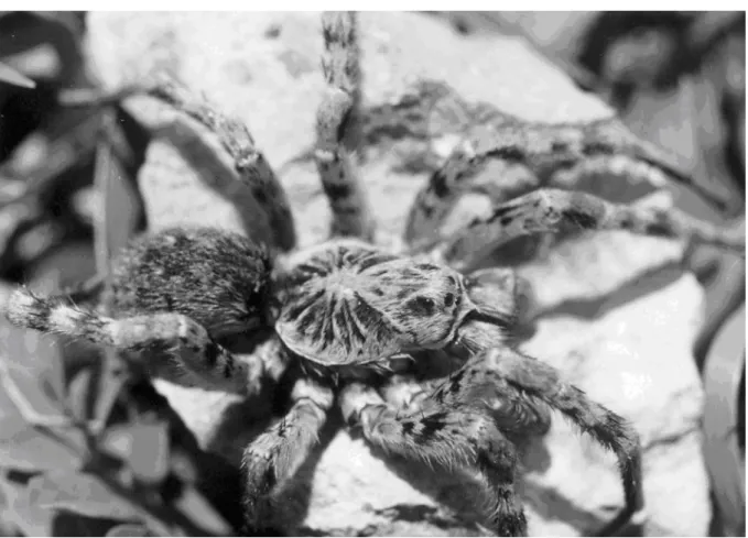
Figure 1. Female L. singoriensis.
L. singoriensis venom apparatus has the same general structure of those from other
spiders, it is situated in the anterior part of the prosoma and composed of a pair of
venom glands, a pair of chelicerae with apical fangs and a pair of canals (or ducts)
that connect the glands through the chelicerae to the tip of the fangs. Like in other
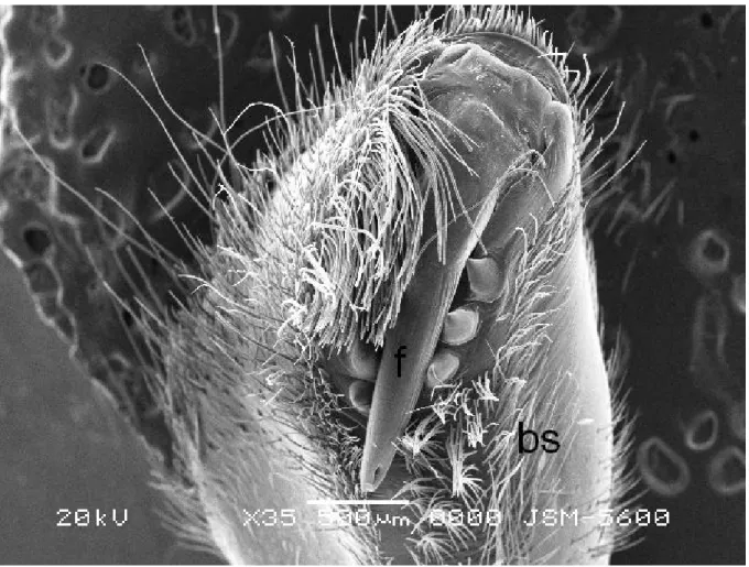
ground-living and burrowing spiders, the chelicerae consist of two parts: basal
segment (paturon) and a movable articulated apical segment (fang). The basal
segments of the chelicerae are very stout and strong, and are covered by hair,
Figure 2. Chelicera of L.singoriensis, the fang (f) and basal segment (bs). The basal
segment is covered by dense hair.
The movable fang rests in a groove of the chelicera basal segment, it looks like an
injection needle and presents the same function (Figures 2 and 3). From the base up
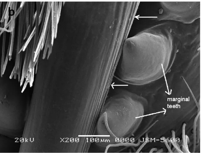
to its apical region, the fang becomes narrower, ending in a quite sharp tip. The pore
is situated on the fang subterminal part, from which the venom is ejected. Parallel
fine grooves, observed running longitudinally on the fang surface, in higher
magnifications appear to form a ridge that constitutes a blade-like structure. In
addition, both sides of cheliceral grooves are equipped with three or four cuticular
Yigit N, et al. Morphological characaterization of the venom apparatus in the wolf spider Lycosa singoriensis
(Laxmann, 1770). J Venom Anim Toxins incl Trop Dis. 2009;15(1):150
Figure 4. The fang, venom pore and marginal teeth (a); blade-like structures (arrows)
in higher magnification (b).
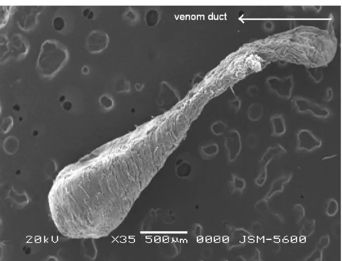
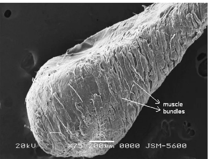
Venom glands, the main components of the venom apparatus, form a pair that is
dorsally located in the prosoma. The glands present similar sizes and have the
appearance of long sacs, with approximately 4 mm, extending from the middle
prosoma to the chelicera base (Figure 5). The distal portion of the venom gland is
wider than the proximal one and there is a large lumen in its center. Furthermore,
each gland is enveloped by a thick muscular layer. Dense muscle bundles are
Yigit N, et al. Morphological characaterization of the venom apparatus in the wolf spider Lycosa singoriensis
(Laxmann, 1770). J Venom Anim Toxins incl Trop Dis. 2009;15(1):152
Figure 6. Muscle bundles that cover the venom gland at higher magnification.
Except for the members of Uloboridae and Archaeidae, most spiders have a venom
apparatus, which does not mean that all are dangerous to humans (6). Among
venomous animals, spiders have proportionally received less attention, since they
are relatively small and produce little amount of venom. Thus, they are generally not
considered as dangerous as snakes or scorpions.
The chelicerae are important for spiders since they are used for defense, seizing
prey, carrying egg cocoons (Lycosidae, Pisauridae), digging soil (Ctenizidae,
Theraphosidae, Barychelidae, Eresidae), transporting small preys (Araneidae) and
making noise (Ammotrechidae, Solifugae) (7, 8). Lycosids are trapdoor spiders that
construct burrows and present large and powerful chelicerae.
In L. singoriensis, both sides of the cheliceral grooves are often armed with cuticular
teeth that act as buttresses for the movable fang. The number and shape of these
marginal teeth are distinct in several species. Spiders whose chelicerae are equipped
with such teeth are able to mash their prey into an unrecognizable mass. Spiders
Yigit N, et al. Morphological characaterization of the venom apparatus in the wolf spider Lycosa singoriensis
(Laxmann, 1770). J Venom Anim Toxins incl Trop Dis. 2009;15(1):154
report severe pain (9). Since L. singoriensis have three or four marginal teeth on both
sides of the cheliceral groove, these structures are probably used to crush skin of
victims, leaving perceivable sings and causing pain.
The tip of the fang is usually sharp in almost all spiders. L. singoriensis fangs have a
venom pore and a blade-like ridge that may facilitate the deep penetration of fangs
into the body of the victim. These same features have been observed previously in
other studied spiders (10-12).
Venom glands of numerous spiders have been investigated by several authors (5, 6,
10-14). The shape and position of venom glands differ among species. In large
tarantulas, venom glands are quite small and lie inside chelicerae (6). In other
spiders, venom glands are two voluminous distinct sacs dorsally located that occupy
the chelicera basal portion up to the prosoma. In L. singoriensis, venom glands are
two lengthy sacs dorsally situated in the prosoma. Regarding shape, they can be
bulbous in Loxosceles intermedia (14); carrot-like in Pelesiophirctus collinus; sac-like
or cylindrical and bilobed in Hetropoda venatoria, Lycosa indagastrix (13) and L.
tarantula (15); and in the form of a long tube in Agelena gracilens and A. labyrinthica
(10, 11).
In L.singoriensis, the venom produced by the glands flows through venom ducts that
pass throughout the chelicerae, and is finally released by the venom pore on the tip
of the fang. The venom discharge process is possible due to the action of a thick
layer of striated muscle bundles that surround the glands. These muscle bundles
spirally cover the glands and end in the first portion of the venom ducts. In many
spiders, including Larinioides ixobolus, Agelena labyrinthica and A. gracilens (10-12),
blocks of muscle bundles spirally encapsulate glands, whereas in other species, like
L. intermedia, external muscular bundles and cell branches develop a web-like
structure (14).
Muscular contractions of the venom glands provide the propulsive force for the
venom expulsion. When a spider bites, its fangs penetrate in the victim and the
venom is injected. In this way, lycosid and geolycosid spiders leave significant fang
REFERENCES
1. Platnick NI. The world spider catalog, version 9.0 [Internet]. American Museum of
Natural History; c2008. Available from:
http://research.amnh.org/entomology/spiders/catalog/INTRO2.html
2. Zyuzin AA. Generic and subfamilial criteria in the systematic of the spider family
Lycosidae (Aranei), with the description of a new genus and two new subfamilies.
Trudy Zool Inst Leningr. 1985;139:41-51.
3. Topcu A, Demir H, Seyyar O. A checklist of the spiders of Turkey. Serket.
2005;9(4):109-40.
4. Isbister GK, Framenau V. Clinical effects of wolf spider bites in Australia. J Toxicol
Clin Toxicol. 2003;41:5.
5. Kovoor J, Munoz-Cuevas A. Comparative histology of the venom gland in a lycosid
and several oxyopid spiders (Araneae). Ekologia. 2000;19(3):129-40.
6. Maretić Z. Spider venoms and their effects. In: Nentwig W, editor. Ecophysiology
of spiders. New York: Springer-Verlag; 1987. p. 142-59.
7. Foelix RF. Biology of spiders. London: Harvard University Press; 1982. 330 p.
8. Jocque R, Dippeneaar-Schoeman AS. Spider families of the world. Tervurem:
Royal Museum for Central Africa; 2007. 336 p.
9. Isbister GK, Framenau VW. Australian wolf spider bites (Lycosidae): clinical effects
and influence of species on bite circumstances. J Toxicol Clin Toxicol.
2004;42(2):153-61.
10. Yigit N, Bayram A, Danisman T, Sancak Z. Functional morphology of the venom
apparatus of Larinioides ixobolus (Araneae: Araneidae). Pakistan J Biol Sci.
2006;9(10):1975-8.
11. Yigit N, Guven T, Bayram A, Cavusoglu K. A morphologic study on the venom
gland of the spider Agelena labyrinthica (Areneae, Agelenidae). Turk J Zool.
2004;28:149-53.
12. Yigit N, Bayram A, Danisman T, Sancak Z. Functional morphology of the venom
apparatus of the funnel spider, Agelena gracilens (Araneae: Agelenidae). Entomol
News. 2007;118(2):161-7.
13. Ridling MW, Phanuel GJ. Functional morphology of the poison apparatus and
histology of the venom glands of three Indian spiders. J Bombay Nat Hist Soc.
Yigit N, et al. Morphological characaterization of the venom apparatus in the wolf spider Lycosa singoriensis
(Laxmann, 1770). J Venom Anim Toxins incl Trop Dis. 2009;15(1):156
14. Santos VLP, Franco CRC, Viggiano RLL, Silveira RB, Cantão MP, Mangili OC,
Veiga SS, Gremski W. Structural and ultrastructural description of the venom gland
of Loxosceles intermedia (brown spider). Toxicon. 2000;38(2):265-85.
15. Maretić Z. Other European araneism. In: Maretic Z, editor. Araneism. Belgrade: