Influence of photobiomodulation on pain perception
during initial orthodontic tooth movement
Influência do fotobiomodulação na percepção da dor durante o movimento
ortodôntico inicial
Welinton Lemos RUMÃOa , Heloísa Cristina VALDRIGHIa, Vivian Fernandes FURLETTIa, Giovana Renata GOUVÊAa, Milton SANTAMARIA-JRa*
aCentro Universitário da Fundação Hermínio Ometto - FHO, Departamento de Ortodontia, Araras, SP, Brasil
How to cite: Rumão WL, Valdrighi HC, Furletti VF, Gouvêa GR, Santamaria-Jr M. Influence of photobiomodulation on pain
perception during initial orthodontic tooth movement. Rev Odontol UNESP. 2020;49:e20200003. https://doi.org/10.1590/1807-2577.00320
Resumo
Introdução: O tratamento com Laser em Baixa Intensidade (LBI) tem sido utilizado para reduzir o
desconforto e a dor desencadeados pelas forças aplicadas durante o tratamento ortodôntico.
Objetivo: Avaliar o feito da aplicação de LBI na percepção da dor na compressão inicial do ligamento
periodontal durante o movimento dentário ortodôntico; e comparar o efeito desta terapia entre os sexos.
Material e método: A amostra foi composta por 30 voluntários, que necessitavam de bandagem dos
primeiros molares inferiores. Após a instalação dos elásticos separadores, aplicou-se o LBI infravermelho na região apical mesial e distal (comprimento de onda 808nm, energia 2J, tempo 20s e fluência de 8,32J/cm2) e
em três ponto na região radicular (comprimento de onda 808nm, energia 1J, tempo 10s e fluência de 4,16J/cm2) no lado irradiado e comparou-se com o primeiro molar contralateral não irradiado (lado
controle), em três tempos: 0hs, 24hs e 48hs. A percepção de dor foi avaliada pela interpretação da Escala Visual Analógica (EVA) em 0hs, 24hs e 48hs após a instalação, com nílvel de significância de 5%.
Resultado: Observou-se que o nível de dor foi significativamente menor (p<0,05) no lado irradiado,
independentemente do sexo e do tempo. O sexo feminino apresentou nível de dor significativamente maior (p<0,05) que o sexo masculino, independentemente do tempo e do lado. Não houve diferença significativa entre os tempos (p>0,05). Conclusão: Concluiu-se que o LBI diminui a percepção de dor inicial em pacientes onde se promoveu a compressão do ligamento periodontal por meio de separação elástica, e que o sexo feminino apresentou maior percepção da sensibilidade dolorosa nos tempos observados.
Descritores: Dor; movimento dentário; fotobiomodulação; Laser em Baixa Intensidade. Abstract
Introduction: Laser in low intensity (LLI) has been used to reduce the discomfort and pain that is
triggered by the forces applied during orthodontic treatment. Objective: To evaluate the effect of LLI application in the pain perception of periodontal ligament initial compression, during orthodontic tooth movement; and to compare the effect of this therapy between men and women. Material and
method: The sample consisted of 30 volunteers, who needed orthodontic band placement on mandibular
first molars. After insertion of the elastic separators, LLI was applied to the mesial and distal apical region (wavelength 808nm, energy 2J, time 20s and fluency of 8.32J/cm2) and at three points on the root region
(wavelength 808nm, energy 1J, time 10s and fluency of 4.16J/cm2) of the first molar (irradiated side) and
compared to the contralateral first molar (non-irradiated side), in three time intervals: 0hs, 24hs and 48hs. Pain perception was evaluated by the Visual Analog Scale (VAS), at 0hs, 24hs and 48hs after insertion, with significance of 5%. Result: The pain level was observed to be significantly lower (p<0.05) on the irradiated side, irrespective of gender and time. Women presented a significantly higher pain level (p<0.05) than men, irrespective of time and side. There were no significant differences between the time intervals (p>0.05). Conclusion: It was concluded that LLI reduced the perception of initial pain in patients in whom compression of the periodontal ligament was promoted by elastic separation, and that women had a greater perception of pain sensitivity in the time intervals studied.
INTRODUCTION
The physical and biological effects of orthodontic tooth movement can be observed early, affecting the extracellular matrix, cells of the alveolar bone, and periodontal ligament, such as granulocytes, fibroblasts, osteoclasts, and osteoblasts. Changes occur in the synthesis and release of cytokines, growth factors, and chemotactic factors1. After 24 hours, pain produced is maintained by the production of prostaglandin in the extracellular medium2,3.
Hyperalgesia is considered a discouraging factor for orthodontic treatment4. Pain perception during tooth movement resulting from the insertion of orthodontic separators, has been evaluated after the use of anti-inflammatory5, analgesic6, irradiation with laser in low intensity (LLI)7 and others therapies8.
Laser therapy has analgesic, anti-inflammatory and tissue repair characteristics9. Initially, it was used in medicine for cutaneous treatment10, and was therefore introduced into Dentistry to be applied - according to its benefits - in the treatment of paresthesias, operative hypersensitivity, and oral lesions such as aphthous ulcers, herpes and cheilitis11. These benefits are related to the capable of penetrating into live tissues, and by selective absorption, produce important biological effects on the inflammatory process and on tissue repair12.
Some therapies can promote stimulation of the periodontal ligament in order to increase the quality and speed of bone remodeling. The LLI, ultrasound and electrical stimulation have been employed for this purpose1,13,14.
The LLI has also been widely used in the healing process of different tissues and during tooth movement15-17. Laser therapy has demonstrated biomodulatory results in its effects on the inflammatory cell decrease and in the improvement of neovascularization18. It has demonstrated biostimulatory effects on the release of cytokines and growth factors in the proliferation process of different cells, improving repair in the final stages of the inflammatory process19,20.
Thus, LLI has been considered an effective alternative for pain control after orthodontic activation, without causing any damage to the orthodontic treatment mechanics21, the application is non-invasive, painless and aseptic. It is capable to cause photochemical reactions in the cells; stimulating collagen production and changing protein synthesis22. Therefore, the aim of this study was to evaluate the perception of pain after LLI application in initial orthodontic movement, and to compare the effect of this therapy between men and women.
METHOD
Sample Selection
This study was approved by the Research Ethics Committee (Report no 43709715.7.0000.5385). All patients read and signed the informed consent document and underwent the routine procedure of tooth separation in orthodontic treatment and authorized the application of laser in low intensity therapy (LLI) for the purpose of controlling painful symptoms, aware that there could be a reduction, increase, or no effect whatever on pain perception.
The sample consisted of 30 patients, with an average age of 28 years old, at the beginning of corrective orthodontic treatment, with the indication of insertion of bands on the lower first molars. The volunteers have mesial and distal contact between the molars, no carious lesions, presence of erupted adjacent permanent second molars, and not be using any type of analgesic or anti-inflammatory agent. The sample size calculation was performed using therapy, sex and
time of treatment as outcomes. For large size effect (f = 0.40), test power of 80% (β = 0.20) and 95% confidence level (α = 0.05), thirty volunteers (n=30) were needed in this study.
Randomization was performed by an external research volunteer who was responsible for generating a random allocation sequence. This sequence was blinded to the patients, who did not know which side received LLI application and which was the non-irradiated side (control side)4. Thus, the experimental design was characterized by a blind split-mouth study.
Elastic separator (MORELLI® - Sorocaba, São Paulo, Brazil) was inserted to promote interdental separation prior to the orthodontic banding procedure, and remained on the mesial and distal surfaces of the mandibular first molars during the 48-hour experimental period of LLI application.
Laser Application Protocol
The laser used was Diode Gallium Aluminum Arsenide (Laser Duo, MMOptics - São Carlos, São Paulo, Brazil), with a wavelength of 808nm and power of 0.1W. The same operator performed the irradiations, on a spot area of 24mm2 (0.24cm2), with the light beam directed perpendicularly and in contact with the mucosa, free of saliva, in relative isolation. The laser was applied on the mesial and distal root apex of the tooth for 20s on each spot; and on three points of application along the mesial and distal thirds of the roots for 10s on each spot.
The applications were performed in three moments: T0, immediately after installation of the elastics, T1 and T2, applied 24 and 48 hours after T0. The LLI was applied, in the mesial and distal apical region (808nm, spot 0.24cm2, 0.1W, 20s, energy 2J and fluency 8.32J/cm2); and in three points along the mesial and distal roots (808nm, spot 0.24cm2, 0.1W, 10s, energy 1J and fluency 4.16J/cm2), which totaled eighth applications, 100s, energy 10J and fluency 41.6J/cm2, in each time (T0, T1 and T2). The Figure 1 shows the experimental design, laser protocol and the timeline of the study.
Figure 1. Representative figure of the laser therapy protocol. (A) Spots of application and energy for each point; (B) Specifications of the laser application protocol; (C) Timeline according to experimental design.
In the non-irradiated tooth, the procedure performed was the same, simulating laser application, to ensure blinding of the study.
Pain Perception
The analgesic effect of LLI application was evaluated by means of the Visual Analog Scale (VAS), delivered to the patients to enable them to quantify their sensitivity to pain in each time interval3,5,7.
The scale was numbered from 0 to 10, corresponding to the pain intensity, differentiating between the left and right sides, in the different time intervals of laser application: initial - 0hs (T0), 24hs (T1) and 48hs (T2). The score established was: 0 corresponded to no pain; 1 and 2 - slight discomfort; 3 and 4 - slight pain; 5 and 6 - moderate pain; 7 and 8 - severe pain, and 9 and 10 - very strong pain (medication to alleviate the discomfort was needed)23.
Statistical Methodology
The data presented asymmetrical distribution, for comparison between the sexes, sides and time intervals. Thus, a generalized linear model was adjusted according to the delineation of repeated measures for the effects of sex, side, time and the interactions. All the analyses were performed by the GENMOD procedure of the software program SAS (SAS Institute Inc., Cary, NC, USA, Release 9.3, 2010), considering the level of significance of 5%.
RESULT
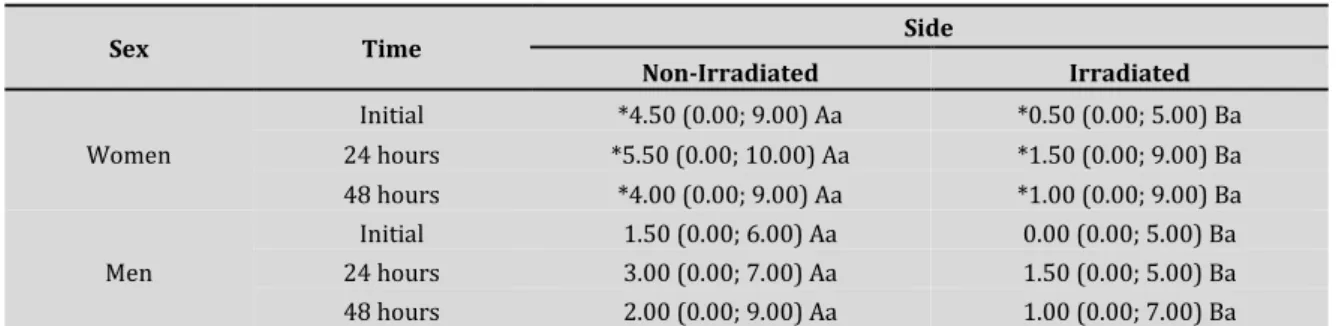
For evaluating the perception of pain, the VAS score data were combined into irradiated and non-irradiated groups, in the different periods of analysis (0hs, 24hs and 48hs). In Table 1 was possible to observe that the level of pain perception was significantly lower on the irradiated side, in men and women in each experimental time of 0hs, 24hs and 48hs (capital letters in the table, p<0.05), showing evidence of the analgesic efficiency of the therapy. But on the same side, the pain perception was similar over time for each sex (lower case letters in the table, p=0.33). Although the perception of pain has decreased, women presented a significantly higher level of pain than the men, irrespective of time and side (p<0.05).
Table 1. Median (minimum; maximum) pain level after placement of the elastic separator and laser application on one of the sides of the mouth, considering the time
Sex Time Side
Non-Irradiated Irradiated Women Initial *4.50 (0.00; 9.00) Aa *0.50 (0.00; 5.00) Ba 24 hours *5.50 (0.00; 10.00) Aa *1.50 (0.00; 9.00) Ba 48 hours *4.00 (0.00; 9.00) Aa *1.00 (0.00; 9.00) Ba Men Initial 1.50 (0.00; 6.00) Aa 0.00 (0.00; 5.00) Ba 24 hours 3.00 (0.00; 7.00) Aa 1.50 (0.00; 5.00) Ba 48 hours 2.00 (0.00; 9.00) Aa 1.00 (0.00; 7.00) Ba Different letters, capital letters in the horizontal and lower case letters in the vertical differ between them, in each sex, p-value<0.05. *Women differ from the men in the same conditions of side and time, p-value<0.05.
In Figure 2, the Box plot presents the pain variation (%) on the irradiated side in comparison with the non-irradiated side in each time interval and sex. Values below zero indicate decreased pain, and above zero indicate increased pain on the irradiated side. It is possible to see in the graph that laser therapy reduces pain in patients in all analyzed periods and in both sexes. Only in women, in period of 48hs, the pain variation was close to zero, indicating that the pain perception was similar on the irradiated and non-irradiated side.
Figure 2. Box plot of percentages of variation in pain level on the irradiated side in comparison with the non-irradiated side in each time interval and sex.
Still, two patients - represented by the circles on the graph - presented discrepant values, with a much higher level of pain on the control side; one woman, in 24hs and one man, in the initial time (Figure 2).
DISCUSSION
Initial compression of the periodontal ligament resulting from the insertion of separator elastics reflects a new experience for those patients without previous orthodontic treatment11. Painful symptom is associated with factors that discourage patients undergoing orthodontic treatment. The period of 2 to 4 days is considered critical after insertion of orthodontic device on the inflammatory process of the periodontal ligament24.
Pain perception may be influenced by sensory, affective, cognitive and behavioral factors; a physiological reaction resulting from stimulus of the nociceptive system. It is a singular experience difficult to be described25. Therefore, in this study, the evaluation of pain perception was possible, because in the same patient one side underwent the therapy with laser, and the other side was the control; a blind split-mouth study. As possible results, we considered the lack of laser therapy influence, the increase, or reduction on the pain perception, resulting from initial compression of the periodontal ligament26.
The period of time that the elastic separator remained in place was 48hs, and the peak painful sensitivity was 24hs after the application of force11. Thus, the experimental period established demanded high analgesic efficiency of the protocol used. The choice with longer periods of evaluation could mask the painful perception, as there would be a natural resolution in the inflammatory process3-5,7.
Hyperalgesia is the result of the inflammatory mediators in the periodontal ligament, such as bradykinin, prostaglandins, histamine and serotonin2. The anti-inflammatory agents such as ibuprofen and acetaminophen are commonly recommended due to their capacity to inhibit prostaglandin level by modulating the synthesis of cyclooxygenases (COX-1 and COX-2)27. With the purpose to avoid the adverse effects of these drugs, new therapies such as LLI have been proposed for pain control28. Laser therapy is non-invasive, painless, antiseptic and easy to handle.
It does not present adverse tissue reactions, if properly employed, and is capable to cause photochemical reactions in the cells, stimulating protein synthesis and collagen production22.
The best biostimulators wavelengths are in the range of 550 to 950nm29, and are capable to inhibit the nerve stimulus, making difficult the occurrence of sensitivity to pain. In this methodology, the laser wavelength of 808nm simulated the action of ATPases, including Na/K ATPase, also known as Sodium (Na) and Potassium (K) pump30. This change favors the entry of Calcium (Ca) into the cell, acting positively on DNA and RNA synthesis. With the maintenance of the Na and K ion concentrations in the extracellular and intracellular medium, respectively, there is no depolarization of the membrane. Thus, one of the pathways of action of laser therapy occurs when the sensitization of the nociceptors is not conducted up to the Central Nervous System (SNC), where it could be interpreted as pain31.
Men and women also demonstrated a different behavior in relation to pain perception. This involves important factors of emotional and psychical order. The sexual steroids, in addition to the differentiation of the cerebral neural circuits between the sexes could influence the pain perception32. This difference between the sexes appeared in this study, in which women were observed to have a higher level of pain perception than the men. Biological factors, such as sexual hormones have been considered one of the main mechanisms that explain these differences. This hypothesis has been supported by the findings conducted with animals and humans33, showing agreement with the results obtained in this study, when women presented a significantly higher level of pain than the men.
The laser application protocol used in this study during the period of major symptomatology revealed that LLI was an efficient therapy for pain control in initial orthodontic tooth movement, and it could be an alternative to the use of analgesics and anti-inflammatory drugs.
CONCLUSION
The study concluded that laser in low intensity diminished the initial pain perception in patients during the compression of the periodontal ligament with elastic separators, in both sexes. However, that the women presented a higher level of pain perception in the experimental period.
REFERENCES
1. Spadari GS, Zaniboni E, Vedovello SAS, Santamaria MP, Amaral MEC, Santos GMT, et al. Electrical stimulation enhances tissue reorganization during orthodontic tooth movement in rats. Clin Oral Investig. 2017 Jan;21(1):111-20. http://dx.doi.org/10.1007/s00784-016-1759-6. PMid:26917494. 2. Krishnan V. Orthodontic pain: from causes to management: a review. Eur J Orthod. 2007
Apr;29(2):170-9. http://dx.doi.org/10.1093/ejo/cjl081. PMid:17488999.
3. Farias RD, Closs LQ, Miguens SAQ Jr. Evaluation of the use of low-level laser therapy in pain control in orthodontic patients: a randomized split-mouth clinical trial. Angle Orthod. 2016 Mar;86(2):193-8. http://dx.doi.org/10.2319/122214-933.1. PMid:26132512.
4. Xiaoting L, Yin T, Yangxi C. Interventions for pain during fixed orthodontic appliance therapy: a systematic review. Angle Orthod. 2010 Sep;80(5):925-32. http://dx.doi.org/10.2319/010410-10.1. PMid:20578865.
5. Al-Balbeesi HO, Bin Huraib SM, AlNahas NW, AlKawari HM, Abu-Amara AB, Vellappally S, et al. Pain and distress induced by eslastomeric and spring separators in patients undergoing orthodontic treatment. J Int Soc Prev Community Dent. 2016 Nov-Dec;6(6):549-53.
http://dx.doi.org/10.4103/2231-0762.195519. PMid:28032047.
6. Azodo CC, Umoh AO. Analgesics prescription in Nigerian dental healthcare services. Niger J Basic Clin Sci. 2013;10(2):86-90. http://dx.doi.org/10.4103/0331-8540.122768.
7. Fujiyama K, Deguchi T, Murakami T, Fujii A, Kushima K, Takano-Yamamoto T. Clinical effect of CO(2) Laser in reducing pain in orthodontics. Angle Orthod. 2008 Mar;78(2):299-303.
http://dx.doi.org/10.2319/033007-153.1. PMid:18251609.
8. Lobre WD, Callegari BJ, Gardner G, Marsh CM, Bush AC, Dunn WJ. Pain control in orthodontics using a micropulse vibration device: a randomized clinical trial. Angle Orthod. 2016 Jul;86(4):625-30. http://dx.doi.org/10.2319/072115-492.1. PMid:26496680.
9. Fernandes-Dias SB, Marco AC, Santamaria M Jr, Kerbauy WD, Jardini MAN, Santamaria MP. Connective tissue graft associated or not with low laser therapy to treat gingival recession. Randomized clinical trial. J Clin Periodontol. 2015 Jan;42(1):54-61. http://dx.doi.org/10.1111/jcpe.12328.
PMid:25363203.
10. Neves LMG, Matheus RL, Santos G, Esquisatto M, Amaral MEC, Mendonça F. Effects of microcurrent application and 670 nm InGaP low-level laser irradiation on experimental wound healing in healthy and diabetic Wistar rats. Laser Phys. 2013 Feb;23(3):035604. http://dx.doi.org/10.1088/1054-660X/23/3/035604.
11. Sun G, Tunér J. Low-level laser therapy in dentistry. Dent Clin North Am. 2004 Oct;48(4):1061-76. http://dx.doi.org/10.1016/j.cden.2004.05.004. PMid:15464564.
12. Silva Neves FL, Silveira CA, Fernandes-Dias SB, Santamaria M Jr, Marco AC, Kerbauy WD, et al. Comparison of two power densities on the healing of palatal wounds after connective tissue graft removal: randomized clinical trial. Lasers Med Sci. 2016 Sep;31(7):1371-8.
http://dx.doi.org/10.1007/s10103-016-1988-6. PMid:27344670.
13. Nimeri G, Kau CH, Abou-Kheir NS, Corona R. Acceleration of tooth movement during orthodontic treatment-a frontier in Orthodontics. Prog Orthod. 2013 Oct;14(1):42.
http://dx.doi.org/10.1186/2196-1042-14-42. PMid:24326040.
14. El-Bialy T, Farouk K, Carlyle TD, Wiltshire W, Drummond R, Dumore T, et al. Effect of low intensity pulsed ultrasound (LIPUS) on tooth movement and root resorption: a prospective multi-center randomized controlled trial. J Clin Med. 2020 Mar;9(3):804. http://dx.doi.org/10.3390/jcm9030804. PMid:32188053.
15. Kawasaki K, Shimizu N. Effects of low-energy laser irradiation on bone remodeling during experimental tooth movement in rats. Lasers Surg Med. 2000;26(3):282-91.
http://dx.doi.org/10.1002/(SICI)1096-9101(2000)26:3<282::AID-LSM6>3.0.CO;2-X. PMid:10738291. 16. Nalcaci R, Cokakoglu S. Lasers in orthodontics. Eur J Dent. 2013 Sep;7(S 01 Suppl 1):S119-25.
http://dx.doi.org/10.4103/1305-7456.119089. PMid:24966719.
17. Yassaei S, Fekrazad R, Shahraki N. Effect of low level laser therapy on orthodontic tooth movement: a review article. J Dent. 2013 May;10(3):264-72. PMid:25512754.
18. Medrado ARAP, Pugliese LS, Reis SRA, Andrade ZA. Influence of lowlevel laser therapy on wound healing and its biological action upon myofibroblasts. Lasers Surg Med. 2003;32(3):239-44. http://dx.doi.org/10.1002/lsm.10126. PMid:12605432.
19. Kipshidze N, Nikolaychik V, Keelan MH, Shankar LR, Khanna A, Kornowski R, et al. Low-power helium: neon laser irradiation enhances production of vascular endothelial growth factor and promotes growth of endothelial cells in vitro. Lasers Surg Med. 2001;28(4):355-64.
http://dx.doi.org/10.1002/lsm.1062. PMid:11344517.
20. Medrado AP, Soares AP, Santos ET, Reis SR, Andrade ZA. Influence of laser photobiomodulation upon connective tissue remodeling during wound healing. J Photochem Photobiol B. 2008 Sep;92(3):144-52. http://dx.doi.org/10.1016/j.jphotobiol.2008.05.008. PMid:18602833.
21. Alazzawi MMJ, Husein A, Alam MK, Hassan R, Shaari R, Azlina A, et al. Effect of low level laser and low intensity pulsed ultrasound therapy on bone remodeling during orthodontic tooth movement in rats. Prog Orthod. 2018 Apr;19(1):10. http://dx.doi.org/10.1186/s40510-018-0208-2. PMid:29658096.
22. Gama SKC, Habib FAL, Monteiro JSC, Paraguassú GM, Araújo TM, Cangussú MCT, et al. Tooth movement after infrared laser phototherapy: clinical study in rodents. Photomed Laser Surg. 2010 Oct;28(Suppl 2):S79-83. http://dx.doi.org/10.1089/pho.2009.2618. PMid:20932152.
23. Doshi-Mehta G, Bhad-Patil WA. Efficacy of low-intensity laser therapy in reducing treatment time and orthodontic pain: a clinical investigation. Am J Orthod Dentofacial Orthop. 2012 Mar;141(3):289-97. http://dx.doi.org/10.1016/j.ajodo.2011.09.009. PMid:22381489.
24. Oliver RG, Knapman YM. Attitudes to orthodontic treatment. Br J Orthod. 1985;12(4):179-88. http://dx.doi.org/10.1179/bjo.12.4.179. PMid:3863673.
25. Patel S, McGorray SP, Yezierski R, Fillingim R, Logan H, Wheeler TT. Effects of analgesics on orthodontic pain. Am J Orthod Dentofacial Orthop. 2011 Jan;139(1):e53-8.
http://dx.doi.org/10.1016/j.ajodo.2010.07.017. PMid:21195257.
26. Karu TI. A suitable model for wound healing: how many times are we to stumble over the same block? Lasers Surg Med. 1999;25(4):283-4.
http://dx.doi.org/10.1002/(SICI)1096-9101(1999)25:4<283::AID-LSM1>3.0.CO;2-3. PMid:10534743.
27. Krishnan V, Davidovitch Z. Cellular, molecular, and tissue-level reactions to orthodontic force. Am J Orthod Dentofacial Orthop. 2006 Apr;129(4):469.e1-32.
http://dx.doi.org/10.1016/j.ajodo.2005.10.007. PMid:16627171.
28. Kyrkanides S, O’Banion MK, Subtelny JD. Nonsteroidal anti-inflammatory drugs in orthodontic tooth movement: metalloproteinase activity and collagen synthesis by endothelial cells. Am J Orthod Dentofacial Orthop. 2000 Aug;118(2):203-9. http://dx.doi.org/10.1067/mod.2000.105872. PMid:10935962.
29. Stolik S, Delgado JA, Anasagasti L, Pérez AM. Effective thermal penetration depth in photo-irradiated Ex vivo human tissues. Photomed Laser Surg. 2011 Oct;29(10):669-75.
http://dx.doi.org/10.1089/pho.2010.2948. PMid:21612514.
30. Kassák P, Sikurová L, Kvasnicka P, Bryszewska M. The response of Na+/K+-ATPase of human erythrocytes to green laser light treatment. Physiol Res. 2006;55(2):189-94. PMid:15910177. 31. Kasai S, Kono T, Yamamoto Y, Kotani H, Sakamoto T, Mito M. Effect of low-power Laser irradiation on
impulse conduction in anesthetized rabbits. J Clin Laser Med Surg. 1996 Jun;14(3):107-9. http://dx.doi.org/10.1089/clm.1996.14.107. PMid:9484084.
32. McCarthy MM, Arnold AP. Reframing sexual differentiation of the brain. Nat Neurosci. 2011 Jun;14(6):677-83. http://dx.doi.org/10.1038/nn.2834. PMid:21613996.
33. Fillingim RB, Gear RW. Sex differences in opioid analgesia: clinical and experimental findings. Eur J Pain. 2004 Oct;8(5):413-25. http://dx.doi.org/10.1016/j.ejpain.2004.01.007. PMid:15324773.
CONFLICTS OF INTERESTS
The authors declare no conflicts of interest.
*CORRESPONDING AUTHOR
Milton Santamaria Júnior, Centro Universitário da Fundação Hermínio Ometto - FHO, Programa de Pós-graduação em Ortodontia, Dr. Maximiliano Baruto, 500, 13607-339 Araras - SP, Brasil, e-mail: [email protected]
Received: January 20, 2020 Accepted: June 8, 2020