Survival outcome among patients with Ewing’s sarcoma of
bones and joints: a population-based cohort study
Zi-Hao Wan
I, Zhi-Hao Huang
II, Liao-Bin Chen
IIIZhongnan Hospital, Wuhan University, Wuhan, Hubei Province, China
INTRODUCTION
Ewing’s sarcoma is a rare cancer that accounts for less than 10% of all malignancies existing in the human body. It stems from primitive neuroepithelial cells, which are able to differentiate into various mesenchymal cells, and has a propensity to metastasize to distant sites at an early stage . This cancer typically occurs in adolescents and young adults, accompanied by a very poor prognosis. It is considered to be a high-grade malignancy, ranking second in the list of malig-nant bone tumors.1-7 It is commonly considered to be an extremely aggressive osteolytic cancer
that usually occurs in the bones of the limbs and pelvis and it can metastasize to distant loca-tions such as bone marrow, the lungs and other soft tissues at an early stage.1,3,7
In the United States, the overall incidence rate of Ewing’s sarcoma is approximately 0.1 case per 100,000 individuals per year, and this rate had not undergone any obvious change over past 30 years. An estimated 90% of these patients are under 20 years old, and the death rate is approx-imately 0.05 cases per 100,000 individuals per year. Additionally, most cases of Ewing’s sarcoma of the bones and joints are found in the limbs, pelvis or spine.4,8
Nonetheless, there is a lack of survival studies on Ewing’s sarcoma arising in the bones and joints and associated prognostic factors, based on up-to-date data on nationwide populations.
OBJECTIVE
The purpose of this study was to demonstrate the survival conditions of patients with Ewing’s sar-coma of the bones and joints and determine independent risk factors associated with their prognosis.
METHODS
The Surveillance, Epidemiology and End Results (SEER) database named “Incidence-SEER 18 Regs Research Data + Hurricane Katrina Impacted Louisiana Cases, Nov 2015 Sub (1973-2013 varying)”
IMD, MSc. Surgeon, Department of Orthopedic
Surgery, Zhongnan Hospital, Wuhan University, Wuhan, Hubei Province, China.
IIMD, MSc. Surgeon, Department of Colorectal
and Anal Surgery, Zhongnan Hospital, Wuhan University, Wuhan, Hubei Province, China.
IIIMD. Surgeon, Department of Orthopedic
Surgery, Zhongnan Hospital, Wuhan University, Wuhan, Hubei Province, China.
orcid.org/0000-0002-4854-4213
KEY WORDS:
Bone and bones. Sarcoma, Ewing. Survival analysis. Retrospective studies.
ABSTRACT
BACKGROUND: The aim here was to elucidate the current survival condition of patients diagnosed with Ew-ing’s sarcoma of the bones and joints and determine independent risk factors associated with the prognosis. DESIGN AND SETTING: Retrospective cohort study based on the Surveillance, Epidemiology and End Results (SEER) database in the United States.
METHODS: We identified 397 patients who were diagnosed with Ewing’s sarcoma of the bones and joints between January 2004 and December 2013. The multivariate Cox proportional hazards model was used to determine factors associated with the risk of death by adjusting for various factors.
RESULTS: The one, two and five-year disease-specific survival rates were 89.08%, 78.08% and 62.47%, re-spectively. The factors related to death were age (≥ 18 years versus < 18 years; hazard ratio, HR = 1.77; 95% confidence interval, CI: 1.38-2.31); tumor site (extremity versus spine and pelvis; HR = 2.03; 95% CI: 1.31-2.62); tumor size (> 10 cm versus ≤ 10 cm; HR = 1.78; 95% CI: 1.34-2.56); and type of treatment (sur-gery alone versus radiotherapy with sur(sur-gery; HR = 0.51; 95% CI: 0.38-0.89; or radiotherapy alone versus radiotherapy with surgery; HR = 1.61; 95% CI: 1.10-2.39; or no treatment versus radiotherapy with surgery; HR = 1.86; 95% CI: 1.23, 2.58).
was selected to perform a population-based search for patients suf-fering from Ewing’s sarcoma of the bones and joints between January 2004 and December 2013.
The SEER Program9 is supported by the National Cancer
Institute of the United States and has provided statistical infor-mation on tumor cases since 1973. It collects data on cases diag-nosed with cancer throughout the United States, with an estimated 28% of the United States population covered. The SEER registry is a validated database that is frequently applied for cancer sur-vival studies. The National Cancer Institute does not require insti-tutional review board approval for SEER studies because it is an unidentified public-use database.
Histological International Classification of Diseases (ICD) codes (ICD-0-3) were used to identify Ewing’s sarcoma (9260/3), (9364/3). Site-specific codes (C40.0-C40.3, C40.8, C40.9, C41.2, C41.4 and C41.8) were used to screen for tumors originating in the extremities, pelvis and spine, while the codes for bones of the skull and face, mandible, rib, sternum, clavicle and associated joints were not included.
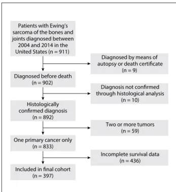
The following primary data were drawn from the database for analysis: age at diagnosis, sex, race, tumor site, tumor size, tumor grade, type of treatment, cause of death and survival in months. Patients diagnosed through either autopsy or the death certificate were excluded. Those who presented secondary malig-nancies at the time of diagnosis or whose diagnoses were not con-firmed by means of histopathological evaluation were also excluded. Cases without complete information were excluded. The inclusion and exclusion procedure is showed in the flow chart of Figure 1.
Well-differentiated and moderately differentiated histological features were classified as low grade while poorly-differentiated and undifferentiated histological types were classified as high grade. The pathological tumor-node-metastasis (pTNM) classification was used in cases of patients who underwent tumor resection, because their tumor gradings could be confirmed through analysis under a microscope. Because the sixth edition of the staging sys-tem of the American Joint Commission on Cancer was used in the SEER database starting in 2004 and the most recent update for this database was in 2013 (which was when the present study began), we chose to use this period (2004 to 2013) for the present study.
The main result from this study was the disease-specific sur-vival (DSS). Descriptive statistics were calculated for all factors. The DSS was calculated by means of the Kaplan-Meier method, and the log-rank test was applied to appraise the deviations. We cal-culated hazard ratios (HRs) and the 95% confidence interval (CI) in the Cox proportional hazards model after adjusting for vari-ous variables. The SEER*Stat application version 8.3.2 (IMS Inc., USA) was used to extract primary data. All statistical analyses was finished in the SPSS software, version 23 (IBM Corp., USA). Differences between groups were taken to be statistically signifi-cant when the P value was less than 0.05.
RESULTS
During the 10-year period, 397 patients diagnosed with Ewing’s sarcoma of the bones and joints were included (249 males and 148 females). Table 1 shows the distribution of the patients’ char-acteristics in the study. The mean age (with SD) at diagnosis was 18.5 (12.4) years. 65.1% of the patients were aged < 18 years. Tumors arising from the extremities accounted for 55.1%. 378 tumors (95.2%) were histologically confirmed to be poor-ly-differentiated or undifferentiated tumors. The mean tumor size at the time of diagnosis was 10.3 cm (5.3). 86 patients (21.6%) received surgery with radiotherapy, 139 (35.1%) underwent sur-gical resection alone, 102 (25.7%) underwent radiotherapy alone and 70 (17.6%) received no treatment.
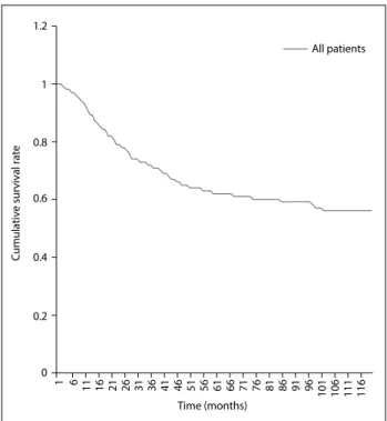
The overall one, two and five-year survival rates after diag-nosis were 89.08%, 78.08% and 62.47%, respectively (Figure 2). The five-year relative survival rates were 78.4%, 66.9%, 47.8% and 44.8% for patients receiving surgery, surgery with radiotherapy, radiotherapy alone and no therapy, respectively (Figure 3). Overall, patients with tumor size ≤ 10 cm had a higher five-year survival rate than did those with tumor size > 10 cm (70.8% versus 52.4%; P < 0.001) (Figure 4). The five-year survival rate were 68.7% and 50.2% for those < 18 and ≥ 18 years (Figure 5).
Table 2 shows the results from univariate and multivariate
Cox proportional hazards analyses. Age ≥ 18 years (HR = 1.77; 95% CI = 1.33-2.01), tumor originating in the spine and pelvis (HR = 2.03; 95% CI = 1.31-2.62), tumor size > 10 cm (HR = 1.78;
Figure 1. Flow chart for selection of the study cohort. Patients with Ewing’s
sarcoma of the bones and joints diagnosed between 2004 and 2014 in the United States (n = 911)
Diagnosed by means of autopsy or death certificate
(n = 9) Diagnosed before death
(n = 902)
Two or more tumors (n = 59) Diagnosis not confirmed through histological analysis
(n = 10) Histologically
confirmed diagnosis (n = 892)
One primary cancer only (n = 833)
Included in final cohort (n = 397)
95% CI = 1.24-2.35), radiotherapy alone (HR = 1.61; 95% CI = 1.10-2.39) and no treatment (HR = 1.86; 95% CI = 1.23-2.58) were associated with increased risk of mortality, while receiving surgery alone (HR = 0.51; 95% CI = 0.38-0.89) was an indepen-dent predictor for longer survival.
DISCUSSION
In our study, we found that the one, two and five-year DSS rates were 89.08%, 78.08% and 62.47%, respectively. These propor-tions were reported to be higher in a previous investigation.10
This difference may reflect that the prognosis of Ewing’s sarcoma originating bones and joints is much worse than that of Ewing’s sarcoma of other parts of human body. Early screening and diag-nosis seem to be more important among people at high risk of having Ewing’s sarcoma of bones and joints.
The development of diagnostic methods based on molecular techniques has had a great effect, because typical chromosomal translocations are commonly detected in Ewing’s sarcoma tissue. The reverse transcription-polymerase chain reaction (RT-PCR)
and fluorescent in-situ hybridization (FISH) are the most frequent measures applied in fusion gene analysis.
It has been reported that the t(11;22) (q24;q12) translocation is found in 85% of cases of Ewing’s sarcoma.11,12 Yang et al.13
fur-ther ascertained that FISH and RT-PCR could be applied as reli-able molecular diagnostic approaches in cases of Ewing’s sarcoma,
Characteristic Total
N %
Patients 397 100
Sex
Female 148 37.2
Male 249 62.8
Age, years
< 18 251 63.2
≥ 18 146 36.8
Mean (with standard deviation) 18.5 (12.4)
-Median 17.8
-Race
White 351 88.4
Black 15 3.8
Other 31 7.8
Tumor site
Extremity 219 55.1
Spine and pelvis 178 44.9
Tumor grade
Low 19 4.8
High 378 95.2
Tumor size
≤ 10 173 43.6
> 10 224 56.4
Mean (with standard deviation) 10.3 (5.3)
-Median 9.6
-Treatment
Surgery with radiotherapy 86 21.6
Surgery alone 139 35.1
Radiotherapy alone 102 25.7
None 70 17.6
Table 1. Characteristics of patients with Ewing’s sarcoma of the bones and joints
All patients
1 1.2
0.8
0.6
0.4
0.2
6
1
11 16 21 26 31 36 41 46 51 56 61 66 71 76 81 86 91 96 101 106 111 116 0
Time (months)
Cumulative survival rate
Figure 2. Kaplan-Meier survival curve for all patients.
Figure 3. Kaplan-Meier survival curves for all patients, stratified according to type of treatment.
Surgery and radotherapy Surgery alone Radiotherapy alone None
1 1.2
0.8
0.6
0.4
0.2
7
1
13 19 25 31 37 43 49 55 61 67 73 79 85 91 97 103 109 115
0
Time (months)
and that FISH displayed features of greater sensitivity and sta-bility. Furthermore, in a meta-analysis on 1,412 cases, Li et al.14
declared that high levels of serum lactate dehydrogenase (LDH) presaged lower DSS among patients with Ewing’s sarcoma. However,
information on these specific molecular indicators is not docu-mented in the SEER database.
The influence of age on survival has always been a matter of debate, with contrary outcomes reported from different studies.
Variable Crude Adjusted*
HR (95% CI) P-value HR (95% CI) P-value
Sex
Female 1 (reference) 1 (reference)
Male 1,47 (1.12-1.93) < 0.05 1.19 (0.98-1.71) 0.074
Age
< 18 years 1 (reference) 1 (reference)
≥ 18 years 1,89 (1.47-2.44) < 0.05 1.77 (1.38-2.31) < 0.05
Race
White 1 (reference) 1 (reference)
Black 1.68 (1.27-2.07) < 0.05 1.52 (1.37-1.86) 0.075
Other 0.63 (0.49-1.53) 0.38 0.91 (0.68-1.74) 0.84
Tumor site
Extremity 1 (reference) 1 (reference)
Spine and pelvis 2.57 (1.74-2.79) < 0.05 2.03 (1.31-2.62) < 0.05 Tumor grade
Low 1 (reference) 1 (reference)
High 2.37 (0.77-4.29) 0.81 - -
-Tumor size
≤ 10 cm 1 (reference) 1 (reference)
> 10 cm 2.01 (1.46-2.77) < 0.05 1.78 (1.34-2.56) < 0.05
Treatment
Radiotherapy with surgery 1 (reference) 1 (reference)
Surgery alone 0.61 (0.42-0.92) < 0.05 0.51 (0.38-0.89) < 0.05
Radiotherapy alone 1.66 (1.24-2.51) < 0.05 1.61 (1.10-2.39) < 0.05
None 2.27 (1.53-3.38) < 0.05 1.86 (1.23-2.58) < 0.05
Table 2. Cox model with hazard ratios and 95% confidence intervals for mortality associated with covariates, among patients with Ewing’s sarcoma of bones and joints
HR = hazard ratio; CI = confidence interval. *Adjusted for sex, age, race, tumor site, tumor size and treatment.
Figure 5. Kaplan-Meier survival curves for all patients, stratified according to age.
Figure 4. Kaplan-Meier survival curves for all patients, stratified according to tumor size.
≤ 10 cm
> 10 cm 1
1.2
0.8
0.6
0.4
0.2
7
1
13 19 25 31 37 43 49 55 61 67 73 79 85 91 97 103 109 115
0
Time (months)
Cumulative survival rates
< 18 years ≥ 18 years 1
1.2
0.8
0.6
0.4
0.2
6
1 11 16 21 26 31
101 106 111 116 36 41 46 51 56 61 66 71 76 81 86 91 96 0
Time (months)
In our study, most of the patients included were young, i.e. under 18 years of age, and the median age of our cohort was 17.8 years at the time of diagnosis. This was almost identical to the results reported previously in the worldwide literature, i.e. that most cases of Ewing’s sarcoma not surprisingly emerged before the second decade of life, and that younger patients were likely to have a bet-ter prognosis.15-17 Regarding the reasons for this phenomenon,
Lee et al.18 and Grevener et al.16 found that fewer cases among adult
patients were treated with chemotherapy. Moreover, elderly patients were more likely to have several comorbidities, including diabe-tes mellitus hypertension or secondary cancers, which made the situation much more complex.
Tumor size was considered to be a prognostic indicator in our study. We found that the mean size was 10.3 cm, which was almost consistent with the results declared in previous studies. We noticed that sizes larger than 10 cm were associated with a negative impact on DSS. However, there is no consensus regarding any critical cut-off size that might indicate a completely different prognosis for this disease.19-23 In a study on 182 patients, Fizazi et al.24 found that
tumor size greater than 10 cm was an independent prognostic factor for survival. Canter25 also recommended that patients with
tumors larger than 10 cm should accept neoadjuvant chemother-apy and investigational therapies, because they were at a high risk of relapse and disease-specific death.
Even so, several studies have asserted that 8 cm might be a more appropriate boundary value. In a retrospective analysis on 220 patients at St. Jude Children’s Research Hospital, Rodriguez-Galindo et al.23 found that neoplasm size larger than 8 cm affected
survival adversely.
In our analysis, tumors arising from the spine and pelvis were an independent factor for poorer survival. The proportion of the patients who accepted surgery was 56.0%, while 47.5% received radiotherapy. We found that surgery alone, radiotherapy alone, and no treatment were independent risk factors.
There are several explanations for this phenomenon. Oberlin et al.26 asserted that it was recommendable that smaller and more
peripheral tumors should be dealt with through surgical resection, while larger and more central unresectable entities should be man-aged with radiotherapy. On the other hand, Granowetter et al.27
pointed out that radiotherapy was not appropriate for patients in whom there was no proof of microscopic remainders of malignant tissue after they underwent operations.
Normally, it is accepted that surgery will provide a decisive par-tial cure. Only when the neoplasm is unresectable or after pallia-tive surgery should radiation therapy be considered. Such patients’ prognoses have been found to be relatively much worse than those of patients who underwent surgery alone.10,28
In a retrospective study on 512 cases, Bacci et al.29 concluded
that surgical resection is more ideal than radiation therapy for
patients with Ewing’s sarcoma of the extremities for whom adequate surgical margins can be achieved. In cases of insufficient surgical margins, high-dose radiotherapy is recommended, while reduced-dose radiotherapy is ineffective.
Furthermore, the main population affected by Ewing’s sarcoma of the bones and joints consists of young people, mostly in their teenage years. For these individuals, excessive exposure to radiation may result in retardation of the development of bones and other organs. This may have side effects of greater severity than those of surgery, which may produce less morbidity. Although surgery is the practice most often used for local control, there are very few randomized controlled trials directly comparing the effects of sur-gery with those of radiotherapy, and the relative positions of these techniques remain contentious.28
In addition, race, sex and tumor grade were not independent factors after adjusting for different variables in our Cox multivar-iate regression model. On the other hand, these valuables were reported to be independent risk factors in relation to other bone cancers in some previous studies.19,20,22,30,31 Further study regarding
whether these factors should be considered as independent risk indicators for the prognosis of patients with Ewing’s sarcoma of the bones and joints is needed.
Our analysis was based on the data documented in the SEER database, which means that we need to acknowledge that there were some limitations relating to our study. Firstly, some variables including data on comorbidities, surgical margins, extent of sur-gical resection, tumor recrudescence and use of targeted therapy in managing this cancer were missing or not recorded in the data-base. Secondly, because of the principle of anonymity in the SEER Program, it was impossible for us to contact the patients in order to gain additional information. Thirdly, it also should not be ignored that because of the existence of confounders, the consequences deduced from a retrospective analysis would normally be of lower methodological grade than those from randomized controlled tri-als. Finally, we were unable to evaluate some specific molecular indicators, such as Ewing’s Sarcoma-Friend leukemia integration 1 transcription factor (EWS-FLI1) and serum LDH, which help in making an early diagnosis and in judging the prognosis.
In spite of these limitations, use of the SEER Program database has significant advantages, in that it provides possibilities for con-ducting studies of this nature based on large populations suffering from rare types of cancer.
CONCLUSION
independent protective factor for better survival. Further inves-tigations combining multiple fields of the gene modulation and molecular mechanism- are expected to elucidate better treatment
strategies for cases of Ewing’s sarcoma of the bones and joints.
REFERENCES
1. Uyeturk U, Helvaci K, Demirci A, et al. Clinical outcomes and prognostic
factors of adult’s Ewing sarcoma family of tumors: single center
experience. Contemp Oncol (Pozn). 2016;20(2):141-6.
2. Biswas B, Bakhshi S. Management of Ewing sarcoma family of tumors:
Current scenario and unmet need. World J Orthop. 2016;7(9):527-38.
3. Delattre O, Zucman J, Melot T, et al. The Ewing family of tumors--a
subgroup of small-round-cell tumors defined by specific chimeric
transcripts. N Engl J Med. 1994;331(5):294-9.
4. Esiashvili N, Goodman M, Marcus RB Jr. Changes in incidence and
survival of Ewing sarcoma patients over the past 3 decades: Surveillance
Epidemiology and End Results data. J Pediatr Hematol Oncol.
2008;30(6):425-30.
5. Riggi N, Stamenkovic I. The Biology of Ewing sarcoma. Cancer Lett.
2007;254(1):1-10.
6. Rodriguez-Galindo C, Spunt SL, Pappo AS. Treatment of Ewing sarcoma
family of tumors: current status and outlook for the future. Med Pediatr
Oncol. 2003;40(5):276-87.
7. Teicher BA, Bagley RG, Rouleau C, et al. Characteristics of human Ewing/
PNET sarcoma models. Ann Saudi Med. 2011;31(2):174-82.
8. ESMo Guidelines Working Group, Saeter G. Ewing’s sarcoma of bone:
ESMO clinical recommendations for diagnosis, treatment and
follow-up. Ann Oncol. 2007;18 Suppl 2:ii79-80.
9. National Cancer Institute. Surveillance, Epidemiology, and End Results
Program. SEER*Stat Installation. Available from: https://seer.cancer.gov/
seerstat/software/. Accessed in 2017 (Oct 6).
10. National Cancer Institute. Ewing Sarcoma Treatment (PDQ®)-Health
Professional Version; 2002. Available from: https://www.cancer.gov/
types/bone/hp/ewing-treatment-pdq. Accessed in 2017 (Sep 29).
11. Delattre O, Zucman J, Plougastel B, et al. Gene fusion with an ETS
DNA-binding domain caused by chromosome translocation in human
tumours. Nature. 1992;359(6391):162-5.
12. Turc-Carel C, Aurias A, Mugneret F, et al. Chromosomes in Ewing’s
sarcoma. I. An evaluation of 85 cases of remarkable consistency of
t(11;22)(q24;q12). Cancer Genet Cytogenet. 1988;32(2):229-38.
13. Yang Y, Wang H, Wei YY, et al. [Application of fluorescence in-situ
hybridization and reverse transcription-polymerase chain reaction in
molecular diagnosis of Ewing’s sarcoma and primitive neuroectodermal
tumor]. Zhonghua Bing Li Xue Za Zhi. 2006;35(6):328-32.
14. Li S, Yang Q, Wang H, et al. Prognostic significance of serum lactate
dehydrogenase levels in Ewing’s sarcoma: A meta-analysis. Mol Clin
Oncol. 2016;5(6):832-8.
15. Arshi A, Sharim J, Park DY, et al. Prognostic determinants and treatment
outcomes analysis of osteosarcoma and Ewing sarcoma of the spine.
Spine J. 2017;17(5):645-55.
16. Grevener K, Haveman LM, Ranft A, et al. Management and Outcome
of Ewing Sarcoma of the Head and Neck. Pediatr Blood Cancer.
2016;63(4):604-10.
17. Davenport JR, Vo KT, Goldsby R, West DC, DuBois SG. Conditional Survival
and Predictors of Late Death in Patients with Ewing Sarcoma. Pediatr
Blood Cancer. 2016;63(6):1091-5.
18. Lee J, Hoang BH, Ziogas A, Zell JA. Analysis of prognostic factors in
Ewing sarcoma using a population-based cancer registry. Cancer.
2010;116(8):1964-73.
19. Ellis MA, Gerry DR, Neskey DM, Lentsch EJ. Ewing Sarcoma of the Head
and Neck. Ann Otol Rhinol Laryngol. 2017;126(3):179-84.
20. Duchman KR, Gao Y, Miller BJ. Prognostic factors for survival in
patients with Ewing’s sarcoma using the surveillance, epidemiology,
and end results (SEER) program database. Cancer Epidemiol.
2015;39(2):189-95.
21. Ahrens S, Hoffmann C, Jabar S, et al. Evaluation of prognostic factors
in a tumor volume-adapted treatment strategy for localized Ewing
sarcoma of bone: the CESS 86 experience. Cooperative Ewing Sarcoma
Study. Med Pediatr Oncol. 1999;32(3):186-95.
22. Karski EE, McIlvaine E, Segal MR, et al. Identification of Discrete Prognostic
Groups in Ewing Sarcoma. Pediatr Blood Cancer. 2016;63(1):47-53.
23. Rodriguez-Galindo C, Liu T, Krasin MJ, et al. Analysis of prognostic
factors in ewing sarcoma family of tumors: review of St. Jude Children’s
Research Hospital studies. Cancer. 2007;110(2):375-84.
24. Fizazi K, Dohollou N, Blay JY, et al. Ewing’s family of tumors in adults:
multivariate analysis of survival and long-term results of multimodality
therapy in 182 patients. J Clin Oncol. 1998;16(12):3736-43.
25. Canter RJ. Chemotherapy: Does Neoadjuvant or Adjuvant Therapy
Improve Outcomes? Surg Oncol Clin N Am. 2016;25(4):861-72.
26. Oberlin O, Deley MC, Bui BN, et al. Prognostic factors in localized Ewing’s
tumours and peripheral neuroectodermal tumours: the third study of
the French Society of Paediatric Oncology (EW88 study). Br J Cancer.
2001;85(11):1646-54.
27. Granowetter L, Womer R, Devidas M, et al. Dose-intensified compared
with standard chemotherapy for nonmetastatic Ewing sarcoma
family of tumors: a Children’s Oncology Group Study. J Clin Oncol.
2009;27(15):2536-41.
28. DuBois SG, Krailo MD, Gebhardt MC, et al. Comparative evaluation of
local control strategies in localized Ewing sarcoma of bone: a report
from the Children’s Oncology Group. Cancer. 2015;121(3):467-75.
29. Bacci G, Longhi A, Briccoli A, et al. The role of surgical margins in
treatment of Ewing’s sarcoma family tumors: experience of a single
institution with 512 patients treated with adjuvant and neoadjuvant
30. Bacci G, Longhi A, Ferrari S, et al. Prognostic factors in non-metastatic
Ewing’s sarcoma tumor of bone: an analysis of 579 patients treated
at a single institution with adjuvant or neoadjuvant chemotherapy
between 1972 and 1998. Acta Oncol. 2006;45(4):469-75.
31. Jawad MU, Cheung MC, Min ES, et al. Ewing sarcoma demonstrates
racial disparities in incidence-related and sex-related differences in
outcome: an analysis of 1631 cases from the SEER database, 1973-2005.
Cancer. 2009;115(15):3526-36.
Sources of funding: None
Conflict of interest: None
Date of first submission: July 30, 2017
Last received: September 11, 2017
Accepted: September 23, 2017
Address for correspondence:
Liao-Bin Chen
Department of Orthopedic Surgery, Zhongnan Hospital, Wuhan
University
169 Donghu Road
Wuhan — China
Tel. + 86-027-67872960