SUMMARY – Tenosynovial giant cell tumor (TGCT) is a benign proliferative lesion of unclear etio-logy. It is predominantly monoarticular and involves the synovium of the joint, tendon sheath, and bursa. TGCT of the temporomandibular joint (TMJ) is rare and aggressive resulting in destruction of surrounding structures. The diagnosis may be suggested by imaging, mainly by the MR features and PET/CT, and confirmed by histopathology. We describe the case of a 50-year-old man who presented with right-sided hearing loss, tinnitus and TMJ pain. Pathology revealed tenosynovial giant cell tumor with chondroid metaplasia. Six years later he developed a recurrence, which was documented to our knowledge for the first time with CT, MR and FDG PET/CT imaging.
Recurrent Temporal Bone Tenosynovial Giant
Cell Tumor with Chondroid Metaplasia:
the Use of Imaging to Assess Recurrence
SOFIA PINA1, MARIA FERNANDEZ2, SILVIA MAYA3, ROBERTO A. GARCIA4, ALI NOOR5,
PUNEET S. PAWHA5, PETER M. SOM5
1 Department of Neuroradiology, Hospital Santo António - CHP; Porto, Portugal 2 Department of Radiology, Virgen de la Salud Hospital; Toledo, Spain
3 Department of Radiology, Valencia Clinical University Hospital; Valencia, Spain
4 Department of Pathology, 5 Department of Radiology, The Icahn School of Medicine at Mount Sinai; New York, USA
Key words: tenosynovial giant cell tumor, temporal bone, CT, MR, PET
Introduction
Tenosynovial giant cell tumor (TGCT) is a benign proliferative lesion of unclear etiol-ogy, which is predominantly monoarticular and involves the synovium of the joint, tendon sheath, and bursa. TGCT of the temporoman-dibular joint (TMJ) is rare with only 58 cases having been reported. It may be clinically mis-interpreted as a parotid mass, may involve the adjacent skull base, and may have variable ex-tension to the temporal bone.
We describe a case of TGCT arising from the temporomandibular articular capsule and extending into the temporal bone, with recur-rence after six years. Histologically, the mass presented areas of classic TGCT with foci of chondroid metaplasia. The mass was com-pletely documented with CT, MR and FDG PET/CT imaging, which to our knowledge is the first such case to be documented with all of these imaging modalities.
Case Report
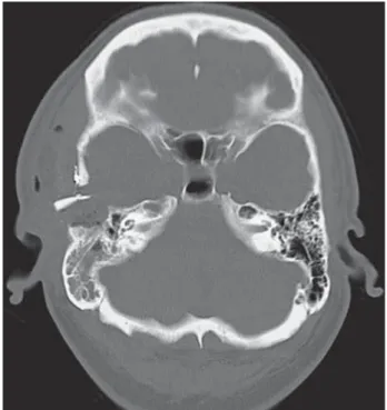
A 50-year-old man with a five-year history of right-sided hearing loss and TMJ pain pre-sented with constant, non-pulsatile tinnitus. The otologic examination at presentation was unremarkable. Imaging studies revealed a de-structive lesion in the right infratemporal fossa and glenoid fossa, extending into the overlying temporal bone. An ill-defined lytic mass within the right temporal bone was documented on CT (Figure 1A), with slightly enhancing soft tis-sue components extending over the roof of the glenoid fossa without gross abnormality of the mandibular condyle. The margins of the lesion had a scalloped appearance. There was exten-sion to the external auditory canal and eroexten-sion along both the anterior and posterior margins of the canal. This mass showed increased F18-FDG uptake in the temporal bone (max SUV of 6.4) and mildly increased FDG uptake in the soft tissue of the external auditory canal (max
A B C
Figure 1 A) Preoperative axial CT scan in bone algorithm shows an ill-defined lytic mass within the right temporal bone. B) Pr-eoperative PET-CT with corresponding area of increased avidity for F18-FDG. C) PrPr-eoperative axial T2-weighted imaging reveals predominantly hypointense right temporal mass.
A B C D E
Figure 2 A) Initial biopsy showing chondroid matrix with “chicken wire” calcification (arrow) resembling chondroblastoma. H&E, original magnification, ×20. B) Resection specimen showing conventional tenosynovial giant cell tumor with mononuclear histio-cyte-like cells, scattered osteoclast-type giant cells and hemosiderin pigment. H&E, original magnification, ×20. C) Tenosynovial giant cell tumor with chondroid metaplasia showing abundant hemosiderin pigment on the left side and chondroid area in the center. H&E, original magnification, ×10. D) Closer view of the interface between classic tenosynovial giant cell tumor and area of chondroid metaplasia. H&E, original magnification, ×20. E) Multinucleated giant cells and a subpopulation of the mononuclear cells are positive for CD68. CD68 immunostain, original magnification, ×20.
Figure 3 Immediate postoperative axial CT scan in bone algo-rithm reveals a surgical bed with smooth bony margins.
SUV of 3.5) (Figure 1B). On MR imaging the soft tissue mass presented a predominantly hypointense signal on T1- and T2-weighted im-ages (Figure 1C). There was no significant ad-enopathy in the neck region.
A biopsy was initially performed through a right preauricular incision, which exposed tu-mor invading the cartilaginous portion of the external auditory canal and the superior and medial aspects of the glenoid fossa. The cap-sule of the condyle was resected, where there was obvious destruction and tumor, as well as the nidus of the origin of the tumor. A tempo-roparietal approach then allowed resection of the remaining temporal bone tumor. The ini-tial biopsy revealed histological features simi-lar to a chondroblastoma of bone, with “chicken wire” calcifications within a chondroid matrix (Figure 2A). Pathological examination of the resection specimen showed tumor involv-ing synovium and bone with classic features of TGCT (Figure 2B), including mononuclear histiocyte-like cells, osteoclast-type giant cells, extensive hemosiderin deposition and areas of chondroid metaplasia (Figure 2C and/or 2D), focally resembling chondroblastoma. The tu-mor cells were diffusely positive for CD68 and focally positive for S100 protein. The final di-agnosis was tenosynovial giant cell tumor with chondroid metaplasia.
Postoperative CT and MR studies showed soft tissue material throughout the operative bed representing a flap and inflammatory op-erative changes. Of note were smooth post sur-gical bony margins (Figure 3). No suspected re-sidual tumor was observed at that time.
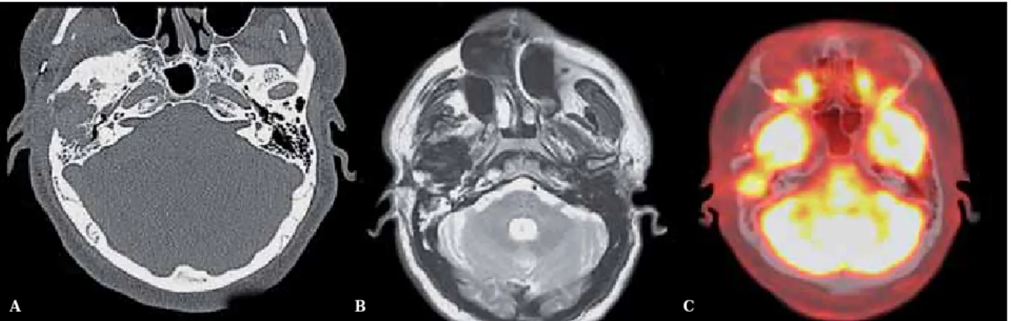
How-ever, MR and CT imaging performed six years after the surgery again showed a lytic lesion with scalloped bony margins extending into the greater wing of the sphenoid bone associated with mildly enhancing soft tissue extending back into the mastoid and middle ear (Figure 4A,B). These features were suspicious for re-current tumor, confirmed on a new PET/CT, which showed a mass with increased 18-FDG uptake in the operative bed of the right tem-poral bone (Figure 4C). Additionally there was a diffuse sclerotic reaction in the greater sphe-noid wing and right mandibular condyle, and there was an enhanced soft tissue thickening around the lateral aspect of external auditory canal, compatible with postoperative changes. The suspected recurrence was biopsy proven and wide surgical removal was performed.
Discussion
Tenosynovial giant cell tumor (TGCT) is de-fined by the World Health Organization as a locally aggressive neoplasm composed of syno-vial-like mononuclear cells admixed with multi-nucleate giant cells, foam cells, siderophages and inflammatory cells. It affects most com-monly the knee and hip in young adults 1.
In-volvement of TMJ is rare. In the current lit-erature there are 58 cases described with one third of them having intracranial involvement
2. Some authors advocate that the majority of
tumors affecting the TMJ are extraarticular, more infiltrative, extend to the temporal bone, and have higher recurrence rates 1,5. By
defi-A B C
Figure 4 A) Axial CT scan in bone algorithm, of 6 years of follow-up, shows a lytic lesion with irregular, ill-defined bony margins. B) Follow-up MR with axial T2-weighted imaging hypointensity, not specific requiring correlation with PET avidity for F18-FDG. C) PET-CT shows increased F18-FDG avidity in the temporal portion of the surgical bed, reflecting recurrence.
nition, the extraarticular form of TGCT con-sists of an infiltrative soft tissue mass, with or without involvement of the adjacent joint 1.
The macroscopic appearance is usually multin-odular, instead of presenting the typical villous patterns seen in the intraarticular forms 1.
Skull base involvement and intracranial extension have been previously described 3-6
as has a case presenting as a middle cranial fossa mass 7. The rare malignant form of TGCT
with metastatic lesions in the lungs and lymph nodes has also been documented 8.
Recurrence is common, may be multiple, with 33-50% developing as the extraarticular form 1. As recurrences are associated with
posi-tive surgical margins, wide excision should be the treatment of choice 1,9. Cai et al. reported a
recurrence rate of 11% for TGCT of the TMJ6. Genetic studies have revealed a translocation involving the genes CSF-1 on chromosome 1 and COL6a3 on chromosome 2 in primary le-sions, as well as recurrences and metastases 1,10.
Clinically, patients usually have symptoms for many years and the most common com-plaints are preauricular swelling, pain or limita-tion of molimita-tion 1,2,9,11. Imaging features of TGCT of
the TMJ consist of a soft tissue mass frequently associated with bone erosion with scalloped margins and cyst formation as better depicted with CT and areas of hyperattenuation due to iron deposition 9. On MR, the nodular masses
usually exhibit low signal intensity on T1- and T2-weighted images, reflecting characteristic hemosiderin deposition 2,9,11. However the MR
appearance may vary depending on the propor-tions of lipid, hemosiderin, fibrous stroma, pan-nus, fluid, and cellular elements6. MR is sensi-tive and specific for the diagnosis of TGCT and also helps in surgical planning. As in our case, avidity on FDG PET/CT has been described 12.
Histopathological features include synovial-lined spaces, blood-filled pseudoalveolar spaces in 10% of the cases, mitotic activity, variable cellularity, with small or large histiocyte-like cells, osteoclast-like multinucleated giant cells and hemosiderin deposits 1,2,13. Giant cells are
positive for CD68 and CD45, and mononu-clear cells are positive for CD681. Interestingly O’Connell et al. document that 67% of TGCT contain dendritic cells expressing S100 protein
14. A minority of cells are neoplastic and
overex-press CFS 12,13. Cartilaginous and osseous
meta-plasia are rare 13.
In our case there was a mixed
immunophe-notype pattern with histiocyte-like and oste-oclast-like giant cells positive for CD68, and also mononuclear cells in chondroid areas positive for S100 protein. This mixed histopa-thology was previously reported by Oda et al. who documented two cases and by Hoch et al. 13 who documented five cases of TGCT
with chondroid metaplasia of the TMJ con-taining hyaline chondroid nodules and dys-trophic calcification in chondroid areas, his-tologically mimicking chondroblastoma. Al-though TGCT of the TMJ is rare, interestingly the TMJ and temporal bone are the most
common sites of skull chondroblastoma 13.
Chondroblastoma should be in the differen-tial diagnosis, as imaging features may over-lap, and the histopathological distinction from TGCT may be hard to establish 13. The
poten-tial relationship to chondroblastoma remains unclear 13. The differential diagnosis of TGCT
on MRI also includes synovial chondromato-sis, tumoral calcium pyrophosphate dihydrate crystal deposition disease, rheumatoid arthri-tis, synovial sarcoma, hemophilia, and syno-vial hemangioma 2,3,9,13. The occurrence of TGCT
with synovial chondromatosis is also reported in the literature 15, but chondromatosis is
char-acterized by the presence of loose bodies (hya-line cartilage) with minimal calcification.
Finally, complete excision with wide margins is the treatment of choice 4. Radiation therapy
may be considered when vital structures are involved and to prevent recurrence 4,8. We
sug-gest that MR and PET/CT, when available, should be strongly considered in the follow-up to monitor tumor recurrence 7.
Conclusion
TGCT of the TMJ with chondroid metapla-sia is benign but locally destructive. Although the definite diagnosis is histological, CT, MR and now FDG PET/CT imaging play an impor-tant role in the preoperative setting, excluding other important etiologies and suggesting the diagnosis of TGCT if hemosiderin deposition is documented. Our case reinforces evidence that TGCT with chondroid metaplasia should be considered in the differential diagnosis of TGCT of the TMJ. The gold standard treatment is complete resection with good long-term out-come. Our case suggests that the best imaging to assess a recurrence may be MR and PET/CT.
References
1 Fletcher CDM, Bridge JA, Hogendorn PCW, et al. World Health Organization classification of tumours of soft tissue and bone. Lyon: International Agency for Research on Cancer (IARC) Press; 2013.
2 Romañach MJ, Brasileiro BF, León JE, et al. Pig-mented villonodular synovitis of the temporomandibu-lar joint: case report and review of the literature. Oral Surg Oral Med Oral Pathol Oral Radiol Endod. 2011; 111 (3): e17-28. doi: 10.1016/j.tripleo.2010.11.019. 3 Tosun F, Carrau RL, Weissman J. Pigmented
vil-lonodularsynovitis of the temporomandibular joint: an extensive case with skull-base involvement. Am J Otolaryngol. 2004; 25 (3): 204-207. doi: 10.1016/j.am-joto.2003.11.006.
4 Day JD, Yoo A, Muckle R. Pigmented villonodular syn-ovitis of the temporomandibular joint: a rare tumor of the temporal skull base. J Neurosurg. 2008; 109 (1): 140-143. doi: 10.3171/JNS/2008/109/7/0140.
5 Herman CR, Swift JQ, Schiffman EL. Pigmented vil-lonodular synovitis of the temporomandibular joint with intracranial extension: a case and literature re-view. Int J Oral Maxillofac Surg. 2009; 38 (7): 795-801. doi: 10.1016/j.ijom.2009.02.013.
6 Cai J, Cai Z, Gao Y. Pigmented villonodularsynovitis of the temporomandibular joint: a case report and the literature review. Int J Oral Maxillofac Surg. 2011; 40 (11): 1314-1322. doi: 10.1016/j.ijom.2011.03.003. 7 Liu Y, Chan J, Chang C, et al. Pigmented villonodular
synovitis of the temporomandibular joint presenting as a middle cranial fossa tumor. J Oral Maxillofac Surg. 2012; 70 (2): 367-372. doi: 10.1016/j.joms.2011.02.031. 8 Yoon HJ, Cho YA, Lee JI, et al. Malignant pigmented
villonodularsynovitis of the temporomandibular joint with lung metastasis: a case report and review of the literature. Oral Surg Oral Med Oral Pathol Oral Ra-diol Endod. 2011; 111 (5): e30-36. doi: 10.1016/j.tri-pleo.2010.11.031.
9 Kim KW, Han MH, Park SW, et al. Pigmented villon-odularsynovitis of the temporomandibular joint: MR findings in four cases. Eur J Radiol. 2004; 49 (3): 229-234. doi: 10.1016/S0720-048X(03)00099-8.
10 Layfield LJ, Meloni-Ehrig A, Liu K, et al. Malignant giant cell tumor of synovium (malignant pigmented vil-lonodular synovitis). Arch Pathol Lab Med. 2000; 124 (111): 1636-1641.
11 Lee JH, Kim YY, Seo BM, et al. Extra-articular pig-mented villonodular synovitis of the temporoman-dibular joint: case report and review of the literature. Int J Oral Maxillofac Surg. 2000; 29 (6): 408-415. doi: 10.1034/j.1399-0020.2000.290603.x
12 Kitapci MT, Coleman RE. Incidental detection of pigmented villonodular synovitis on FDG PET. Clin Nucl Med. 2003; 28 (8): 668-669. doi: 10.1097/01. rlu.0000079430.82897.b8.
13 Hoch BL, Garcia RA, Smalberger GJ. Chondroid tenosynovial giant cell tumor: a clinicopathologi-cal and immunohistochemiclinicopathologi-cal analysis of 5 new cases. Int J Surg Pathol. 2011; 19 (2): 180-187. doi: 10.1177/1066896910381899.
14 O’Connell JX, Fanburg JC, Rosenberg AE. Giant cell tumor of tendon sheath and pigmented villonodu-lar synovitis: immunophenotype suggests a synovial cell origin. Hum Pathol. 1995; 26 (7): 771-775. doi: 10.1016/0046-8177(95)90226-0.
15 Cai XY, Yang C, Chen MJ, et al. Simultaneous pig-mented villonodular synovitis and synovial chondro-matosis of the temporomandibular joint: case report. Int J Oral Maxillofac Surg. 2009; 38 (11): 1215-1218. doi: 10.1016/j.ijom.2009.06.014.
Sofia Pina, MD
Neuroradiology Fellowship Serviço de Neurorradiologia Hospital de Santo António Centro Hospitalar do Porto Largo Prof. Abel Salazar 4099-001 Porto 4099-001 Portugal
Tel.: +351962502494 E-mail: sofiapina@mail.com