401
Vieira SC et al. Breast hemangioma mimicking metastasis at PET-CT
Radiol Bras. 2011 Nov/Dez;44(6):401–402
Breast hemangioma mimicking metastasis at PET-CT
*
Hemangioma de mama simulando metástase no PET-CT
Sabas Carlos Vieira1, Jucélia Saraiva e Silva2, Eveline Brandão Madeira3, Júlio César Queiroz de França3, Sebastião Nunes Martins Filho3
Breast hemangioma is a rare benign tumor that presents either absent or low 18
F-fluoro-2-deoxy-D-glucose (FDG) uptake at positron emission tomography (PET). The authors report the case of a breast nodule pathologically compatible with hemangioma in a woman whose PET-scan has demonstrated increased FDG uptake (simulating a malignant tumor). A brief review of factors leading to false positive and false negative PET results is also undertaken.
Keywords: PET/CT; FDG-18
F; Hemangioma; Breast.
Hemangioma de mama é um tumor benigno raro que apresenta pouca ou nenhuma captação de 18
F-flúor-2-deoxi-D-glicose (FDG) na tomografia por emissão de pósitrons (PET). Relatamos um nódulo mamário compatível,
patologica-mente, com hemangioma, em uma mulher cuja PET scan demonstrou captação elevada de FDG (simulando tumor
maligno). Também fizemos breve revisão das causas que levam a resultados falso-positivos e falso-negativos pela PET.
Unitermos: PET/CT; FDG-18
F; Hemangioma; Mama.
Abstract
Resumo
* Study developed at Universidade Federal do Piauí (UFPI), Teresina, PI, Brazil.
1. MD, Oncologist and Breast Specialist, President of Socie-dade Brasileira de Mastologia, Full Professor, Discipline of On-cology at Faculdade de Medicina da Universidade Federal do Piauí (UFPI), Teresina, PI, Brazil.
2. MD, Pathologist, Physician at Department of Pathology and Medical Practice, MedImagem, Teresina, PI, Brazil.
3. Graduate Students of Medicine, Universidade Federal do Piauí (UFPI), Teresina, PI, Brazil.
Mailing Address: Eveline Brandão Madeira. Rua Fidalma Mar-tins de Carvalho, 4355, Bl-06, ap. 204, Ininga. Teresina, PI, Brazil, 64048-480. E-mail: [email protected]
Received January 14, 2011. Accepted after revision June 21, 2011.
Vieira SC, Silva JS, Madeira EB, França JCQ, Martins Filho SN. Breast hemangioma mimicking metastasis at PET-CT. Radiol Bras. 2011 Nov/Dez;44(6):401–402.
0100-3984 © Colégio Brasileiro de Radiologia e Diagnóstico por Imagem CASE REPORT
The final histopathological study revealed the presence of a benign vascular neoplasm measuring 1.6 × 1.5 × 0.5 cm, consisting of thin-walled, ectatic, congested blood vessels inserted in the conjunctival stroma, and absence of atypias, compatible with capillary hemangioma.
DISCUSSION
Capillary hemangiomas are benign vas-cular tumors characterized by proliferation of capillary vessels(4). Breast hemangiomas
primarily affect post-menopausal women and may increase in size in the setting of hormone replacement therapy(5).
Several imaging methods are available for detection, diagnosis and decision mak-ing on the approach to be adopted in the setting of breast diseases. However, mam-mography still remains as the most relevant imaging technique for breasts. In order to standardize mammographic findings re-porting, the BI-RADS classification was developed, subdividing imaging findings into five classes as follows: negative, be-nign, probably bebe-nign, suspicious and highly suspicious of malignancy (6).
Some studies have reported the finding of breast hemangiomas as well circum-scribed, macrolobular lesions that may con-The present study reports a case of
breast hemangioma in a woman whose PET/CT scan has demonstrated increased FDG uptake (simulating a malignant tu-mor). A brief review of factors leading to false positive and false negative PET re-sults is also undertaken.
CASE REPORT
At clinical examination, a female, 63-year-old patient presented a nodule measur-ing about 1.5 cm in the junction of the lower quadrants of her right breast. The nodule had partially defined contours and fibroelastic consistency. The axilla was clinically negative. The patient presented a previous history of surgery for a colon tu-mor, with adjuvant chemotherapy and that later progressed with development of pul-monary metastasis.
Mammography demonstrated a partially delimited, lobulated nodular lesion in the right breast, and ultrasonography revealed a solid lesion measuring 1.8 × 0.7 cm, that was isoechoic in relation to the breast tis-sue, classified as BI-RADS 4.
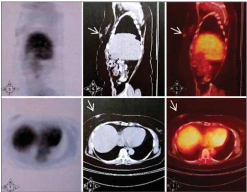
The patient was submitted to FDG-PET/ CT that demonstrated increased FDG up-take by the breast nodule (Figure 1). Such finding determined the nodule resection.
INTRODUCTION
Hemangiomas are benign vascular tu-mors, rarely found in the breast, which present low 18F-fluoro-2-deoxy-D-glucose (FDG) uptake at positron emission tomog-raphy (PET), being differentiable from malignant tumors(1).
Positron emission tomography with FDG has been utilized to differentiate ma-lign from benign lesions, since this imag-ing modality can detect the glucose me-tabolism that is generally greater in malig-nant than in benign tumors(2). However,
402
Vieira SC et al. Breast hemangioma mimicking metastasis at PET-CT
Radiol Bras. 2011 Nov/Dez;44(6):401–402 tain calcification(7,8). However, such
find-ings are nonspecific, which may explain the significant number of hemangiomas classified either as BI-RADS 3 or 4 and the non differentiation from fibroadenomas or cysts(8).
PET/CT detects glycolytic hyperactiv-ity of malignant cells through FDG (a glu-cose analogue) uptake. Such uptake is caused by an increase in the number of glu-cose transporter proteins and in hexokinase and phosphofructokinase levels which pro-mote glycolysis(3). Once phosphorylated at
FDG-6-phosphate by hexokinase, struc-tural changes prevent FDG to be catabo-lized or transported back into the extracel-lular space, being selectively accumulated within tumor cells(2).
False-negative results may be observed at PET scan in tumors with low glycolytic activity such as adenomas, low-grade lym-phomas and small-sized lesions(9).
Addi-tionally, in cases of disease adjacent to sites of physiological uptake (heart, kidneys,
bladder and liver), FDG-PET should be supplemented with other imaging modali-ties to confirm the results(3). FDG-PET scan
performed within up to one month follow-ing chemotherapy may present decreased sensitivity because of the reduced number of metabolically active tumor cells, which is not always predictive of a good re-sponse(2).
On the other hand, false-positive results may be observed in infectious and inflam-matory diseases where activated macroph-ages and neutrophils show increased FDG accumulation, since they utilize glucose as a source of energy for chemotaxis and ph-agocytosis(10). It has been suggested that
inflammatory cells utilize more glucose under hyperglycemic than under eugly-cemic conditions and, therefore, lesions containing such cells are most frequently interpreted as malignant lesions under hy-perglycemic conditions(3).
Accumulation of FDG in hemangiomas may be related to blood retention in the Figure 1. Positron emission tomography/computed tomography (FDG-PET/TC) demonstrating increases FDG uptake in a breast (arrows).
lesion, resulting in focal ischemia. Then, the secondary hypoxia may accelerate the anaerobic glycolysis, leading to a high FDG uptake(11).
CONCLUSION
Despite its significant contribution to the evaluation, diagnosis and treatment of cancer patients, FDG-PET/CT may present false-positive and false-negative results. Therefore, the understanding of the causes of false results is critical to avoid equivo-cal diagnoses.
REFERENCES
1. Sakurai K, Haram M, Ozawa Y, et al. Thoracic hemangiomas: imaging via CT, MR, and PET along with pathologic correlation. J Thorac Im-aging. 2008;23:114–20.
2. Selzner M, Hany TF, Wildbrett P, et al. Does the novel PET/CT imaging modality impact on the treatment of patients with metastatic colorectal cancer of the liver? Ann Surg. 2004;240:1027– 34; discussion 1035–6.
3. Chang JM, Lee HJ, Goo JM, et al. False positive and false negative FDG-PET scans in various tho-racic diseases. Korean J Radiol. 2006;7:57–69.
4. Courcoutsakis NA, Hill SC, Chow CK, et al. Breast hemangiomas in a patient with Kasabach-Merritt syndrome: imaging findings. AJR Am J Roentgenol. 1997;169:1397–9.
5. Mariscal A, Casas JD, Balliu E, et al. Breast he-mangioma mimicking carcinoma. Breast. 2002; 11:357–8.
6. Vieira AV, Toigo FT. Classificação BI-RADS: categorização de 4.968 mamografias. Radiol Bras. 2002;35:205–8.
7. Siewert B, Jacobs T, Baum JK. Sonographic evaluation of subcutaneous hemangioma of the breast. AJR Am J Roentgenol. 2002;178:1025–7.
8. Mesurolle B, Sygal V, Lalonde L, et al. Sono-graphic and mammoSono-graphic appearances of breast hemangioma. AJR Am J Roentgenol. 2008;191: W17–22.
9. Carter KR, Kotlyarov E. Common causes of false positive F18 FDG PET/CT scans in oncology. Braz
Arch Biol Technol. 2007;50(special number):29– 35.
10. Shim SS, Lee KS, Kim BT, et al. Focal parenchy-mal lung lesions showing a potential of false-positive and false-negative interpretations on in-tegrated PET/CT. AJR Am J Roentgenol. 2006; 186:639–48.