UNIVERSIDADE DA BEIRA INTERIOR
Ciências da Saúde
Purification of HPV16 E6/E7 DNA plasmid-based
vaccine using a modified monolithic support
Alexandra Sabrina Antunes Soares
Dissertação para obtenção do Grau de Mestre em
Ciências Biomédicas
(2º ciclo de estudos)
Orientadora: Profª. Doutora Ângela Sousa
Coorientadora: Profª. Doutora Fani Sousa
ii
“You've got to find what you love. And that is as true for
your work as it is for your lovers. Your work is going to
fill a large part of your life, and the only way to be truly
satisfied is to do what you believe is great work. And the
only way to do great work is to love what you do. If you
haven't found it yet, keep looking. Don't settle. As with
all matters of the heart, you'll know when you find it.
And, like any great relationship, it just gets better and
better as the years roll on. So keep looking until you find
it. Don't settle.”
iii
To the most important people of my life,
whom I love very much,
My parents…
iv
Acknowledgments
First, I specially thank my supervisors Professor Ângela Sousa e Professor Fani Sousa for believing in me and my ability to develop this work, and for all the patience, help and knowledge they transmitted to me along the past year.
To Professor Doctor João Queiroz, rector from University of Beira Interior, I would like to express my gratitude for making possible the development of this investigation project.
I would also like to express my gratitude to the people at the Biotechnology and Biomolecular Sciences group of the Health Sciences Research Centre at University of Beira Interior for all their help, with a special acknowledge to Patricia Pereira and Marta Silva for their friendship, complete availability and support.
I would like to thank my friends for all the coffee, long talks, patience and full support, especially to Cátia, Elisabete, Pedro, Rui, Stéphanie and Vanessa.
To my family, especially my godparents, Maria de Lurdes e José Orlando, my grandmother Carminda, my “sister” Andrea and my brother Óskar for all their love and support in the most difficult times.
Finally, I will be eternally grateful to my parents for all the love, sacrifices, good advice and support they gave me all my life.
v
Abstract
Human papillomavirus (HPV) is one of the most common sexually transmitted diseases in the world and has been associated with several human cancers, like cervical cancer. Thus, effective vaccination against Human papillomavirus represents an opportunity for the control of this cancer. The development of therapeutic Human papillomavirus vaccines is required to facilitate the control and eliminate on of a preexisting Human papillomavirus infection. In the last years, the expansion of efficient plasmid DNA purification processes has fostered therapeutics applications like gene therapy and DNA vaccination. Recently, the application of chromatographic operations based on affinity interactions between plasmid DNA or impurities with specific amino acids immobilized in stationary phases has demonstrated good results in the supercoiled plasmid DNA purification. Despite of selectivity achieved with these ligands, conventional matrices present limitations such as the low binding capacity and diffusivity for plasmid DNA samples. Owing to bottlenecks associated to conventional matrices, monolithic supports have emerged as interesting alternatives due to the versatility of their structural characteristics. The research work present in this thesis describes a new strategy that combines the selectivity of arginine as affinity ligand with the versatility of the epoxy-based monoliths to efficiently purify the supercoiled HPV-16 E6/E7 plasmid from other plasmid isoforms and Escherichia coli impurities present in clarified lysate. Additionally, breakthrough experiments were designed to compare the dynamic binding capacity of plasmid DNA to the conventional arginine-agarose matrix with the modified monolithic support. The dynamic binding capacity obtained for the arginine-epoxy monolith was significantly higher than the capacity achieved in the arginine conventional support. Quality control tests indicated that the plasmid sample resultant from the purification step presented a purity degree approximately 100% and an homogeneity higher than 97% of supercoiled isoform, with an extremely reduced level of impurities (RNA, proteins, genomic DNA and endotoxins). Overall, given that the plasmid DNA final product meets regulatory specifications, this combined support can be the key to obtain an adequate non-viral vaccine against a Human papillomavirus infection.
Keywords
Affinity chromatography, arginine ligand, Human papillomavirus, DNA vaccines, modified monolithic support, supercoiled plasmid DNA
vi
Resumo
O Papiloma Vírus Humano é um vírus sexualmente transmissível que está relacionado com o desenvolvimento de vários cancros, como o cancro do colo do útero. Este tipo de cancro é a segunda maior causa de morte em mulheres, afetando mundialmente cerca de meio milhão de mulheres, das quais aproximadamente 274 mil morrem. A evidente associação que existe entre o Papiloma Vírus Humano e o cancro do colo do útero torna o Papiloma Vírus Humano um alvo interessante para o desenvolvimento de vacinas, no sentido de prevenir ou tratar o desenvolvimento do cancro. Atualmente, já são comercializadas duas vacinas preventivas, a Gardasil da Merck e a Cervarix da GlaxoSmithKline. Apesar de ambas mostrarem ser eficazes e seguras, possuem algumas limitações como elevado custo, não protegem contra todos os tipos de Papiloma Vírus Humano e trata-se de vacinas exclusivamente preventivas. Assim o desenvolvimento de vacinas terapêuticas pode ser uma estratégia promissora para colmatar estas falhas.
A utilização do DNA plasmídico (pDNA) como uma vacina não viral tem-se tornado numa potencial estratégia terapêutica para prevenir ou tratar determinadas doenças de forma menos invasiva e segura comparando com os vetores virais. O mecanismo de actuação desta vacina baseia-se na expressão de proteínas antigénicas que desencadeiam uma resposta imunológica, evitando a progressão da doença . A preparação destas vacinas de DNA plasmídico requer o desenvolvimento de processos de produção e purificação que permitam obter grandes quantidades de plasmídeo na sua forma biologicamente ativa (isoforma superenrolada (sc)), cumprindo os requisitos das agências reguladoras no que diz respeito ao grau de pureza. Da ocorrência natural de complexos proteínas-DNA em sistemas biológicos sugeriu o desenvolvimento de uma estratégia de cromatografia de afinidade, utilizando matrizes convencionais de agarose com determinados aminoácidos imobilizados que reconhecem especificamente a isoforma superenrolada do DNA plasmídico. No entanto, as matrizes convencionais apresentam algumas limitações quando comparadas com os suportes monolíticos que possuem excelentes propriedades de transferência de massa e elevada capacidade de ligação para moléculas de grandes dimensões como o DNA plasmídico.
Desta forma, o trabalho apresentado nesta tese consistiu na modificação de um monolito de epoxy por imobilização de aminoácidos de arginina, conjugando assim a selectividade deste ligando com a versatilidade do monolito de epoxy, tendo como finalidade purificar a isoforma sc do pDNA HPV-16 E6/E7. Numa fase inicial, foram realizados vários ensaios com amostras de DNA plasmídico pre-purificadas com o kit comercial, no sentido de avaliar e confirmar a presença dos ligandos de arginina, comparando o comportamento cromatográfico do monolito modificado com o mesmo suporte não modificado. Os resultados comprovaram que o monolito modificado reconhece especialmente o DNA plasmídico, permitindo a separação das isoformas superenrolada e circular aberta através de um gradiente por passos de NaCl. O mesmo não aconteceu com o monolito de epoxy não modificado, pois não ocorreu qualquer tipo de
vii
interacção do plasmídeo com o suporte. Posteriormente foi realizado um estudo de capacidade de ligação dinâmica para completar a caracterização do monolito com ligandos de arginina e comparar com a coluna convencional de arginina-argarose. Os resultados comprovaram que, para as mesmas condições de caudal e concentração de pDNA HPV-16 E6/E7, o monolito possui uma capacidade de ligação significativamente superior em comparação com a coluna convencional. Uma vez conseguida a separação das isoformas e a caracterização do monolito modificado com a arginina, a segunda fase do trabalho consistiu na purificação da isoforma sc do pDNA HPV-16 E6/E7 a partir de um lisado complexo de
Escherichia coli. Os testes de controlo de qualidade revelaram que a amostra de sc de pDNA
HPV-16 E6/E7, resultante da purificação com o monolito de epoxy modificado, apresentava um grau de pureza de aproximadamente 100% e uma homogeneidade superior a 97%. Para além disso, constituintes do hospedeiro como RNA e proteínas não foram detectados na amostra purificada e a quantidade de DNA genómico e endotoxinas estavam a baixo dos valores referenciados pelas agências reguladoras como a Food and Drug Delivery.
Em suma, a combinação de ligandos de aminoácidos com suportes monolíticos pode ser uma solução promissora para obtenção de uma vacina não-viral baseada na isoforma sc do pDNA HPV-16 E6/E7, com o grau de pureza requerido para futuras aplicações terapêuticas contra a infecção por Papiloma Vírus Humano.
Palavras-chave
Cromatografia de afinidade, ligando de arginina, monolito de epoxy modificado, Papiloma Vírus Humano, Vacinas de DNA
viii
Table of Contents
Chapter I - Introduction
... 1 Introduction... 2 1.Human papillomavirus ... 4 1.1.Molecular biology of HPV ... 51.1.1.Human papillomavirus E6 oncoprotein ... 7
1.1.2.Human papillomavirus E7 oncoprotein ... 8
1.2.Preventive HPV vaccines ... 9
1.3.Therapeutic HPV vaccines ... 10
2.Gene Therapy and DNA Vaccines ... 11
2.1.DNA vaccination ... 11
2.2.Viral and Non-viral vectors ... 13
2.2.1.Viral delivery systems ... 14
2.2.2.Non-viral delivery system ... 15
3.Plasmid DNA as non-viral vector ... 17
3.1.Plasmid DNA manufacturing ... 17
3.2.Downstream process ... 18
3.3.Plasmid DNA Purification ... 19
3.3.1.Size-exclusion chromatography (SEC) ... 20
3.3.2.Anion-exchange chromatography (AEC) ... 20
3.3.3.Hydrophobic interaction chromatography (HIC) ... 20
3.3.4.Affinity chromatography (AC) ... 21
3.4.Monolithic Technology... 23
Chapter II -
Material and Methods
... 242.1. Materials... 25
2.1.1. Plasmid DNA ... 25
2.2. Methods ... 26
2.2.1. Plasmids and bacterial growth conditions ... 26
2.2.2. Alkaline lysis with Qiagen Kit ... 26
ix
2.2.4. Affinity chromatography ... 27
2.2.5. Agarose gel electrophoresis ... 28
2.2.6. Dynamic binding capacity ... 28
2.2.7. Plasmid quantification ... 29
2.2.8. Protein quantification ... 30
2.2.9. Genomic DNA quantification ... 30
2.2.10. Endotoxin quantification ... 31
Chapter III -
Results and discussion
... 323.1. Plasmid amplification in recombinant E. coli and electrophoresis optimization ... 33
3.2.Separation of sc pDNA from oc pDNA ... 37
3.3.Dynamic binding capacity ... 42
3.4.sc pDNA purification from the clarified E. coli lysate ... 44
3.5.Analytical parameters for plasmid quality assessment ... 52
Chapter IV - Conclusions and futures perspectives
... 58x
List of Figures
Chapter I – Introduction
Figure 1. Structure and genome organization of the HPV-16. ... 5
Figure 2. Effect of the interaction between E6 protein and p53 tumor suppressor. ... 8
Figure 3. Effect of the interaction between the E7 protein and pRB protein... 9
Figure 4. DNA vaccines stimulate the induction of both cellular and humoral responses ... 13
Figure 5. The three stages necessaries to obtain a pure supercoiled plasmid DNA ... 18
Figure 6. Principle of affinity chromatography. ... 21
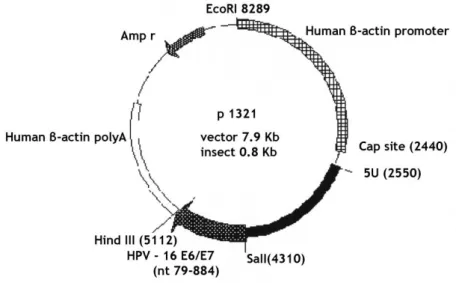
Chapter II - Material and Methods Figure 7. Plasmid HPV-16 E6/E7 backbone. ... 25
Figure 8. Reference curve plasmid DNA standards (1-100 µg/ml). ... 29
Figure 9. Reference curve Bovine Serum Albumin standards (0.01-0.1 mg/ml). ... 30
Figure 10. Reference curve E. coli DH5α genomic DNA standards (0.005 - 50 ng/µL). ... 31
Figure 11. Reference curve endotoxins standards (0.01-0.1 EU/mL). ... 31
Chapter III – Results and Discussion Figure 12. Agarose gel electrophoresis analysis of HPV-16 E6/E7 plasmid sample (stained with 0.25 µg/mL of GeenSafe). ... 34
Figure 13. Agarose gel electrophoresis analysis of HPV-16 E6/E7 plasmid sample, after four days at room temperature (stained with 0.25 µg/mL of GeenSafe) ... 35
xi
Figure 14. Agarose gel electrophoresis analysis of HPV-16 E6/E7 plasmid sample (0.5 µg/mL of ethidium bromide). ... 35
Figure 15. Agarose gel electrophoresis analysis of plasmid sample at different agarose percentages and Greensafe volumes ... 36
Figure 16. Evaluation of the non-immobilized epoxy monolith role in the oc and sc plasmid isoforms separation. ... 37
Figure 17. Evaluation of epoxy-monolith modification by immobilization of arginine amino acid. ... 38
Figure 18. Separation of pDNA isoforms with arginine-epoxy monolith. Elution was performed by stepwise gradient increasing NaCl concentration (600 mM and 2M) ... 40
Figure 19. Separation of pDNA isoforms with arginine-epoxy monolith. Elution was performed by stepwise gradient increasing NaCl concentration (560 mM and 2M) ... 41
Figure 20. Breakthrough experiments with epoxy monolith and conventional arginine-agarose matrix. ... 43
Figure 21. Chromatographic profile of the E. coli lysate sample injection in the arginine-epoxy monolith. Elution was performed by stepwise gradient increasing NaCl concentration (560 mM and 1M) ... 45
Figure 22. Chromatographic profile of the E. coli lysate sample injection in the arginine-epoxy monolith. Elution was performed by stepwise gradient increasing NaCl concentration (0M, 560 mM and 1M) ... 46
Figure 23. Chromatographic profile of the E. coli lysate sample injection in the arginine-epoxy monolith. Elution was performed by stepwise gradient increasing NaCl concentration (0M, 700 mM and 1M) ... 47
Figure 24. Chromatographic profile of the E. coli lysate sample injection in the arginine-epoxy monolith. Elution was performed by stepwise gradient increasing NaCl concentration (0M, 730 mM and 1M) ... 48
Figure 25. Chromatographic profile of the E. coli lysate sample injection in the arginine-epoxy monolith. Elution was performed by stepwise gradient increasing NaCl concentration (0M, 690 mM, 770 mM and 1M) ... 49
xii
Figure 26. Chromatographic profile of the E. coli lysate sample injection in the arginine-epoxy monolith. Elution was performed by stepwise gradient increasing NaCl concentration (600 mM, 740 mM and 1M) ... 50
Figure 27. Chromatographic profile of the E. coli lysate sample injection in the arginine-epoxy monolith. Elution was performed by stepwise gradient increasing NaCl concentration (600 mM, 740 mM, 795 mM and 1M) ... 51
Figure 28. Analytical chromatographic profiles of different pDNA-containing samples recovered throughout the purification process ... 54
xiii
List of tables
Chapter I – Introduction
Table 1. Human papillomavirus (HPV) grouping, according to their risk to cause cervical
cancer ... 4
Table 2. A brief description of the functions of human papillomavirus open-reading frames .. 7
Table 3. Disadvantages of current licensed human papillomavirus vaccines – Cervarix® and Gardasil® ... 10
Table 4. Advantages of DNA vaccines ... 12
Table 5. Advantages and disadvantages of main viral vector groups ... 15
Table 6. Advantages and limitations of several non-viral gene delivery systems ... 16
Table 7. Affinity chromatography methods for plasmid DNA purification ... 22
Chapter III – Results and Discussion Table 8. Summary of binding and elution of oc and sc plasmid isoforms in arginine-epoxy monolithic chromatography. ... 38
Table 9. Comparison of the dynamic binding capacity between arginine-epoxy monolith and conventional arginine-agarose matrix.. ... 44
Table 10. HPLC analysis of concentration, purity and recovery yield of sc HPV-16 E6/E7 pDNA isolated by arginine-epoxy monolith. ... 55
Table 11. Protein, gDNA and endotoxins quantification in several steps of the isolation and purification processes to obtain the sc pDNA from the clarified E. coli lysate. ... 56
xiv
List of Acronyms
AC Affinity chromatography
AEC Anion-exchange chromatography
AP 1 Activator protein 1
APC Antigen-presenting cells
ATPase Adenosine triphosphate
BCA Bicinchoninic acid
BSA Bovine Serum Albumin
ºC Celsius
CDI CarbonylDiImidazole
cDNA Complementary DNA
Cys Cysteines
CR Conserved Regions
CTL Cytolytic T lymphocyte
DC Dendritic cell
DNA Desoxirribonucleic acid
E6AP E6-associated protein
EAEMP European Agency for the Evaluation of Medical Products
E. coli Escherichia coli
EDTA Ethylene-diamine tetraacetic acid
EGF Epidermal growth factor
FDA Food and Drug Administration
g gram
gDNA Genomic DNA
h Hours
H2SO4 Sulfuric acid
H-bond Hydrogen bond
HIC Hydrophobic interaction chromatography
HCl Chloride acid
HPV Human Pappilomavirus
HSIL High-grade squamous intraepithelial lesion
kDa kilo dalton
kbp kilo base pairs
KH2PO4 Monopotassium phosphate
K2HPO4 Dipotassium phosphate
KRF 1 Keratinocyte-specific transcriptional factor 1
L liter
LAL Limulus amebocyte lysate
xv
ln Linear
MHC Major histocompatibility complex
min minutes
mL milliliter
mm millimeter
mRNA Messenger RNA
NaCl Sodium chloride
NaOH Sodium hydroxide
NF-I/CTF Nuclear factor
(NH4)2SO4 Ammonium sulfate
nm Nanometer
oc Open circular
OD600 Optical density at 600 nm
ORF Open reading frame
pb Base pairs
PCR Polymerase chain reaction
PDGF Platelet-derived growth factor
pDNA Plasmid DNA
Rb Retinoblastoma
RNA Ribonucleic acid
rRNA ribosomal RNA
rpm Revolutions per minute
S. cerevisiae Saccharomyces cerevisiae
sc Supercoiled
SDS Sodium dodecylsulfate
SEC Size-exclusion chromatography
siRNA Small interfering RNA
SV40 Simian virus 40
TAE Tris, acetic acid, EDTA
Tris Tris(hydroxymethyl) aminomethane
UV Ultraviolet
V volt
VLP Virus-like particles
W/W Mass/mass
Chapter I
Introduction
2
Introduction
Human papillomavirus (HPV) is one of the most common sexually transmitted diseases in the world and has been identified as an etiological factor for several important cancers, including anogenital cancers, and a subset of head and neck cancers [1]. Among HPV-associated cancers, cervical cancers have the most significant morbidity, being the second largest cause of the cancer deaths in women worldwide [2]. Infection with HPV is a key cause of cervical cancer because the HPV DNA has been detected in 99.7% of patients with cervical cancer [1]. More than 200 HPV genotypes have been identified and are classified into low or high-risk types, depending on their propensity to cause cervical cancer, being the HPV-16 and HPV-18 the most high risk types associated with the cervical cancer [2]. The clearly association between infection with high-risk HPV and cervical cancer showed that HPV serves as an ideal target for development of preventive and therapeutic vaccines. Recently, two licensed prophylactic HPV vaccines, Gardasil (bivalent vaccine comprising HPV types 16 and 18) and Cervarix (quadrivalent vaccine comprising HPV types 6, 11, 16 and 18) have been shown to be safe and capable to generating significant protection against specific HPV types [3]. However, prophylactic HPV vaccination does not have therapeutic effect against established HPV infections and HPV-associated lesions [3]. In this way, the development of therapeutic HPV vaccines can be a promising strategy to accelerate the control of cervical cancer and treat currently infected patients [4].
DNA vaccines have emerged as attractive and potentially effective strategies for treatment of inherent and acquired diseases. The most advantage of this novel vaccine technology is the ability to inducing both humoral and cellular immunity [5]. The underlying principle of DNA vaccines consists in the administration of therapeutic genes for specific proteins that induce the immune responses against pathogen [6]. Central to the concept of DNA vaccination is the ability to deliver exogenous nucleic acids to eukaryotic cells of various tissues. However, the success of DNA vaccination is largely dependent on the development of a vector or vehicle that can selectively and efficiently deliver therapeutic genes into these cells. On the other hand therapeutic genes can be transported by viral vectors but this delivery system has presented higher toxicity and immunogenicity degree [7, 8]. Thus, plasmid DNA (pDNA)-based vaccines are more safe, stable, easy production and prolonged action if compared to virus-based vaccines, becoming the most attractive gene-transfer system to be used as biopharmaceutical product [2].
The successful implementation of clinical approaches using plasmid DNA-based strategies requires the continuous improvement of production and purification processes of pDNA [9]. In this way, different chromatographic techniques, including size-exclusion, anion exchange, hydrophobic interaction, reversed phase and affinity chromatography have been applied for supercoiled (sc) pDNA isolation from the remaining host impurities, with more or less success [10]. Recently, a new affinity chromatography approach using some amino acids as specific binding agents have been efficiently applied in the purification of the sc pDNA from the
3
clarified Escherichia coli (E. coli) lysate [11-13]. Despite of the selectivity achieved with these amino acids, these conventional matrices present some limitations, such as the low capacity of available supports for pDNA and low diffusivity of pDNA samples [14]. Therefore the monolithic stationary phase has been extensively investigated for pDNA purification. This new support has attracted increasing attention because of its easy preparation, structural and functional properties and high performance compared to conventional columns [14].
Hence, the purpose of this thesis is to develop and implement a new strategy for sc (supercoiled) pDNA HPV-16 E6/E7 isoform purification. The affinity purification strategy is based on the combination of the specificity and selectivity achieved with arginine ligand with the advantages provided by a monolithic support. In addition, the characterization of the sc pDNA sample obtained is also envisioned since it should be free from the host contents in compliance with all regulatory requirements and capable of inducing gene expression with high efficiency.
Thus, the work described in this thesis may be divided in three main stages:
- First comprises the biosynthesis of pDNA HPV-16 E6/E7 in E. coli recombinant organism;
- The second stage consists on the implementation of an monolithic affinity purification strategy with immobilized arginine amino acids to separate the sc and oc (open circular) plasmid isoforms;
- The third stage consists on the purification of the sc plasmid isoform from the clarified E. coli lysate;
The final research topic consist in analytical characterization of the sc pDNA recovered the arginine-epoxy monolith.
4
1. Human papillomavirus
Human Papillomavirus infection represents the most common sexually transmitted disease in the world [15]. HPV has been identified as an etiological factor in several important cancers, including a subset of anal, vaginal, vulval, penile, head and neck cancers [1]. Apart from these cancers, cervical cancer is the second largest cause of cancer deaths in women worldwide and HPV infection represents the most important risk factor in its development [16]. HPV DNA has been detected in 99.7% of patients with cervical cancer [15]. It is estimated that half a million of new cases are diagnosed by year and approximately 270.000 of those patients die [4, 17].
Human Papillomavirus are members of the Papovaviridae family [18]. More than 200 HPV types have been identified and are classified based on DNA sequence analysis, it being each one associated with infection in specific epithelial sites [2, 19]. Thus, the HPV types can be divided in two main HPV genera, Alpha and Beta papillomavirus [20]. Beta papillomavirus are typically associated with cutaneous infection, whereas Alpha papillomavirus mainly infect the mucosal/genital areas [21]. Alpha HPV are further subdivided into low, intermediate or high-risk depending on their propensity to cause cervical cancer [18], as it is presented in Table 1. However, Alpha HPV also include some HPV types mostly found in cutaneous lesions, such as HPV-2, which cause common warts [22]. More than 30 HPV types are known to infect cervical epithelium, associated with high-grade squamous intraepithelial lesions (HSILs), which are precursors of cervical cancer [4, 20]. These HPV types are classified as high-risk HPV, being 70% of the cervical cancers attributable to HPV-16 and HPV18 [23]. HPV-16 is considered the most significant high-risk HPV type, being responsible for approximately 50% of cervical cancer [24]. On the other hand, the low-risk HPV type is associated with benign genital warts and it is rarely associated with cervical cancer [25]. Since HPV is present in the majority of cervical cancers, the HPV appearing and infection can be controlled through an adequate vaccination.
Table 1. Human papillomavirus grouping, according to their risk to cause cervical cancer (adapted from
[18]).
HPV groups HPV types
High Risk HPV-16, HPV-18, HPV-45, HPV-56
Intermediate risk HPV-31, HPV-33, HPV-35, HPV-51, HPV-52, HPV-58
5
1.1. Molecular biology of HPV
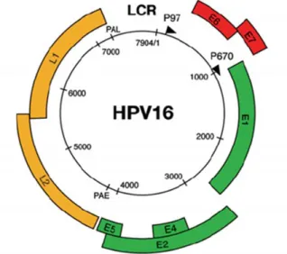
Although there are several differences in the DNA sequence of all HPV types, their structure and genomic organization are similar. Human Papillomavirus are small, non-enveloped with a diameter of 55-60 nm and have an icosahedral capsid composed by 72 capsomers [26, 27]. They contain a circular double-strand, covalently closed DNA of around 8000 base pairs (pb) [7904 bp for HPV16 (GenBank® accession number NC_001526)] in the form of chromatin-like complex with cellular histones [18, 20, 28]. The viral genome is organized in three regions (Figure 1): the long control region (LCR) and two protein-encoding regions (early and late gene regions) [26]. All viral genes are encoded on one strand of HPV genomic DNA.
Figure 1. Structure and genome organization of the HPV-16. The HPV-16 genome (7904 bp) is shown as a
black circle with the early (p97) and late (p670) promoters marked by arrows. Early ORFs (E1, E2, E4 and E5) are represented in green, while the other early ORFs (E6 and E7) are in red. The late ORFs (L1 and L2) are represented in yellow. All the viral genes are encoded on one strand of the double-stranded circular DNA genome. The long control region is showed between yellow and red regions (adapted from [20]).
Long control region represents approximately 10% of the genome and unlike other two regions does not encode for proteins [26, 29]. Indeed, the LCR contains numerous binding sites for many different transcriptional repressors and transcriptional activators, including activator protein 1 (AP 1), keratinocyte-specific transcriptional factor 1 (KRF 1), nuclear factor (NF-I/CTF) and virally derived transcriptional factors encoded by early and late regions [27, 29]. Thus, LCR is responsible by transcription control from early and late regions and controls the production of viral proteins and infectious particles. LCR also regulates the viral gene expression, suggesting that it may play a critical role in determining the range of hosts for specific HPV types [27].
The late gene region encodes 40% of the HPV genome and contains two separate open reading frames (ORFs), L1 and L2 that are structural components of the viral capsid [2, 26]. The L1
6
gene encodes the major (55 kDa) capsid protein and forms the main architectural structure of the icosahedral viral capsid [21, 26]. This protein is highly conserved among different papillomavirus species [29]. On the other hand, the L2 gene encodes the minor (70 kDa) capsid protein and it has more sequence variations than L1 protein [26, 27]. Both capsid proteins are only expressed in terminally differentiated squamous epithelial cells [28]. The expression of L1 and L2 proteins is tightly regulated and associated with differentiation of infected epithelial cells [2].
Finally, the early gene region is the major segment of the viral genome encoding 50% of the total genome [26]. This region is downstream of the LCR region and is composed of six ORFs, E1, E2, E4, E5, E6 and E7 [27]. The E genes encode nonstructural proteins that regulate virus transcription and replication and are expressed in nonproductively infected cells and transformed cells. Depending upon the HPV type, the E4, E5 and E7 genes usually encode a single polypeptide, whereas the E1, E2 and E6 genes may be expressed as several related polyproteins through differential splicing [26]. Thereby, early proteins play a critical role in regulation of viral DNA replication (E1 and E2), viral RNA transcription (E2), cytoskeletal reorganization (E4) and cellular transformation (E5, E6 and E7) [2]. In Table 2 are summarized the functions of each viral protein.
The E1 gene encodes polypeptides of 68 and 27 kDa required for extrachromosomal DNA replication and conclusion of the viral life cycle [26, 27]. The 68 kDa protein encodes adenosine triphosphate (ATPase) and helicase activity that binding to a specific sequence in LCR region will initiate DNA replication [26]. The E2 gene also encodes two proteins that together with E1 protein are responsible for regulation of extrachromosomal DNA replication. One of the E1 and E2 proteins inhibits the transcription of the early region whereas the other protein increases the transcription of the early region [29]. By acting as transcriptional activators and repressors due to the binding to a specific sequence in LCR region, E2 proteins regulate virus transcription and genome replication [30].
Such as L1 and L2 capsid protein, the E4 protein is expressed late in virus replication when complete virions are being assembled [31]. The E4 protein is associated with cellular membranes and accumulates in the cell cytoplasm [32]. Despite this protein does not seem to possess transforming proprieties, it has an important role for the virus maturation and replication [32].
The functional role of E5 protein is less known because this protein is not expressed in most HPV-positive cancers, suggesting that the E5 protein is not essential in maintaining the malignant transformation of the host cells [29]. When present, the E5 protein interacts with cell membrane receptors, such as epidermal growth factor (EGF) and platelet-derived growth factor (PDGF) and this may stimulate cell proliferation in HPV-infected cells [27]. Therefore, the E5 protein possesses a weak transforming activity in HPV-infected cells [27].
7
Table 2. A brief description of the functions of human papillomavirus open-reading frames (adapted
from [26])
Genes Function
L1 Major capsid protein
L2 Minor capsid protein
E1 Viral DNA synthesis
E2 Transcription regulatory protein
E4 Disrupts cytokeratins, late protein
E5 Interacts with growth factor receptors
E6 Transforming protein, binds and initiates p53 degradation
E7 Binds retinoblastoma gene family members (Rb1, p107, p103)
1.1.1. Human papillomavirus E6 oncoprotein
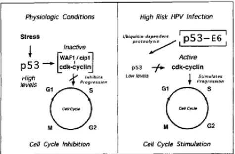
Papillomavirus E6 protein consists in 150 amino acids with approximately 18 kDa and it is localized in the nuclear matrix and non-nuclear membrane fraction [21, 33]. At the structural level, the E6 protein is composed by two zinc finger domains and in the base of each zinc finger are two motifs containing two cysteines (Cys-X-X-Cys, where x is any amino acid), which are conserved throughout all HPV types [34]. In the second zinc finger domain exists an LXXLL motif, which is required to mediate interactions between the E6 protein and other proteins, namely the cellular E3 ubiquitin-ligase known as E6-associated protein (E6AP) [21]. E6 protein can form a complex with E6AP that is required for the association of E6 and p53 proteins, resulting in loss of p53 activity within cells [35]. In this case, the p53 degradation occurs through a ubiquitin-dependent mechanism, in contrast to the large T antigen of simian virus 40 (SV40) and E1B protein of adenovirus 5, which will inactive the p53 protein by sequestration into a complex [36]. This mechanism has been reported for the high-risk HPV-16 and 18, while in the case of low-risk HPV, E6 also binds p53 but with reduced efficiency, it being not capable of inducing p53 degradation [35].
P53 tumor suppressor can sense damage or potential damage in the cellular DNA and invoke a protective response (by blocking the cell cycle or inducing apoptosis in the affected cell), it being extremely important to maintain the genome integrity [37]. When the p53 protein detects damage in the cellular DNA, it activates the WAF1/Cip1 gene, which is an inhibitor of cyclin-dependent kinases. High cellular levels of WAF1/Cip1 inhibit cyclin-dependent kinases, which are essential for progression through the cell cycle. Thus, the cell cycle stops in the G1
phase in order to repair the damage in the DNA and maintain genome integrity [38]. Meanwhile, inactivation or mutation of p53 function leads to continued replication of cells with damaged DNA promoting mutation, chromosomal instability and carcinogenesis of the host genome (as it is showed in Figure 2) [26].
8
Figure 2. Effect of the interaction between E6 protein and p53 tumor suppressor. (Left) In normal
physiologic conditions when p53 detects damage in the cellular DNA, it activates WAF1/Cip1, which inhibits cyclin-dependent kinases and consequently stops the cell cycle in G1 phase. (Right) When an
infection occurs with a high-oncogenic risk HPV type, the presence of E6-p53 complexes promotes the p53 inactivation, leading to uncontrolled cell replication (adapted from [27]).
1.1.2. Human papillomavirus E7 oncoprotein
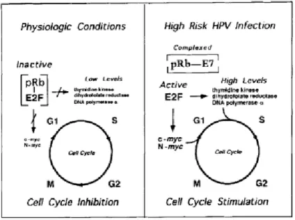
Papillomavirus E7 protein consists in 98 amino acids that were localized to the nuclear matrix [34]. This protein is composed by three functional domains designated by Conserved Regions (CR1, 2 and 3), knowing that CR1 and CR2 domains are structural and functionally related to the E1A protein of adenovirus and the large T-antigen of simian virus 40 [39]. The CR1 domain plays a crucial role in cellular transformation independent of pRb-binding, whereas the CR2 domain exits Leu-X-Cys-X-Glu (LXCXE) motif act as a pRb-binding site [21, 40]. Thus, E7 proteins form complexes with the retinoblastoma (Rb) protein, which is related with proteins p107 and p130, being negative regulators of cell growth [26].
In physiological conditions, the Rb protein forms complexes with transcription factors, such as E2F, which regulate the expression of cellular genes that function in DNA synthesis. The Rb protein binds to E2F transcription factor, becoming phosphorylated by one or more cyclin-dependent kinases, and together with the hypophosphorylated Rb are responsible to block cell cycle progression in G0 and G1 phases [41]. Other Rb-related proteins, p107 and p130,
also form complexes with E2F and regulate the cell cycle progression [27]. In the case of progression from G1 into S phase, E2F-Rb complexes are dissociate and the free E2F
stimulates the transcription of the genes required for transition into S phase [41]. In contrast, the E7 protein binds to Rb, p107 and p130 proteins and releases free E2F transcription factors in the cells, which can stimulate the transcription of the E2F-dependent genes necessary for DNA replication and cellular cycle progression [27, 42] (as it is show in Figure 3). The ability
9
of E7 protein for disrupt E2F-Rb complexes has been reported to high-risk HPV types, while the low-risk HPV-E7 protein have demonstrated a low Rb, binding affinity [27, 42].
Figure 3. Effect of the interaction between the E7 protein and pRB protein (Left) In normal physiologic
conditions, the pRB protein forms complexes with E2F by phosphorylation of the pRB with cyclin-dependent kinases and consequently the Rb stays hypophosphorylated leading to the cell cycle stop in G0 or G1 phases. (Right) When the infection with a high-oncogenic risk HPV type occurs, the presence of
E7-pRB complexes releases free E2F, which stimulate the transcription of the genes responsible for DNA replication and cell cycle progression (adapted from [27]).
1.2. Preventive HPV vaccines
As the HPV viral DNA has been detected in more of 99% patients with a cervical cancer, the vaccination with fragments of HPV genomic DNA can be a potential solution for this pathology, it being essential a basic understanding of the HPV biology. Two vaccination types can be used against the cervical cancer. Preventive vaccines prevent the infection by generating neutralizing antibodies to block HPV viral infection and therapeutic vaccines eliminate the infection by inducing a virus-specific T cells-mediated response [2].
Recently, two HPV preventive vaccines (quadrivalent and bivalent) were developed and have been licensed for use in many countries. The quadrivalent vaccine of HPV L1 virus-like particles (VLP) was called Gardasil, developed by Merck (NJ, USA) and approved by the USA Food and Drug Administration (FDA). This vaccine is produced in Saccharomyces cerevisiae (S.
cerevisiae) and protects against four of the most medically relevant HPV genotypes: HPV-16
and HPV-18 for cervical cancer, and HPV-6 and HPV-11 for benign genital warts [1, 24]. The bivalent vaccine HPV L1 VLP was called Cervarix and developed by GlaxoSmithKline (GlaxoSmithKline Biologicals, Rixensart, Belgium). This vaccine uses an insect cell expression system and protects against two HPV genotypes, HPV-16 and HPV-18 [1, 24].
10
Both Gardasil and Cervarix vaccines are based on the use of HPV VLP to generate neutralizing antibodies against the major capsid protein, L1. Despite of L1 is not expressed in the infected basal cells, this protein has been largely studied as target for the preventive vaccine development. These vaccines only induced the humoral response, by promoting the production of specific antibodies. Moreover, the ability of HPV-16 L1 VLP vaccination to generate memory B cells has been reported. In this way, memory B cells provide a rapid response of antibodies upon secondary exposures [43]. Although Gardasil and Cervarix vaccines have been applied with some success as preventive vaccines, they also present some limitations, such as high manufacturing cost, type-restricted protection and no present therapeutic effects which are displayed in Table 3.
Table 3. Disadvantages of current licensed human papillomavirus vaccines – Cervarix® and Gardasil®
(adapted from [44]).
Limitation Impact
Cost of manufacturing, trials and intellectual property leads to high vaccine price
Developing countries that do not have the economic means to afford the vaccine continue to bear the burden of cervical cancer
Type restriction
Vaccine does not protect against all HPV types causing cervical cancer, thus screening must continue for HPV-vaccinated women
No therapeutic value No effect on individuals with pre-existing HPV infection
1.3. Therapeutic HPV vaccines
Because of limitations from current HPV vaccines previously mentioned, the development of therapeutic vaccines to facilitate the control of cervical cancer and others HPV-associated malignances becomes increasingly necessary. The choice of target antigen is the key of the therapeutic vaccines design. HPV early proteins are potential target antigens because they are involved in the regulation of virus transcription and replication and can be expressed in both nonproductively infected cells and in transformed cells. Particularly, HPV E6 and E7 viral genes are mainly responsible for malignant transformation [4].
11
2. Gene Therapy and DNA Vaccines
Millions of people die each year from hereditary, degenerative and infectious diseases, or cancer [45]. These diseases are considered untreatable by conventional clinical methods. In last decades, the determination of relationships between genes and the maintenance and regulation of the organism, and the decoding of the Human Genome in 2000, led to the conclusion of the existence of wide range of diseases depending on the gene [46, 47]. Thus, it emerged the opportunity for the development of therapeutic strategies based on the gene intervention such as gene therapy and DNA vaccination [48].
The Human Gene Therapy is defined as the introduction of genetic material that encodes a desired gene into human target cell with the aim of correcting phenotypic or genotypic abnormalities or providing new functions to the cells [49, 50]. This therapeutic strategy may be divided into somatic and germ gene therapies [49]. However, only somatic cells are targeted for treatment because genetic modifications in somatic cells affect the individual patient, while alterations in germ cells have potential to affect the future generations [49]. DNA vaccination is other innovative human DNA therapeutic strategy based on the gene intervention for the treatment of inherited and acquired diseases [51]. DNA vaccination is based on the administration of therapeutic genes in human target cells nucleus [52]. When administrated, therapeutic gene is transcribed and translated in the cytoplasm of the cell and the resultant protein will induce the immune response [52, 53].
2.1. DNA vaccination
As previously mentioned, millions of people die at each year due to incurable diseases that often provide drug resistance. In this way, the research of more stable and efficient vaccines has been increasingly important to treat some infectious pathologies. DNA vaccines are new therapeutic strategies that consist in the synthesis of DNA vector-encoded therapeutic genes for specific proteins, which induce the immune responses against the pathogen [6]. DNA vaccination might provide several important advantages over conventional vaccines, such as facilitated development and production (more advantages are presented in Table 4) [54].
12
Table 4. Advantages of DNA vaccines (adapted from [55]).
Advantages of using DNA vaccines compared with conventional protein vaccines
Immunogenicity Can induce both humoral and cellular immune responses
Low effective dosages (micrograms) in animal models
Safety Unable to revert into virulence unlike live vaccines
Efficacy does not require the use of toxic treatments unlike some killed vaccines
Engineering Plasmid vectors are simple to manipulate and can be rapidly tested
Combination approaches are easily adapted
Manufacture Low manufacturing cost
Produced at high frequency in bacteria and easily isolated
Stability More temperature-stable than conventional vaccines
Long shelf-life
Mobility Ease of storage and transport
Likely not to require a cold chain
DNA vaccination has become an attractive immunization strategy due to the ability to induce strong and long-lasting cellular and humoral immune responses with high efficiency [5]. The DNA vaccine encoding therapeutic genes enters the cell nucleus, where the gene initiates the transcription, followed by protein production in the cytoplasm [6]. These proteins generate peptides that have been processed and presented by professional antigen-presenting cells (APCs), like macrophages and dendritic cells (DCs), in the context of major histocompatibility complex (MHC) class II molecules that then are exhibited on the surface of the cell [56]. Specific helper T cells (CD4+ T cells) recognize this antigen peptide/MHC Class II molecule
complex and are activated [45]. CD4+ T cells play a central role in immune response. CD4+ T
cells secrete cytokines that have a myriad activities including, depending up on the cytokine, promoting B cell survival and antibody production and helping cytolytic T lymphocyte (CTL) responses [56]. In order to induce cytotoxic T lymphocyte response, CD8+ T cells recognize
peptides derived from endogenous proteins presented in the context of MHC class I molecules [54]. The presence of co-stimulatory molecules produced by activated CD4+ T cells is another
requirement for the activation of CD8+ T cells, resulting in a potent antitumor effect against
antigen-expressing tumors [55, 57]. On the other hand, DNA vaccines are capable to induce humoral immune response. In this way, B cells recognize antigens that are either present extracellularly, or exposed extracellularly by being transmembrane proteins inducing a strong antibody response to the respective proteins [56]. Figure 4 illustrate, the simplified form, the generation of both cytotoxic and helper T-cells responses and antibody generation by the DNA vaccines.
On the other hand, DNA vaccines are highly effective at inducing long-lived memory responses, so that upon subsequent exposure to the pathogen a rapid and specific immune response is mounted to prevent and/or clear infection [58].
13
Figure 4. DNA vaccines stimulate the induction of both cellular and humoral responses. Professional
antigen presenting cells take up an exogenous antigen into its endolysosomal degradation pathway, which convert proteins in peptides. These peptides are present in association with MHC class II molecules. After, CD4+ T cells recognize the peptide/MHC Class II molecule complex and are activated
to produce cytokines. These cytokines help B cells activate into antibody producing cells, and help cytolytic T lymphocyte responses. Despite the action of this cytokines in cytolytic T lymphocyte responses, the recognition of the peptide/MHC class I molecules by the CD8+ T cells is necessary. For
antibody responses, B cells recognize and respond to antigens that can present in transmembrane of the APC or present extracellularly (adapted from [45]).
2.2. Viral and Non-viral vectors
Gene medicines or nucleic acid drugs can be categorized on the basis of their therapeutic relevance as gene inhibitors or gene inductors [59]. Gene inhibitors (i.e. oligonucleotides, ribozymes, DNAzymes, aptamers, and small interfering RNAs (siRNAs)) are potent drugs that interfere with mRNA leading to silencing of the defective genes [60]. On the other hand, gene inductors (cDNA (complementary DNA) and plasmids containing transgenes) can be divided into gene vaccines and gene substitutes [59]. Gene vaccines are antigens of specific
pathogens encoding either the genes or RNA that have the ability to induce T cell-mediated
response and humoral immune response, as well as production of antibodies [61]. Gene substitutes are transcriptionally fully competent genes introduced into cells to reimburse deficiency of a specific protein or its insufficient protein production [60].
14
Central to the concept of gene therapy is the ability to deliver exogenous nucleic acids to wide variety of cells, tissues, and organs [50]. However, one of the problems associated to DNA vaccination is to develop the delivery vehicle of therapeutics genes that brings together 3 major criteria: (1) it should protect the gene against degradation by nucleases in intercellular matrices, (2) it should bring the gene across the plasma membrane and into the nucleus of target cells, and (3) it should have no detrimental effects [62]. Two major groups of vehicles for delivering genetic material exist, the viral and non-viral vectors [63].
2.2.1. Viral delivery systems
Viruses have numerous biological proprieties that allow them to recognize and enter in eukaryotic cells, expressing their genes and consequently infecting the host cell. These proprieties led to the use of viruses as gene delivery vectors [64]. Viral vectors encapsulate the viral sequences that are required for the assembly of viral particles, the packaging of the viral genome into the particles and the therapeutic gene [46]. Table 5 represents the main viral vectors (Retroviruses, Lentiviruses, Adenoviruses and Adeno-associated virus) and their advantages and disadvantages.
15
Table 5. Advantages and disadvantages of main viral vector groups (Adapted from [47]).
Vector Advantages Disadvantages
Adenovirus
High transfection efficiency
Transfects proliferating and nonproliferating cells
Substantial clinical experience
Strong immune responses
Insert size limit of 7.5 kbp (kilo base pairs)
Difficult to manufacture and quality control
Poor storage characteristics
Short duration of expression
Retrovirus Fairly prolonged expression High transfection efficiency Substantial clinical experience Low immunogenicity
Low transfection efficiency in-vivo
Insert size limit of 8 kbp ex-vivo
Transfects only proliferating cells
Difficult manufacture and quality control
Safety concerns (mutagenesis)
Lentivirus Transfects proliferating and non-cells Transfects haematopoietic stem cells
Very difficult manufacture and quality control
Poor storage characteristics
Insert size limit of 8 kbp
No clinical experience
Safety concerns (origins in HIV)
Adeno-associated virus
Efficient transfection of wide variety of cell types
in-vivo
Prolonged expression
Low immunogenicity
Difficult manufacture and quality control
Insert size limit of 4.5 kbp
Limited clinical experience
Safety concerns (mutagenesis)
In general, viral vectors are very efficient gene delivery vectors, however this delivery system also presents some disadvantages. These include safety concerns (mutagenesis and carcinogenesis), induction of immune responses (which abolishes the therapeutic gene, if consecutive administrations are made), the low DNA amount that can be loaded and the high commercial cost [63, 65]. Considering these limitations, the use of non-viral DNA-based therapy has emerged as a convincing approach in medical science.
2.2.2. Non-viral delivery system
Non-viral vectors are being developed to overcome problems associated with viral gene delivery, such as capacity for insertional mutagenesis [66]. In fact, non-viral vectors are less pathogenic and may have reduced toxicity by comparing to the existing viral vector, being safer and easier to manufacture under low cost. On the other hand, non-viral vectors also include as advantage the capacity of delivering larger genetic units without limited size.
16
Non-viral delivery systems for gene transfer can be classified into physical (carrier-free gene delivery) and chemical categories (synthetic vector-based gene delivery). Physical approaches employ a physical force that permeates the cell membrane and facilitates intracellular gene transfer. On the other hand, chemical approaches use synthetic or naturally occurring compounds as carriers to deliver nucleic acids into the target cells [60]. Although a significant progress has been made in the basic science and applications of various non-viral gene delivery systems, the non-viral approaches also present some limitations. In Table 6, briefly discuss the advantages and limitations of the non-viral gene delivery systems
Table 6. Advantages and limitations of several non-viral gene delivery systems (adapted from [62]).
Method Advantages Disadvantages
Physical methods
Needle injection Simplicity and safety Low efficiency
Gene gun Good efficiency Tissue damage in some applications
Electroporation High efficiency
Limited working range; need for surgical procedure for
nontopical applications
Hydrodynamic delivery
High efficiency, simplicity, effectiveness for liver gene delivery
Extremely effective in small animals; surgical procedure may be needed for localized gene delivery
Ultrasound Good potential for site
specific gene delivery Low efficiency in vivo
Chemical methods
Cationic lipids
High efficiency in vitro; low to medium high for local and systemic gene delivery
Acute immune responses; limited activity in vivo
Cationic polymers
Highly effective in vitro; low to medium high for local and systemic gene delivery
Toxicity to cells; acute immune responses
Lipid/polymer hybrids
Low to medium-high efficiency in vitro and
in vivo; low toxicity
17
3. Plasmid DNA as non-viral vector
In therapeutic applications, plasmid DNA is used as vector or vehicle that can selectively and efficiently deliver a gene to target cells and induce the production of relevant proteins. In the last years, the use of plasmid-DNA-based delivery vectors has gained popularity (14% of all trials in 2004 and 18% in 2007), being the most popular non-viral system used in clinical trials [67]. This fact is probably due to the low toxicity and immunogenicity when compared with viral vectors [68].
Plasmids are circular, double stranded DNA molecules with a size range of 0.8 to 120 kilo base pairs, being considered very large biomolecules when compared to proteins. Plasmid DNA structure is divided into hydrophilic backbone (sugar and phosphate group) and hydrophobic interior of double helix (planar bases stacked on each other) [10]. For pH higher than 4, the phosphate groups are negatively charged, resulting in a negative DNA molecule [10, 69]. The supercoiled plasmid isoform is considered the most appropriate and active form for therapeutic applications due to its functional, compact and undamaged structure. However, other isoforms can be generated from the sc pDNA isoform by either single strand nick (open circular isoform) or double strand nick (linear isoform) [70]. The existence of different pDNA isoforms depends on factors such as DNA sequence, supercoiling stress or unfavorable environment conditions (extreme pH or high temperature) [10]. High temperatures promote the extended thermal motion, which leaded a gradual unwinding of the double DNA helix [10, 69]. In this way, several studies of transfection efficiency have been performed to investigate the biological effects of the pDNA topology and have revealed higher transfection efficiency with sc plasmid isoform [13, 71]. On the other hand, the application of plasmid DNA naked for transfection of eukaryotic cells results in lower transfection efficiency than comparing to viral vectors. Estimates indicate that only one in every thousand plasmid molecules presented to the cells can perform an efficient transfection [68]. The use of chemical or physical methods has significantly improved the gene delivery efficiency [72]. Hence, the best option is to develop adequate strategies to produce high copy number of sc pDNA, to obtain this plasmid isoform from the host avoiding its degradation, to eliminate the host impurities by an efficient purification method and to combine with a good physical or chemical-gene delivery system.
3.1. Plasmid DNA manufacturing
The simplicity of production methods is also one of the advantages of non-viral pDNA vectors for clinical applications instead of viral vectors. Over the years, several effective techniques for pDNA manufacturing have been developed, although the major challenge remains to achieve scalable and economical means of producing large quantities of sc pDNA fulfilling the requirements of regulatory agencies such as the Food and Drug Administration and the
18
European Agency for the Evaluation of Medical Products (EAEMP). In general, the manufacturing process starts by the plasmid vector construction and selection of appropriate bacterial host, followed by the choice and optimization of the fermentation conditions and subsequent isolation and purification steps [68], as it is presented in Figure 5.
Figure 5. Three necessary stages to obtain a pure supercoiled plasmid DNA (adapted from [68]).
The design and engineering of pDNA is an important step to ensure the successfully pDNA manufacturing and to obtain the maximum degree of transfection. The basic structure of the most plasmid DNA applied in gene therapy and DNA vaccination includes: (1) an origin of replication for efficient propagation in an adequate host such as Escherichia coli, (2) an antibiotic resistance gene for growth selection, (3) a strong promoter to drive expression in eukaryotic cells, (4) a polyadenylation termination sequence, and (5) a gene insert coding for the antigen of interest [58].
Posteriorly, the choice of bacterial strain and selection of fermentation conditions are important factors to enable the production of large quantities of sc pDNA under stable conditions [68].
3.2. Downstream process
After fermentation, the specific plasmid content is only about 3% W/W of the E. coli extract [73]. Thus, a sequence of unit operations is essential for the release of plasmids from the host cells and their separation from other cellular constituents [58]. Although this downstream
19
process is critical for impurities elimination, also it is considered the critical step of the pDNA manufacturing process [74].
The first step in the pDNA downstream processing consists in E. coli cells recovery from the broth by centrifugation or microfiltration [74]. Thereafter, plasmid molecules can be released from host cells by several disruption techniques, although the alkaline lysis is the most applied technique [69]. Bearing this in mind, the alkaline lysis is based on cell disruption by using a buffer containing sodium hydroxide (NaOH) as alkali-promoted hydrogen bond disruption, followed by the release of all intracellular components (plasmid DNA, RNA, genomic DNA (gDNA), endotoxins and proteins). NaOH with high pH levels promotes the denaturation of genomic DNA, cell wall material and most of the cellular proteins. Peculiarly, sc pDNA isoform also unwinds as a consequence of the NaOH, but if the pH is lower than 12.5 prevents the separation of the complementary strands [69]. Following the lysis step, a precipitate containing cell debris, denatured gDNA and proteins is formed by adding the neutralizing solution with potassium acetate and finally the precipitate is removed by centrifugation [68].
Although most gDNA and proteins have been denatured and precipitated during alkaline lysis, the clarified sample resultant from alkaline lysate still contains proteins, RNA, gDNA, endotoxins and less than 1% (w/w) of pDNA. Concentration and clarification steps should be also performed to remove proteins, endotoxins and host nucleic acids and increase the plasmid mass fraction [69, 74]. The reduction of RNA and proteins are usually achieved by precipitation with a chaotropic salt, such as ammonium sulfate [69].
3.3. Plasmid DNA Purification
The purification of pDNA for research or clinical applications requires efficient technologies to obtain a final product constituted only by the biologically active supercoiled isoform (higher than 97%), according to guidelines established by regulatory agencies [68, 75]. Liquid chromatography is the central operation to separate the sc pDNA isoform from the others plasmid isoforms such as open circular (oc), linear (ln) and denatured isoforms together with the removal of host remaining impurities [75]. In addition, the physical and chemical similarities between impurities and pDNA, such as negative charge (RNA, gDNA and endotoxins), hydrophobicity (endotoxins) and molecular mass (gDNA and endotoxins) represent one of the major bottlenecks in the sc pDNA obtaining process [69]. Several chromatographic methods based on properties such as size, charge, hydrophobicity or affinity has been developed with more or less success in sc pDNA purification [10].
20
3.3.1. Size-exclusion chromatography (SEC)
Size-exclusion chromatography allows the plasmid fractionation and purification from other molecules present in a clarified lysate based on differences of their molecular size. Smaller molecules, such as RNA and endotoxins, have greater ability to penetrate inside the pore space, retarding the movement through the column. While, larger molecules ,such as gDNA and different forms of pDNA, travel around the particles of the packing material and are first eluted [76]. Thus, SEC can be used with high productivity in separation of pDNA from RNA and the other smaller impurities. However, the capacity of SEC to differentiate plasmid isoforms and isolate sc pDNA in one single step is limited [10].
3.3.2. Anion-exchange chromatography (AEC)
The simplest explanation of anion-exchange chromatography of nucleic acids is based on the attraction of oppositely charged molecules. Indeed, nucleic acids are considered polyanionic molecules that will interact with positively charged ligands immobilized in the stationary phase [68]. Variations in the salt concentration have been explored for binding/elution of different nucleic acids. According to general principles of anion-exchange, the binding should preferably occur at low salt concentration. After binding, the different nucleic acids should elute by increasing order of overall net charge, which in turn is dependent on the chain length and conformation of several molecules [69]. With an optimized gradient, the separation of supercoiled and open circular forms of pDNA in an anion-exchange column can be possible, because sc isoform is more compact and has a higher charge density than the open circular pDNA form. However, due to the existence of physical and chemical similarities between impurities and pDNA, like chemical composition and structure (gDNA and RNA) or charge (endotoxins), their separation can in some cases be insufficient [10].
3.3.3. Hydrophobic interaction chromatography (HIC)
Hydrophobicity is another physiochemical property that can also be explored in the purification of the sc pDNA isoform. This technique recognizes differences in hydrophobicity between double-strand and single-strand nucleic acid and endotoxins. In this way, the sample binding is achieved by column loading at a high salt concentration. The elution of different species is achieved by increasing order of hydrophobicity and by reducing the salt concentration, which weakens hydrophobic interactions. HIC technique showed to be efficient in the separation of pDNA from the RNA and endotoxins, nonetheless the ability to separate pDNA isoforms is weak. Moreover, the need for application of high salt concentrations is also view as a disadvantage [10].
21
3.3.4. Affinity chromatography (AC)
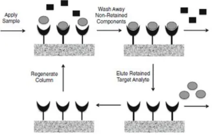
Affinity chromatography is the unique technique that uses specific binging agent to recognize and purify biomolecules based on their biological function or chemical structure [75]. Affinity chromatography has become a popular method for biomolecules purification due to advantages of eliminating additional steps, increasing yields and improving process economics. However this chromatographic method presents some limitations mainly in regard to the biological origin of ligands that tend to be fragile and associated to low binding capacity [10]. In this way, synthetic ligands, combining the selectivity of natural ligands with high capacity and durability of synthetics systems, have been developed [10].
Affinity chromatography separates biomolecules on the basis of reversible interactions between the target biomolecule and its specific ligand immobilized in the stationary phase (as it is showed in Figure 6). Thus, a sample containing the target biomolecule is injected onto the stationary phase, with appropriate pH and ionic strength in the buffer, allowing the binding between the target biomolecule and the specific ligand [77]. After binding, the elution steps can be performed either specifically, with competitive ligand, or non-specifically by changing buffer conditions, such as pH, ionic strength or polarity depending on the characteristics of the biomolecules [10].
Figure 6. Principle of affinity chromatography. Firstly, a sample containing the target molecule is
injected onto the column. After binding of molecules with specificity for immobilized ligands, a wash was applied for elution of the unbound species. Finally, alterations in elution buffer lead to elution of bounded species (adapted from [77]).
Several affinity chromatography types have been utilized in the sc pDNA purification with more or less success, among which, immobilized metal-ion affinity chromatography, triple-helix affinity chromatography, polymyxin B affinity chromatography, protein-DNA affinity
![Table 1. Human papillomavirus grouping, according to their risk to cause cervical cancer (adapted from [18])](https://thumb-eu.123doks.com/thumbv2/123dok_br/18901452.935342/19.892.139.813.889.977/table-human-papillomavirus-grouping-according-cervical-cancer-adapted.webp)
![Table 2. A brief description of the functions of human papillomavirus open-reading frames (adapted from [26])](https://thumb-eu.123doks.com/thumbv2/123dok_br/18901452.935342/22.892.146.799.152.348/table-brief-description-functions-papillomavirus-reading-frames-adapted.webp)

![Table 6. Advantages and limitations of several non-viral gene delivery systems (adapted from [62])](https://thumb-eu.123doks.com/thumbv2/123dok_br/18901452.935342/31.892.142.797.407.977/table-advantages-limitations-viral-gene-delivery-systems-adapted.webp)
![Figure 5. Three necessary stages to obtain a pure supercoiled plasmid DNA (adapted from [68])](https://thumb-eu.123doks.com/thumbv2/123dok_br/18901452.935342/33.892.157.772.242.567/figure-necessary-stages-obtain-pure-supercoiled-plasmid-adapted.webp)