UNIVERSIDADE DE LISBOA FACULDADE DE CIÊNCIAS DEPARTAMENTO DE BIOLOGIA ANIMAL
Characterization of a new genetically modified Plasmodium
berghei parasite – development of a pre-erythrocytic malaria
vaccine
Mestrado em Biologia Humana e Ambiente
Carolina Morgado de Andrade
Dissertação orientada por:
Doutor António Mendes (Instituto de Medicina Molecular, Faculdade de Medicina, Universidade de Lisboa)
Dedico esta dissertação à minha família. Por tudo o que sou hoje, devo-o a vocês.
Abstract
Malaria is an infectious disease caused by a protozoan parasite of the genus Plasmodium and transmitted to humans and other animals by infected female Anopheles mosquitoes. Although there are five species of Plasmodium that can infect humans, the majority of the deaths are caused by P. falciparum and P. vivax. Unfortunately, there is still no licensed vaccine against malaria. Whole-organism pre-erythrocytic malaria vaccines have been shown to elicit sterile immunity in both humans and rodents. They induce effective CD8+ T cell responses, required for sterilizing immunity in malaria. Nevertheless, current whole-organism pre-erythrocytic malaria vaccination approaches, which rely on the use of human-infective sporozoites, present risks and limitations that cannot be overlooked.
According to the 2013 World Health Organization guidelines, a malaria vaccine should be available for use and licensed by 2030, with at least 75% efficacy and capable of protecting against both P. falciparum and P. vivax. Here, we propose a vaccination approach where a genetically modified rodent P. berghei parasite is used as a platform to deliver immunogenic antigens against the two major human infective Plasmodium species: P. falciparum and P. vivax – the PbTriCS parasite. Our results show that PbTriCS development during the sporogonic stage and in vitro hepatic stage is not impaired, having similar infectivity to wild-type parasites. Although our results in C57BL/6J mice have shown a reduced fitness by PbTriCS parasites in establishing an in vivo liver infection, we demonstrated by ELISA assays that this parasite is able to trigger humoral responses against P. falciparum and P. vivax CS peptides.
We conclude that PbTriCS has the potential to be a good malaria vaccine candidate as it is able to elicit strong antibody responses against both P. falciparum and P. vivax. Further studies should be pursued to functionally demonstrate the protective capacity of the immune response induced through immunization with PbTriCS.
Keywords: Malaria vaccine, P. berghei, P. falciparum, P. vivax,
Resumo
A malária é uma doença infecciosa endémica em 97 países, causada por um protozoário do género Plasmodium e transmitida por um mosquito fêmea do género Anopheles. Existem cinco espécies capazes de causar infecção em seres humanos, P. falciparum, P. vivax, P. malariae, P. ovale e P. knowlesi. Destas, P. falciparum é o parasita predominante no continente africano e é o responsável pelo maior número de mortes. A Organização Mundial de Saúde (OMS) estima que em 2013 a malária foi responsável pela morte de 584 mil pessoas, sendo que destas, 453 mil correspondem a crianças abaixo dos cinco anos.1 4 5
O ciclo de vida do parasita da malária inicia-se quando um mosquito infectado se alimenta de sangue num hospedeiro humano, depositando na sua pele centenas de esporozoítos – a forma do parasita residente nas glândulas salivares do mosquito e capaz de infectar o fígado. Os esporozoítos entram rapidamente na circulação sanguínea chegando ao fígado, onde atravessam os sinusoides hepáticos e diversos hepatócitos, até invadirem um hepatócito com formação dum vacúolo parasitóforo, onde o parasita reside durante todo o seu desenvolvimento hepático14. A fase hepática da infeção por Plasmodium é uma etapa obrigatória do seu ciclo de vida, ainda que clinicamente silenciosa. No fígado, o parasita prolifera, diferenciando-se numa forma replicativa, a forma exo-eritrocítica. Em humanos, a infecção por P. vivax ou P. ovale pode dar origem a formas hepáticas dormentes, os hipnozoítos, que podem causar recaídas na doença, meses ou anos após a infecção.4 5 A fase hepática termina com a libertação de vesículas cheias de merozoítos – os merosomas – para a corrente sanguínea.16 Na corrente sanguínea os merosomas libertam os merozoítos que infectam os eritrócitos, iniciando assim o estádio eritrocítico da infeção. 4 16 Os merozoítos infetam os eritrócitos em ciclos de invasão, crescimento intracelular, multiplicação e re-infeção de novos eritrócitos.18 As células-filhas dos merozoítos podem desenvolver-se em formas assexuadas ou sexuadas do parasita. As primeiras continuam o ciclo de invasão e multiplicação nos eritrócitos ao passo que as segundas originam gametócitos. É na fase sanguínea da infecção que a sintomatologia da malária aparece.19 13 Aquando da ingestão de sangue por uma fêmea Anopheles num humano infectado, há a entrada de gametócitos presentes no sangue para o intestino médio do mosquito. Aqui, os gametócitos diferenciam-se em gâmetas, que se fundem, dando origem a um zigoto diplóide que inicia a meiose. Os zigotos transformam-se numa forma móvel, o oocineto que migra e penetra o epitélio do intestino médio do mosquito, onde se transforma numa forma séssil, o oócisto. O oócisto sofre replicação mitótica produzindo esporozoítos, que após a ruptura do oócisto ficam livres na hemolinfa indo invadir as glândulas salivares do mosquito. O parasita está então pronto para iniciar um novo ciclo num novo hospedeiro.19
Atualmente, as medidas de controlo de malária envolvem o controlo do vetor e o tratamento da doença. Infelizmente, não se encontra atualmente
licenciada nenhuma vacina contra a malaria, a qual será sempre uma ferramenta essencial para a erradicação desta doença. Em 2013 a OMS lançou orientações em relação ao desenvolvimento / licenciamento de uma vacina para a malária segundo as quais 2030 deverá haver uma vacina contra ambas as espécies P. falciparum e P. vivax e ter pelo 75% de eficácia. Tradicionalmente, as estratégias de vacinação contra a malária incluem: as vacinas pré-eritrocíticas (têm como alvo o esporozoíto e a fase hepática da infeção); as vacinas eritrocíticas (têm como alvo a fase assexuada do parasita no sangue); e as vacinas que bloqueiam a transmissão (têm como alvo os gametócitos/gâmetas do parasita e/ou outros estádios no mosquito).25 As vacinas pré-eritrocíticas são até ao momento as mais promissoras, podendo dividir-se em dois tipos: as de subunidade e as de organismo inteiro. As vacinas de subunidade baseiam-se no uso de uma pequena porção do parasita, uma proteína ou um péptido, para evocar uma resposta imune. Por seu lado, as de organismo inteiro baseiam-se no uso do parasita todo, administrado numa forma atenuada, ou sob o tratamento com antimaláricos. Atualmente, o candidato a vacina mais avançado, e o único que chegou a estudos clínicos fase 3, é uma vacina de subunidade, a RTS,S, que usa a proteína circumsporozoite (CS) de P. falciparum para evocar respostas imunes.32 No entanto, a sua eficácia é de cerca de 30% e diminui ao longo do tempo.33
As vacinas de organismo inteiro mostraram ser as mais eficazes visto que foi possível induzir respostas imunes em roedores e em humanos capazes de prevenir uma infecção subsequente por Plasmodium.27 28 37 Além disso, as vacinas de organismo inteiro são capazes de espoletar respostas imunes mediadas por células T CD8+.38 39 No entanto, as vacinas de organismo inteiro apenas são contra uma espécie de Plasmodium e baseiam-se no uso de esporozoítos capazes de infetar humanos, o que representa riscos e limitações.
O PrudêncioLab do Instituto de Medicina Molecular, tem vindo a investigar o uso do P. berghei como uma estratégia de vacinação contra a malária. P. berghei é um dos parasitas de malária em roedores e em trabalhos anteriores ficou provada a sua capacidade de infectar células do fígado humano, bem como a sua incapacidade de se reproduzir em eritrócitos humanos, impedindo-o de causar a doença. Assim, P. berghei é uma abordagem de vacinação mais segura em relação a outros candidatos a vacina pré-eritrocítica de organismo inteiro. O PrudêncioLab caracterizou um P. berghei geneticamente modificado (GM) que expressa a proteína CS de P. falciparum sob o controlo do promotor UIS4 de P. berghei. Os resultados deste trabalho mostram que este parasita GM é capaz de suscitar uma resposta imune contra uma infeção causada por P. falciparum.
Tendo em conta as diretrizes da OMS, o PrudêncioLab criou recentemente um novo parasita P. berghei GM que expressa simultaneamente as proteínas CS de P. falciparum e P. vivax em loci neutros, o parasita PbTriCS. Este projeto de mestrado teve como objetivo a caracterização deste parasita, avaliando a sua capacidade de desenvolvimento e infeção em mosquitos
Anopheles, in vitro em células Huh7, e in vivo em ratinhos C57BL/6J. Para concluir, avaliaram-se as respostas humorais contra péptidos das CS de P. berghei, P. falciparum e P. vivax. espoletadas pela imunização com PbTriCS. A primeira parte da caracterização consistiu em avaliar o desenvolvimento do parasita em mosquitos Anopheles. Para tal, contaram-se os números de oócistos no intestino médio e os números de esporozoítos presentes nas glândulas salivares. Comparados os resultados com os de PbGIMO, a linha inicial não-transfetada e caracterizada como correspondendo ao P. berghei selvagem, não foram encontradas diferenças significativas entre parasitas, permitindo concluir então que a inserção de dois genes em loci neutros não tem impacto no desenvolvimento do parasita no mosquito. Procedemos então à análise do movimento de gliding dos esporozoítos numa superfície de vidro, não tendo sido encontradas diferenças significativas no número de círculos relativamente ao PbGIMO e concluíndo também que as 3 CS são expressas no esporozoíto.
Na segunda parte do trabalho, caracterizámos o parasita in vitro através da infeção da linha celular de hepatoma humano Huh7. Concluímos, através da de microscopia de imunofluorescência, que PbTriCS possui uma infetividade semelhante a PbGIMO, sendo o seu desenvolvimento normal, às 48 após a infeção. Foi-nos possível também observar que todas as CS estão a ser corretamente expressas. Decidimos então avançar para a caracterização da infeção in vivo em ratinhos C57BL/6J às 46 horas após a infeção tendo-se observado por qRT-PCR uma redução na infeção do fígado por PbTriCS comparativamente a PbGIMO. A análise de secções de fígado por microscopia de imunofluorescência mostrou que esta redução de infeção se deve a um menor número de parasitas no fígado ao invés de um menor desenvolvimento dos mesmos. No entanto, todas as CS estão a ser corretamente expressas in vivo em PbTriCS. Para entender esta observação, realizámos um ensaio de atravessamento celular, tendo-se concluído que o atravessamento por PbTriCS é cerca de 50-60% inferior ao de parasitas PbGIMO. Isto foi indiretamente confirmado pela observação experimental que o número de parasitas PbTriCS capaz de estabelecer uma infeção no fígado de ratinhos C57BL/6J é menor que PbGIMO.
Por último, na terceira parte do trabalho, utilizamos o parasita PbTriCS para imunizar ratinhos C57BL/6J e, por ELISA, foi possível concluir que o soro dos ratinhos imunizados possui anticorpos capazes de reconhecer os péptidos das CS de P. berghei, P. falciparum e P. vivax.
Em conclusão, apesar do parasita PbTriCS ter uma menor capacidade de estabelecer uma infeção hepática in vivo em comparação com PbGIMO, os parasitas capazes de infetar o fígado não têm o seu desenvolvimento comprometido e expressam corretamente as várias proteínas CS, permitindo que uma imunização com PbTriCS induza a produção de elevados títulos de anticorpos contra a CS de P. falciparum e P. vivax. Embora mais estudos devam ser feitos, nomeadamente ao nível de respostas imunes e de eficácia, podemos concluir que o parasita PbTriCS constitui um bom candidato a vacina contra P. falciparum e P. vivax.
Palavras-chave: Vacina para a malária; P. berghei; P. falciparum; P. vivax;
Acknowledgments / Agradecimentos
Quero começar por agradecer ao Miguel a oportunidade que me deu de desenvolver a minha dissertação de mestrado no PrudêncioLab. Acho que sempre deixei claro em todos os e-mails o quanto admirava (e admiro ainda mais) o projeto e a motivação que tinha em fazer a tese no teu laboratório. Permitiste-me concretizar um sonho ao aceitares-me como estudante de mestrado. Foi um ano incrível, em que aprendi mais do que poderia antecipar, e tudo se deve a teres-me aceitado no teu laboratório. Nunca conseguirei expressar o quão grata me sinto.
Ao António, por ter sido o melhor orientador que poderia ter tido. Pela autonomia que cedo me incutiste, responsabilizando-me na independência que tive nas experiências e, ao mesmo tempo, me ensinares tanto. Agradeço-te por teres puxado imenso por mim e nunca duvidares das minhas capacidades, mesmo quando eu duvidava. Obrigada!
À Prof.ª Deodália, por me ter incentivado, conduzindo-me à frequência deste mestrado, pela dedicação aos seus alunos e pelo carinho. Um obrigada pela paciência e um pedido de desculpa pelas visitas frequentes ao seu gabinete num "verdadeiro estado de nervos". Obrigada!
A todos os membros do laboratório, por fazerem do PrudêncioLab o laboratório mais espetacular do IMM! Quero agradecer pela paciência, pelos conselhos, pelo carinho com que me acolheram e alcunha carinhosa que me puseram. Ao Jorge, por me ter feito questionar tudo e ensinar a ser minuciosa em relação às minhas experiências. Admiro-te como cientista e desejo-te a maior sorte nesta nova fase da tua vida. Não tenho dúvidas que serás incrível! À Marta, por tanto que não seria possível enumerar aqui. És uma pessoa com uma energia incrível e contagiante. Obrigada por todo o apoio! À Patrícia - embora o negues, és das pessoas mais genuinamente boas que conheço! Obrigada por teres sempre uma palavra e um abraço querido. À Filipa, um pedido de desculpa pelas perguntas parvas e um obrigada por tornares aquele laboratório numa animação e alegria constantes. À Margarida, por todo o carinho, simpatia e palavras amigas. To Marija, thank you for keeping me company in all the late night experiments and the advices you gave me when I arrived that I will never forget. Good luck in this new adventure! À Joana, não só por teres criado o nosso parasita mas também pelos conselhos que me deste e por me teres ensinado muito nos primeiros dias de laboratório. Boa sorte no teu doutoramento, estou certa que serás brilhante! Ao Miguel Duarte, obrigada pelos conselhos, por me ensinares e desejares o melhor durante este ano. Não foi fácil seguir-te como estudante de mestrado do PrudêncioLab. Aos novos estudantes do PrudêncioLab, Raquel e João, que em tão pouco tempo já tenho muito a agradecer pela boa disposição e palavras de incentivo, na fase final e mais exigente deste projeto. Desejo-vos muito boa sorte neste ano. Vai ser um ano incrível e não podiam estar melhor entregues!
Também gostaria de agradecer ao FigueiredoLab por serem uns vizinhos impecáveis e, em especial, ao Francisco, à Leonor, ao Fabien e ao Fábio Bento por todos os conselhos, sugestões, dicas, motivação, etc. Obrigada! Por último, e não menos importante, ao MotaLab em especial à Ana, à Margarida, ao João, to Aparajita and Debanjan for being always available to questions and support. A huge thank you!
À minha família, por todo o apoio incrível que me deram ao longo dos meus 25 anos. Por nunca terem duvidado de mim e sempre me apoiarem em tudo, por terem ficado tão ou mais felizes que eu em ter conseguido fazer a tese no laboratório que mais desejava. Aos meus pais, pela minha educação, pelas boleias tardias neste ano, pela ética na vida e no trabalho, que aprendi convosco, e por tudo. Ao Leonardo, à Rita e à Gisela por serem os melhores irmãos que é possível ter. To Jacinta for all the advices concerning investigation. Ao Carlos, por seres o único leigo, além do Leo, que percebe muito razoavelmente o que aqui está escrito e por todos os conselhos que me deste ao longo da minha vida académica. E ao Nuno, por todo o interesse, paciência e carinho. Tudo o que sou hoje devo-o a vós. E quem me conhece sabe o orgulho que tenho na minha família. Obrigada por tudo. Aos meus amigos por todo o apoio e por serem o meu porto de abrigo.
Ao Bernardo, por tudo. Por me teres dado espaço para me dedicar à tese, por me ajudares nos momentos mais difíceis. Por acreditares em mim e estares sempre ao meu lado. Obrigada.
A todos os que não estão aqui mencionados, mas que me ajudaram e apoiaram nesta fase, seja com um sorriso ou uma palavra amiga, obrigada.
INDEX
ABSTRACT I RESUMO II ACKNOWLEDGMENTS / AGRADECIMENTOS VI LIST OF FIGURES X LIST OF TABLES XI ABBREVIATIONS XII 1. INTRODUCTION 1 1.1MALARIA:KEY FACTS 1 1.2EPIDEMIOLOGY 11.3PLASMODIUM LIFE CYCLE 2
1.4MALARIA CONTROL MEASURES 5
1.5WHOMALARIA VACCINE TECHNOLOGY ROADMAP 5
1.6MALARIA VACCINES 6
1.6.1.PRE-ERYTHROCYTIC MALARIA VACCINES 7
1.7RODENT MODELS OF PLASMODIUM INFECTION 12
1.7.1PLASMODIUM BERGHEI AS A MODEL TO STUDY MALARIA 12
1.7.2PLASMODIUM BERGHEI AS A VACCINATION APPROACH 12
1.8CIRCUMSPOROZOITE PROTEIN AS THE IMMUNODOMINANT ANTIGEN IN
PLASMODIUM 13
1.9GENETIC TRANSFORMATION OF PLASMODIUM BERGHEI 14
1.9.1PLASMODIUM BERGHEI EXPRESSING PLASMODIUM FALCIPARUM CS 14
1.9.2PLASMODIUM BERGHEI EXPRESSING PLASMODIUM VIVAX CS 15
1.9.3P. BERGHEI SIMULTANEOUSLY EXPRESSING P. FALCIPARUM AND P. VIVAX CS
15
2. AIMS 18
3. MATERIALS & METHODS 20
3.1PARASITE LINES 20
3.2GENOTYPING 20
3.3SPOROGONIC STAGE 22
3.3.1MOSQUITOES AND PLASMODIUM BERGHEI LIFE CYCLE MAINTENANCE 22
3.3.2OOCYSTS 22
3.3.3SPOROZOITES 22
3.4.IN VITRO CHARACTERIZATION OF PBTRICS 23
3.4.1IMMUNOFLUORESCENCE MICROSCOPY 23
3.4.2GLIDING MOTILITY ASSAY 24
3.4.3CELL TRAVERSAL ASSAY 24
3.5IN VIVO CHARACTERIZATION OF PBTRICS 24
3.5.1MICE 24
3.5.2LIVER INFECTION 24
3.6.1IMMUNIZATIONS 27
3.6.2ELISA 27
3.7STATISTICAL ANALYSIS 28
4. RESULTS 30
4.1GENOTYPING OF THE PBTRICS PARASITE 30
4.2SPOROGONIC DEVELOPMENT OF PBTRICS 32
4.2.1PBTRICS OOCYSTS DEVELOPMENT IN MOSQUITO’S MIDGUTS 32
4.2.2INFECTIVITY OF PBTRICS SPOROZOITES TOWARDS MOSQUITO SALIVARY
GLANDS 32
4.3GLIDING MOTILITY OF PBTRICS PARASITES 34
4.4HEPATIC INFECTIVITY OF PBTRICS PARASITES 35
4.4.1IN VITRO HEPATIC INFECTIVITY & DEVELOPMENT OF PBTRICS PARASITES 35
4.4.2IN VIVO HEPATIC INFECTION OF PBTRICS PARASITES 36
4.4.3IN VITRO &IN VIVO PBTRICS PROTEIN EXPRESSION 38
4.5CELL TRAVERSAL ABILITY OF PBTRICSPARASITES 41
4.6THE IMPACT OF CELL TRAVERSAL DEFICIENCY ON IN VIVO HEPATIC INFECTION 41
4.6.1.INFECTION OF MACROPHAGE DEPLETED MICE 41
4.6.2EARLY LIVER INFECTION 43
4.7HUMORAL RESPONSES ELICITED BY IMMUNIZATION WITH PBTRICS 43
5. DISCUSSION 46
LIST OF FIGURES
Figure 1: Transmission of P. falciparum, P. vivax and co-endemicity of P.
falciparum and P. vivax worldwide between high and low transmission. 2
Figure 2: Life-cycle of the Plasmodium parasite. _____________________ 3
Figure 3: RTS,S particle composition. ______________________________ 8
Figure 4: Schematic representation of Pb(PfCS@UIS4) through the GIMO
transfection method. _______________________________________ 15
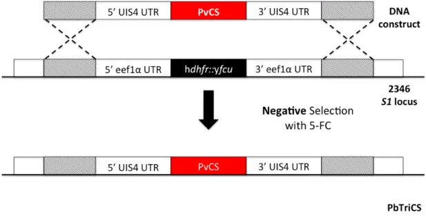
Figure 5: Schematic representation of PbTriCS transfection through the
GIMO transfection method. __________________________________ 16
Figure 6: Genotyping of PbTriCS parasites ________________________ 31
Figure 7: Sporogonic development of PbTriCS ______________________ 33
Figure 8: Gliding motility assay of PbTriCS _________________________ 34
Figure 9: PbTriCS infectivity and development in Huh7 in comparison with
PbGIMO parasites ________________________________________ 36
Figure 10: In vivo liver Pb infection load measured by qRT-PCR ________ 37
Figure 11: In vivo infectivity and development of PbTriCS _____________ 37
Figure 12: Immunofluorescence microscopy of EEFs in Huh7 hepatoma cell
line at 48 hours post-infection ________________________________ 39
Figure 13: Immunofluorescence microscopy of EEFs in C57BL/6J mice livers
at 46 hours post-infection ___________________________________ 40
Figure 14: FACS analysis of the percentage of traversed cells, normalized to
control. _________________________________________________ 41
Figure 15: Macrophage depletion and infection load at 46 hours post-infection
_______________________________________________________ 42
Figure 16: In vivo liver Pb infection load at 6 hours post-infection measured
by qRT-PCR. _____________________________________________ 43
Figure 17: Humoral responses assessed by ELISA determination of IgG titer
LIST OF TABLES
Table 1: PCR reaction components mixture ________________________ 21
Table 2: PCR reaction program for 5' integration, 3' integration and selectable
marker __________________________________________________ 21
Table 3: PCR reaction program for gene of interest (PvCS@UIS4) ______ 21
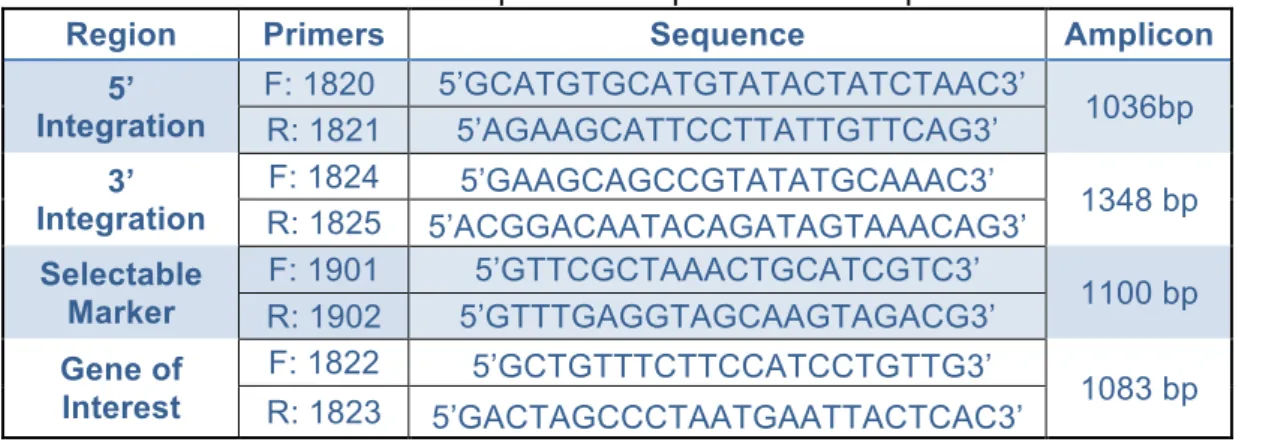
Table 4: Primers used and correspondent sequences and amplicons ____ 21
Table 5: cDNA reaction mixture __________________________________ 25
Table 6: cDNA thermo-cycler reaction program _____________________ 25
Table 7: qRT-PCR components mixture ___________________________ 26
Table 8: qRT-PCR reaction program ______________________________ 26
Table 9:Primers used in qRT-PCR analysis ________________________ 26
Table 10: Number of sporozoites in midguts, hemolymph and salivary glands
per mosquito at days 16, 18, 21 and 24 expressed as mean ± standard error of the mean. _________________________________________ 34
ABBREVIATIONS
2346 PbTriCS motherline kb Kilo Base Pairs
5-FC 5-fluorocytosine p.i. post-infection
bp Base Pairs PbCS Plasmodium berghei
Circumsporozoite Protein
BSA Bovine Serum Albumin PBS Phosphate-Buffered Saline Solution cDNA Complementary
Deoxyribonucleic acid PCR
Polymerase Chain Reaction
CS Circumsporozoite PfCS Plasmodium falciparum Circumsporozoite Protein
DNA Deoxyribonucleic acid PvCS Circumsporozoite Protein Plasmodium vivax
dNTP Deoxyribonucleotide triphosphate qRT-PCR
Quantitative Real-Time Polymerase Chain
Reaction
EEF Exo-Erythrocytic Form RAS Radiation Attenuated
Sporozoites
FACS Fluorescence-Activated
Cell Sorting RBC Red Blood Cell
GAP Genetically Attenuated Parasites RNA Ribonucleic Acid
GOI Gene of Interest RPM Rotations per Minute hdhfr human dihydrofolate reductase RPMI Roswell Park Memorial Institute medium HEPES 4-(2-hydroxyethyl)-1-piperazineethanesulfonic acid SM Selectable Marker HPRT Hypoxanthine Guanine Phosphoribosyl Transferase TR Targeting Region
Huh7 Human Hepato Cellular
Carcinoma Cells UTR Untranslated Region
i.p. Intraperitoneal Injection WT Wild-Type
i.v. Intravenous Injection yfcu
yeast cytosine deaminase and uridyl phosphoribosyl
INTRODUCTION
[Type the abstract of the document here. The abstract is typically a short summary of the contents of the document. Type the abstract of the document here. The abstract is typically a short summary of
1. INTRODUCTION
1.1 Malaria: Key facts
Malaria is a mosquito-borne disease, which is endemic in 97 countries, affecting 3.3 billion people, 1.2 billion of whom are at high risk of contracting malaria.1 The World Health Organization estimated 198 million cases of malaria in 2013 (95% uncertainty interval, 124-283 million), 584 thousand of which resulted in death (95% uncertainty interval, 367-755 thousand), including 453 thousand deaths of children under the age of five.1 Malaria has high rates of mortality as well as morbidity, affecting the economy of endemic regions, where malaria and poverty are closely related, making this disease a major obstacle to the economic development of affected regions.23
Malaria is caused by an apicomplexa parasite of the genus Plasmodium, which is transmitted by female mosquitoes of the genus Anopheles, the definitive host of Plasmodium, where sexual reproduction takes place.4 There are five species of Plasmodium that may cause the disease in humans: P. falciparum, P. vivax, P. malariae, P. ovale and P. knowlesi. P. falciparum and P. vivax are the most common.4 The former may lead to cerebral malaria and is responsible for the majority of the cases of malaria-associated death. The latter not only is responsible for 47% of malaria cases outside the African continent, but it may also form dormant parasites, termed hyponozoites, which may cause disease relapses. 15
1.2 Epidemiology
P. falciparum and P. vivax represent the greatest malaria-related public health concerns since they are responsible for the highest incidence rates.1 Although P. falciparum is responsible for the majority of deaths, it is predominant mostly in the African continent whereas P. vivax is more widespread since this species can develop in its vector can endure higher altitudes and cooler climates.1 Despite P. vivax being more widespread than P. falciparum, it has a lower transmission due to the predominance of the Duffy-negative genotype among the African continent.1 (Figure 1) P. vivax was thought to require the Duffy antigen to invade human erythrocytes, and in its absence, invasion is prevented. 6 7 8 However, recent studies have described P. vivax infection in Duffy negative individuals.9 There are 3 alleles that characterize the Duffy blood group: FYA, FYB and FYO and four different phenotypes: Fy (a+ b+), Fy (a+ b-), Fy (a- b+) and Fy (a- b-). The latter is predominant in African populations and individuals carrying this phenotype are resistant to a P. vivax infection. 1011
Other genetic traits are known to provide protection and genetic resistance to malaria infection, such as sickle-cell anemia, thalassemia, hemoglobinophaties, glucose-6-phosphate dehydrogenase (G6PD) deficiency and O allele from the ABO blood group. 12
Malaria-associated symptoms can vary between mild and severe: the symptoms begin with fever, headache, fatigue, muscle aches, nausea, vomiting, anemia and a palpable spleen a few days after the start of the disease. In children, a palpable liver is present whereas jaundice is notable in adults. The disease may progress to severe malaria where the symptoms include acute pulmonary edema and kidney injury, generalized seizures and coma and if not treated, death. 13 For a long period of time, it was believed that an infection with P. vivax was benign and was not responsible for the same numbers of mortality as P. falciparum. However, recent reports have shown that P. vivax infection is able to cause severe malaria, respiratory distress and coma, and has similar mortality rates to P. falciparum. 714
Figure 1: Transmission of P. falciparum, P. vivax and co-endemicity of P. falciparum
and P. vivax worldwide between high and low transmission.
Map created using World Health Organization Malaria Report 2014 based on individual country profiles. 1
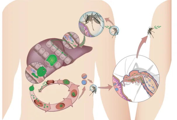
1.3 Plasmodium life cycle
The life cycle of Plasmodium (Figure 2) parasite begins when an infected female Anopheles mosquito takes a blood meal injecting hundreds of sporozoites into the skin of the mammalian host. Sporozoites, the liver-infective forms of Plasmodium parasites that reside in the mosquito’s salivary glands, eventually reach a blood vessel through a mode of locomotion known as gliding motility. After entering the bloodstream, the sporozoites reach the liver within minutes where they cross the liver sinusoids (fenestrated blood
vessels lined with endothelium and Kupffer cells), and traverse several hepatocytes by a process of migration known as cell traversal where they disrupt the hepatocytes plasma membrane without invading them. 15 Ultimately, sporozoites productively invade a hepatocyte with the formation of a parasitophorous vacuole (PV) where the parasite will reside during its liver stage development. The PV membrane (PVM) is the main interface between the parasite and the host. Inside the PV the parasite proliferates and dedifferentiates into a replicative form, the exo-erythrocytic form (EEF). Infection by human parasites P. vivax and P. ovale may produce cryptic liver-stage forms, known as hypnozoites, that can stay dormant for months or even years, before resuming development, eventually causing disease relapses.45
Figure 2: Life-cycle of the Plasmodium parasite.
Parasite transmission occurs when an infected female Anopheles mosquito takes a blood meal on a susceptible vertebrate host. The mosquito injects sporozoites into the skin, which enter the blood stream eventually reaching the liver. In the liver, sporozoites traverse several hepatocytes until infecting one. P. vivax and P. ovale may develop into dormant forms, the hypnozoites. The liver stage of infection ends with the release of merozoites that invade erythrocytes initiating the blood stage of infection and causing malaria symptoms. Plasmodium differentiates into gametocytes, male (microgamete) and female (macrogamete) and the mosquito upon a blood meal ingests them. In the mosquito’s midgut, microgametes penetrate the macrogametes generating zygotes. The zygotes become motile and elongated and are called ookinetes. These penetrate the mosquito’s midgut wall and will develop into oocysts. The oocysts mature and sporozoites develop within. Upon oocysts rupture, they release sporozoites that will migrate to the mosquito’s salivary glands. Upon the mosquito’s next blood meal, sporozoites are injected into the host’s skin and the cycle continues.
The EEF enters schizogony, undergoing rapid growth, where the parasite exceeds the size of an uninfected hepatocyte by 10 times,16 and DNA
replication. The liver stage of infection is a clinically silent to the host, yet obligatory, step in the lifecycle of the parasite, and culminates with the release of merozoite-filled vesicles, the merosomes, into the bloodstream. It is estimated that each merosome packs up from a few to thousands of merozoites. 16 In the bloodstream, the merosomes eventually burst in the microvasculature of the lungs, 17 releasing the merozoites, Plasmodium erythrocyte-infective forms, which will then initiate the blood stage infection. The liver stage of Plasmodium infection requires distinct time for each developmental stage in rodent and human malaria. The full liver stage infection takes 5 to 15 days for human-infective Plasmodium species, whereas in parasites infecting rodent hosts the parasite only takes 2 days to complete the process. 416.
Once in the blood, the parasite undergoes successive cycles of invasion, intracellular growth, multiplication and re-invasion, beginning with merozoites invading erythrocytes (red blood cells – RBC) by attaching to their surface, entering and enclosing themselves inside. Inside the RBCs, the parasite acquires a biconcave disc form known as ring stage. The parasite then rounds up and becomes a trophozoite, actively feeding, growing and modifying the RBC. The next parasite form is the schizont, where the parasite nucleus will divide to form approximately 16 nuclei. Merozoites then become visible at the RBC periphery, each one containing a nucleus. The blood stage cycle may start all over again when the RBC breaks down and the merozoites are released into the bloodstream. 18 These daughter merozoites from individual schizonts develop into either asexual stage or sexual stage parasites. The former will continue the cycle by invading new RBCs whereas the latter originate microgametocytes (males) or macrogametocytes (females). Gametocyte development starts identically to the asexual erythrocytic cycle, as the sexually committed merozoite invades a new RBC and grows into a trophozoite, later transforming into gametocytes instead of developing into schizonts. 19 It is in the blood stage of infection that the malaria-associated symptoms appear. 13
Gametocytes and erythrocytic parasite stages are ingested when a female Anopheles mosquito takes a blood meal from an infected host. The ingested blood will go to the mosquito midgut lumen where mosquito-specific factors and environmental factors will trigger parasite gametogenesis, transforming gametocytes into extracellular gametes. Microgametocytes undergo three mitotic replication producing eight daughter genomes by a process called exflagellation, where the motile microgametes fertilize the macrogametes that have already emerged from the host erythrocyte. Fusion of the micro and macrogametes forms diploid zygotes that initiate meiosis without nuclear or cellular division. Zygotes then transform into a motile-form called ookinete that will migrate and penetrate the midgut epithelium shifting from the lumen to the midgut basal lamina, where it transforms into a sessile spherical form, the oocyst. After the oocyst is formed, mitotic replication begins and continues throughout the period of sporogony that occurs within the oocyst, resulting in 1x10³ to 1x10⁴ sporozoites being produced by oocyst. During this period, the oocyst increases in size and eventually the oocyst capsule starts to fragment and small holes appear through which sporozoites escape leaving the
collapsed oocyst capsule on the external surface of the midgut. Sporozoites are then free in the mosquito haemolymph where some, but not all, will successfully invade the mosquito’s salivary glands by gliding motility with the formation of a parasitophorous vacuole 19.
1.4 Malaria control measures
Since there is still no licensed vaccine for malaria, the control of Plasmodium populations relies on the control of its vector Anopheles spp. Vector control measures include insecticide-treated nets (ITNs) and indoor residual spraying (IRS). 20 Although these measures were able to protect the population at risk of contracting malaria, insecticide resistance is spreading. 1 This is of great concern since it may lead to resurgence of malaria in non-immune populations, leading to an increase in mortality and morbidity. 21 The World Health Organization (WHO) recommends the use of Artemisinin-based Combination Therapies (ACTs) for the treatment of P. falciparum and P. vivax. In areas where there is no documented resistance to chloroquine this should be used for the treatment of P. vivax. Artemisinin-based monotherapy should be avoided since it leads to parasite resistance against artemisinin. 1 Currently, the only drug available against dormant liver-stage hypnozoites is primaquine, which can prevent relapses in P. vivax and P. ovale infections. However, primaquine can lead to acute haemolytic anemia in G6PD-deficient individuals. 22
Throughout history, eradication programs that relied mostly in vector control have proven to be inefficient, such those against malaria and yellow fever. On the other hand, eradication campaigns based on vaccination successfully eradicated smallpox globally and eradicated polio in some regions. 23
1.5 WHO Malaria Vaccine Technology Roadmap
In 2013, the WHO issued some guidelines in the Malaria Vaccine Technology Roadmap concerning the licensing of malaria vaccines.
As stated: 24 By 2030, license vaccines targeting Plasmodium falciparum and Plasmodium vivax that encompass the following two objectives, for use by the international public health community:
• Development of malaria vaccines with protective efficacy of at least 75 percent against clinical malaria suitable for administration to appropriate at-risk groups in malaria- endemic areas.
• Development of malaria vaccines that reduce transmission of the parasite and thereby substantially reduce the incidence of human malaria infection. This will enable elimination in multiple settings. Vaccines to reduce transmission should be suitable for administration in mass campaigns.
1.6 Malaria Vaccines
Naturally acquired immunity (NAI) against blood stage malaria occurs in individuals in endemic areas and protects them from severe disease and death when exposed to Plasmodium. 25 In areas where malaria is hyper and holoendemic, meaning that transmission is high and near every individual is infected, NAI provides protection to adults and older children from severe mortality and morbidity, as the disease becomes asymptomatic in these individuals, but little is known about how this protection occurs. 26 Although an asymptomatic status is achieved among adults, sterile immunity against infection is never accomplished. 26
Studies in the late 1960s by Nussenzweig et al. showed that radiation attenuated sporozoites could induce protective immunity to infection in mice.27 In the early 1970s, Clyde et al. showed that immunization of naïve volunteers with irradiated P. falciparum sporozoites led to high levels of immunity. 28 This work was the first proof that the development of a vaccine is indeed possible and that the scientific community should continue to focus on the search for a suitable malaria vaccine. 29
Traditionally, there are three Plasmodium vaccination approaches that depend on the lifecycle stage that is the target for the host’s immune response. These include pre-erythrocytic malaria vaccines, which target the sporozoite and liver stage prior to blood infection; erythrocytic malaria vaccines, which target the asexual blood stage of the parasite; and transmission-blocking malaria vaccines, which target the gametocyte/gamete stage of the parasite, thus blocking transmission between the host and the mosquito. 25
• Pre-erythrocytic vaccines, if proven efficacious, should not only prevent disease by preventing blood infection but also block the transmission to the mosquito. 29 Additionally, the pre-erythrocytic stage is ideal for vaccination since only a small number of sporozoites are injected by the mosquito, in contrast with thousands of merozoites that are released into the blood. Due to the clinically silent nature of the pre-erythrocytic stage of the parasite lifecycle, this is an excellent target to elicit immune responses. 30
• Erythrocytic vaccines, although targeting the asexual blood stages of the parasite, do not prevent infection, instead reducing clinical symptoms or severe illness. 29 The most advanced erythrocytic vaccine candidates target antigens that coat the surface of merozoites or antigens involved in invasion of erythrocytes. 29 Examples of these candidates include MSP119 that targets the merozoite surface protein 1 (MSP1) therefore inhibiting erythrocyte invasion and AMA1, apical membrane antigen 1, which is thought to prevent erythrocyte invasion and is shown to inhibit erythrocyte invasion in vitro and it may also impair hepatocyte invasion. Studies and trials with these vaccine candidates have shown discouraging results in pursuing erythrocytic vaccines against malaria. 29
• Transmission-blocking vaccines are based on antibodies that target antigens unique to gametocytes or gametes in order to interrupt development of the parasite in the mosquito, therefore blocking transmission from the host to the mosquito. 29 There are some transmission-blocking vaccines that advanced into clinical trials, namely Pfs25 for P. falciparum and Pvs25 for P. vivax. These vaccines target a protein expressed on the surface of the zygote and the ookinete. 25 These vaccines are thought of as “altruistic vaccines” 25 because the vaccinated individuals are not protected from infection nor disease. 29 Thus, in order to specify the efficacy of such vaccine candidate, an entire community must be vaccinated to determine if transmission of the parasite wears down. 25
A desirable vaccine candidate should produce protective immune responses that outweigh the responses in naturally acquired immunity to malaria. Not only should these immune responses act rapidly, but they should also be long lasting and entirely prevent infection. 29 This thesis will present some more extended information on pre-erythrocytic malaria vaccines as they are the main focus of the thesis work.
1.6.1. Pre-erythrocytic malaria vaccines
As mentioned above, in the early 1970s Clyde et al. have shown the potential of a pre-erythrocytic vaccine to produce sterile immunity in naïve volunteers against P. falciparum, making the pre-erythrocytic stage the only stage demonstrated to be capable of eliciting sterilizing immunity. 31
Pre-erythrocytic malaria vaccines can be divided into two groups, depending on the strategy employed to elicit the immune responses: sub-unit and whole-organism vaccines.
1.6.1.1. Sub-unit pre-erythrocytic malaria vaccines
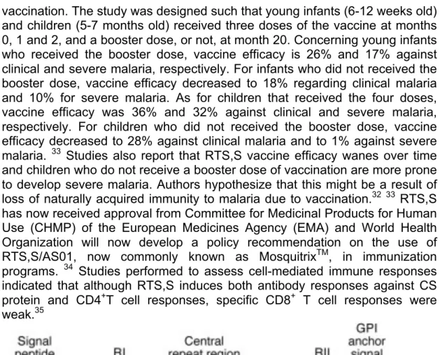
Pre-erythrocytic subunit malaria vaccines use a small portion of the parasite, usually a protein or a peptide, as the antigenic basis to elicit an immune response whereas pre-erythrocytic whole-organism malaria vaccine uses the whole parasite, either attenuated or under antimalarial therapy. Concerning the sub-unit malaria vaccines, RTS,S/AS01, created in 1987, is the most advanced malaria vaccine candidate to date and the first malaria vaccine candidate to undergo large-scale phase 3 clinical trials in Africa. 32 RTS,S/AS01 targets the pre-erythrocytic stage of P. falciparum eliciting antibody responses to its circumsporozoite (CS) protein.32 RTS,S/AS01 is composed of the P. falciparum CS protein central repeat region (R) and T cell epitopes (T) fused with hepatitis B surface antigen (S) and co-expressed in Saccharomyces cerevisiae (S) (Figure 3). RTS,S is formulated with AS01 adjuvant to enhance immune response to antigens and previous studies have confirmed that RTS,S/AS01 is able to provide protective immunity. 2931 33 Final results from phase 3 clinical trials were published in July 2015 and reported on the efficacy and safety of the vaccine 3 years after first
vaccination. The study was designed such that young infants (6-12 weeks old) and children (5-7 months old) received three doses of the vaccine at months 0, 1 and 2, and a booster dose, or not, at month 20. Concerning young infants who received the booster dose, vaccine efficacy is 26% and 17% against clinical and severe malaria, respectively. For infants who did not received the booster dose, vaccine efficacy decreased to 18% regarding clinical malaria and 10% for severe malaria. As for children that received the four doses, vaccine efficacy was 36% and 32% against clinical and severe malaria, respectively. For children who did not received the booster dose, vaccine efficacy decreased to 28% against clinical malaria and to 1% against severe malaria. 33 Studies also report that RTS,S vaccine efficacy wanes over time and children who do not receive a booster dose of vaccination are more prone to develop severe malaria. Authors hypothesize that this might be a result of loss of naturally acquired immunity to malaria due to vaccination.32 33 RTS,S has now received approval from Committee for Medicinal Products for Human Use (CHMP) of the European Medicines Agency (EMA) and World Health Organization will now develop a policy recommendation on the use of RTS,S/AS01, now commonly known as MosquitrixTM, in immunization programs. 34 Studies performed to assess cell-mediated immune responses indicated that although RTS,S induces both antibody responses against CS protein and CD4+T cell responses, specific CD8+ T cell responses were weak.35
Figure 3: RTS,S particle composition.
HBsAg: Hepatitis B surface antigen;36
According to the 2013 WHO Malaria Vaccine Technology Roadmap a malaria vaccine should be available for used and licensed by 2030 have at least 75% efficacy. Therefore, the modest results from RTS,S/AS01 phase 3 clinical trials do not meet WHO guidelines and the search for a more effective vaccine continues. However, the effort and time required in clinical trials and the protection by vaccination in malaria-endemic areas are to be admired. 2425
1.6.1.2 Whole-organism pre-erythrocytic malaria vaccines
Whole-organism pre-erythrocytic malaria vaccines rely on the use of the whole parasite, either attenuated or under antimalarial therapy, to elicit an immune response. 29
Early work on malaria vaccines during the 1940s by Russell and Mohan with fowls immunized with P. gallinaceum and later by Freund et al. first with ducks immunized with P. lophurae and then with rhesus monkeys with P. knowlesi has shown that a whole-organism malaria vaccine can accomplish good protective efficacy against the parasite. 37 As previously mentioned, later work by Nussenzweig et al. showed that protective immunity could be induced in mice, and studies by Clyde et al. have shown that 90% of naïve human volunteers were immunized against P. falciparum after immunization with infected and irradiated mosquitoes. 37
Whole-organism vaccines can be divided into 3 categories according to the method used to attenuate the parasite in order to elicit an immune response. The parasite can be attenuated, either by genetic manipulation or radiation, or it can be delivered live to volunteers taking antimalarial chemoprophylaxis.
a) Radiation-attenuated sporozoites (RAS)
Radiation-attenuated sporozoites prevail as the “gold standard” after studies in rodents and humans showed its sterilizing protection potential against Plasmodium infections.27 28 38 RAS are incapable of division and differentiation. They are able to enter the hepatocytes but do not develop inside these cells. Nonetheless, they are able to trigger potent CD8+ T cell responses, unlike heat-killed sporozoites that induce very low CD8+ T cell responses. 39 40 Studies have shown that these CD8+ T cell responses are required for sterilizing immunity in malaria but it remains unclear how these cells are able to reduce the liver-stage parasite burden. 41 Immunizations with irradiated sporozoites have shown that two months after the immunization, the CS antigen continues to be presented. Importantly, this non-replicating antigen persists and is able to elicit immune responses by priming naïve T cells to acquire effector functions in order to clear the parasite in the liver long after immunization. 39
RAS immunization has been the focus of the efforts by the US-based company, SanariaTM, who have developed PfSPZ, a malaria vaccine candidate that targets P. falciparum and consists of P. falciparum aseptic, purified and cryopreserved sporozoites attenuated by irradiation– metabolically active and non-replicative.42 One downside of this vaccine is the route of administration. In animal models the common route is intravenous as it is the closest to the proboscis mosquito bite. However, an undetermined number of sporozoites are injected per mosquito and studies relying in mosquito bite are harder to standardize.29 43 Therefore, other routes of administration were evaluated and it has been shown that subcutaneous and intradermal sporozoite administration led to a suboptimal immunogenicity and protective efficacy.44 For this reason, more recent clinical trials of this vaccine
candidate used either mosquito bites or intravenous injection as a method of immunization.4045
Although this strategy of malaria vaccination is able to elicit strong immune responses, it needs around 1000 irradiated P. falciparum infected mosquitoes to achieve that since the parasite it is not able to fully develop in the liver 42 whereas using CPS vaccination approach (see below) only 12-15 mosquitoes are required per immunization.46 Moreover, this vaccination approach requires the availability of biosafety level III laboratories, since P. falciparum infected mosquitoes represent a risk of infection to humans. Another downside of this vaccination approach is that it only delivers antigens against one species of Plasmodium, not being able to meet the WHO Malaria Vaccine Technology Roadmap guidelines.
b) Genetically attenuated parasites (GAP)
Genetically attenuated malaria parasites were firstly developed in 2005 by deleting essential genes for liver-stage development of Plasmodium through the method of gene targeting that allows, by homologous recombination, the inactivation or modification of a gene. 47 48 49 50 If this strategy is to be used as a method of sporozoite attenuation, the gene targeted for deletion has to be essential in liver-stage development in order for the parasite to be arrested in the liver and be unable to proceed with blood stage infection. However, this gene cannot be essential in blood stage, sexual and mosquito stage development, since blood stage parasites are required to the current transgenesis methods, and a normal development in the mosquito is needed for the production of sporozoites. It is also essential that the GAP lack drug-resistance markers, therefore meeting regulatory concerns in genetically attenuated organisms in vaccines.30 It is also important that the GAP is created by double crossover recombination instead of single crossover recombination, making the parasite incapable of reverting to the wild-type phenotype and complete liver-stage development. 30
One example of this vaccination strategy includes the deletion of UIS (upregulated in infective sporozoites) genes – essential to early liver-stage development. The UIS3 gene was successfully deleted in P. berghei and immunizations in rodents with this genetically attenuated parasite have proved to be capable of sterile immunity when challenged with wild type sporozoites and no breakthroughs were observed. However, this immunity is only active against pre-erythrocytic stages since parasitaemia was observed in mice when challenged with blood-stage parasites. 49 UIS4 gene has also been deleted in P. berghei and immunizations in mice have shown that this parasite is also capable of sterile immunity. However, when higher doses of sporozoites were administered intravenously (50,000 sporozoites vs 1,000 sporozoites) mice became infected with blood-stage parasites and genotyping confirmed that the blood-stage parasites were UIS4 knockout, therefore the parasite did not revert to wild-type. 48
Another GAP vaccine candidate relies on the double knockout of p52 and p36 genes that are shown to be essential in establishing the parasitophorous vacuole, therefore the deletion of these genes results in early liver-stage
developmental arrest. 51 Initial studies carried out this double knockout parasite in a rodent model demonstrated that this model of a GAP vaccine is capable of inducing sterile protection and no breakthroughs were reported. 52 Later, a double knockout with human parasite P. falciparum orthologous genes was performed 52 and human trials were performed in order to test safety and immunogenicity of this vaccine candidate. 51 Volunteers were first exposed to 5 mosquito bites and then to 200 mosquito bites employing mosquitoes infected with the double knockout P. falciparum parasite developed immune responses against the repeat region of the CS protein, whereas volunteers exposed to only 5 mosquito bites did not. However, 12 days after the second mosquito bite, one volunteer presented blood stage parasites. After genotyping, it was shown that these parasites were indeed the double knockout parasite and therefore the parasite did not revert to the wild type. As recently shown in rodent model of malaria, p52 and p36 mutants are rarely capable of establishing an infection in a hepatocyte without the formation of a parasitophorous vacuole, which could explain the presence of blood stage parasites in one of the volunteers. Although this study showed promising results in terms of elicited antibody responses, investigation should proceed in optimizing multiple gene deletion responsible for essential steps in the liver stage development.51
Although this vaccination approach has the major advantage of a homogenous population of parasites where the genetic identity and phenotype can be defined in order to elicit optimal immune responses comparable with irradiated attenuated sporozoites immunizations, 30 as rodent studies and early human trials show, it is not risk-free since rare events may occur where the parasite is able to complete is life-cycle.
c) Chloroquine Chemoprophylaxis with Sporozoites (CPS)
Sterile immunity against Plasmodium was shown to be achieved by immunizing rodents with P. berghei while treating them with chloroquine, a drug known to kill asexual blood-stage parasites but not liver-stage parasites.53
Studies in humans have also shown that sterile immunity can be accomplished employing a drug-sensitive strain of P. falciparum, under chloroquine prophylaxis. Moreover, these studies have demonstrated to be more efficient in terms of protection that RAS immunizations since the latter requires a significantly higher number sporozoites to elicit sterile protection. 46 54 Thirty-two months after the initial immunization, volunteers were re-challenged with infected P. falciparum mosquitoes and four out of six volunteers were negative for blood-stage parasites, while the other two had a delay in parasitemia.55 It has been shown in studies using two groups of immunized individuals either by P. falciparum sporozoite inoculation under chloroquine treatment or with P. falciparum infected erythrocytes under chloroquine treatment that sterile protection is achieved by immune responses against the pre-erythrocytic stage only. The group inoculated with P. falciparum infected erythrocytes under chloroquine treatment skipped all the pre-erythrocytic stage of Plasmodium life-cycle and therefore was susceptible against a sporozoite challenge, whereas the group immunized with P.
falciparum sporozoites under chloroquine treatment were all protected against a sporozoite challenge. Nonetheless, none of the groups were protected against a challenge with P. falciparum infected erythrocytes nor was a difference in the period of parasitaemia appearance.56
Even though this immunization strategy is capable of eliciting sterile protection, it does not represent a possible strategy to be broadly implemented as it relies on the administration of fully infectious P. falciparum sporozoites. Nonetheless, it is a remarkable model to study immune responses against malaria.46
1.7 Rodent models of Plasmodium infection
1.7.1 Plasmodium berghei as a model to study malaria
Studying malaria in humans, although desirable, bears a lot of limitations. The ability to study the entire life-cycle is hindered by the lack of access to post-mortem organs and tissue samples. Even if access to these tissues is possible, they represent only the end stage of a complex life-cycle, and access to the events that led to this stage, such as the pre-erythrocytic stage or the study of cerebral malaria, is very limited. Moreover, it is not possible to control the immune responses in such studies. Therefore, the study of malaria has relied on the use of rodents for decades and such studies greatly contributed to a better understanding and knowledge of the parasite biology and host-parasite interactions. 5758
There are several species of Plasmodium known to infect rodents – P. berghei, P. yoelii, P. chabaudi and P. vinckei – and several of strains available in each species.4 57 Regarding rodent-infectious Plasmodium parasites, P. berghei is widely used to study severe malaria clinical outcomes such as cerebral malaria, placental malaria, malaria-associated acute lung injury, acute kidney injury, severe malarial anemia and liver injury. 57 Although the scope of disease manifestations may vary between species and hosts, this must be seen has a reflection of the diversity in human disease and immunity.5758
1.7.2 Plasmodium berghei as a vaccination approach
Several vaccine candidates began with studies in rodent models of malaria, and even though they were not able to anticipate the success in human trials, they reliably conclude which ones were not. 58
Recently, the Prudêncio Lab at Instituto de Medicina Molecular has been working on a new whole-organism pre-erythrocytic malaria vaccine using a genetically modified P. berghei parasite as a method of presenting the immunogenic antigens of human infective Plasmodium spp. to the human immune system (see section 1.9.1). In order to accomplish the development of this new vaccine candidate, three premises had to be validated regarding P. berghei infectivity and immunogenicity: (1) Using in vitro human hepatoma
cell lines, ex vivo cultures of human primary hepatocytes and in vivo liver-humanized mouse models (LHmice) it was demonstrated that P. berghei can infect and develop within human liver cells. Therefore, is able to present antigens that will generate the immune response required from a pre-erythrocytic vaccine; (2) P. berghei is not known to cause human pathology, thereby ensuring higher safety levels than previous whole organism vaccine approaches. Moreover, using blood-humanized mice (BHmice), it was experimentally demonstrated for the first time that P. berghei parasites have very low infectivity of RBCs, and even though some merozoites are able to infect human erythrocytes, they are incapable of multiplying and therefore causing disease; (3) A genetically modified P. berghei expressing the selected antigen is able to elicit an immune response capable of protecting against a subsequent P. falciparum infection (Unpublished data).
1.8 Circumsporozoite Protein as the immunodominant antigen
in Plasmodium
The circumsporozoite protein (CSP) is the most abundant protein in Plasmodium sporozoites. In the sporozoite surface, the CSP forms a dense coat and has been shown that plays a role in gliding motility. In the oocyst, CSP is essential for sporozoite formation and it is believed to be required to establish infection in the host’s liver.59 60 The structure of CSP is highly conserved among Plasmodium species infecting rodents, primates and humans.60 CSP is composed by a central repeat region, differing through Plasmodium species. This central repeat region is flanked by two highly conserved domains: Region I and region II. Region I is a 5-aminoacid sequence at the N-terminus and region II has a thrombospondin repeat at C-terminus. 60 P. vivax CSPs may differ around the world between VK210 and VK247. These two types of CSPs diverge in the central repeat region. 61
Studies with transgenic mice expressing P. yoelii CS gene under constitutive promoter control have shown that the circulating CS is able to inhibit the infectivity of live sporozoites when injected intravenously or by mosquito bite. The results from these studies show that CSP is an immunodominant protective antigen in humoral responses against Plasmodium infections. 62 Moreover, in a study where irradiated sporozoites immunized WT mice were compared with CSP-Tg/JhT(-/-) BALB/c irradiated sporozoites immunized mice, T-cell responses were evaluated by IFN- γ ELISPOT of mouse splenocytes. CSP-Tg/JhT(-/-) BALB/c mice are transgenic for the CS and unable to make antibodies. The authors reported that CD8+ T-cell responses to non-CSP antigens were 20-100 times lower than those against CSP.63 As mentioned above, the most advanced malaria vaccine candidate until date, RTS,S is based on the CSP of P. falciparum.
1.9 Genetic transformation of Plasmodium berghei
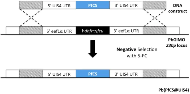
In 2011, Lin, J. et al. developed the gene insertion/marker out (GIMO) method for transgene expression in rodent malaria parasites. This method allows the simultaneous introduction of heterologous proteins and removal of a drug-selectable marker. 64
In this report, the authors designed and constructed a mother line (GIMOPBANKA for P. berghei) where a hdhfr::yfcu selectable marker is stably integrated into the non-essential 230p locus present in chromosome 3. The gene of interest is integrated into the same locus by double cross-over homologous recombination, replacing the hdhfr::yfcu selectable marker cassette. These parasites can be negatively selected – as the motherline expressing the yfcu gene will not survive the 5-FC treatment. 5-FC is the drug 5-fluorocytosine that will kill all parasites expressing yfcu. 64
1.9.1 Plasmodium berghei expressing Plasmodium falciparum CS
Previous studies performed in the Prudêncio Lab with a transgenic P. berghei expressing P. falciparum CS under the control of P. berghei UIS4 promoter (Pb(PfCS@UIS4)), generated by the GIMO method (Figure 4), showed that this parasite has salivary gland and hepatic infectivities similar to wild-type P. berghei (Unpublished data), overcoming the limitations of a previously assayed transgenic parasite, where the endogenous CS was replaced by P. falciparum CS (Pb(PfCS)), which was shown to have low mosquito salivary gland and hepatic infectivities. 65
As previous stated, this transgenic P. berghei parasite is able to i) infect human hepatocytes in vivo; ii) unable to cause a human blood-stage infection that leads to pathology; iii) immunizations with Pb(PfCS@UIS4) parasites elicits immune responses against a P. falciparum infection.
Further studies, using a non-natural host species, the New Zeeland White rabbit, as a model of P. berghei infection demonstrated that P. berghei is able to infect rabbit primary hepatocytes ex vivo and rabbit livers in vivo. However, blood-stage forms of the parasite were never detected in circulation in the rabbit. In order to assess infectivity of the merosomes developed in rabbit primary hepatocytes, merosomes were collected from the culture supernatant and injected into naïve mice. After around 7 days post-infection, mice were positive for blood-stage parasites (Unpublished data).
Humoral responses were analyzed in both mice and rabbits after immunizations with Pb(PfCS@UIS4) and results show high titers of anti-PfCS antibodies in the serum of these animals. Moreover, serum from mice immunized with Pb(PfCS@UIS4) is able to recognize and bind with high avidity to P. falciparum sporozoites and is capable to functionally inhibit P. falciparum hepatic invasion (Unpublished data).
Cellular responses were analyzed by ELISPOT assay with splenocytes of Pb(PfCS@UIS4) immunized mice and results showed that P. berghei/
Pb(PfCS@UIS4) elicit cross-species cellular immune responses against P. falciparum, which are not dependent on the presence of the PfCS immunogen on the P. berghei parasite.
Figure 4: Schematic representation of Pb(PfCS@UIS4) through the GIMO
transfection method.
1.9.2 Plasmodium berghei expressing Plasmodium vivax CS
In 2013, a transgenic P. berghei expressing P. vivax CS under the control of the P. berghei UIS4 promoter (Pb(PvCS@UIS4)) was generated by Inês Albuquerque, employing the GIMO method. This parasite was further characterized by Miguel Duarte and, similarly to Pb(PfCS@UIS4), Pb(PvCS@UIS4) was shown to have normal sporogonic development and hepatic infectivity similarly with the wild-type P. berghei (Unpublished data). Even though this characterization showed positive results, no immunization studies were yet performed with this parasite.
1.9.3 P. berghei simultaneously expressing P. falciparum and P.
vivax CS
Due to the success of Pb(PfCS@UIS4) and preliminary studies with Pb(PvCS@UIS4) having shown to being identical in terms of sporogonic, in vitro and in vivo development to the wild-type , and in compliance with the World Health Organization guidelines, the Prudêncio Lab decided to generate a P. berghei transgenic parasite simultaneously expressing CS of P. falciparum and P. vivax in non-essential loci under the control of P. berghei UIS4 promoter – PbTriCS.
Therefore, at the Leiden University Medical Center, the hdhfr::yfcu selectable marker was stably integrated into the non-essential S1 locus present in