Vol.56, n.6: pp. 962-970, November-December 2013
ISSN 1516-8913 Printed in Brazil BRAZILIAN ARCHIVES OF
BIOLOGY AND TECHNOLOGY
A N I N T E R N A T I O N A L J O U R N A L
Expression of Costimulatory Molecules in Antigen-Activated
Peritoneal Macrophages Treated with either Ovalbumin or
Palmitoyl-Ova Conjugates
Flávia Márcia Oliveira
1*, Valéria Ruiz-de-Souza
2, Maria Aparecida Campana-Pereira
3and
Cristiano Machado Gontijo
31Departamento de Educação em Saúde; Universidade Federal de Sergipe; Lagarto - SE - Brasil. 2Departamento de
Morfologia; Universidade Federal de Minas Gerais; Belo Horizonte - MG - Brasil. 3Departamento de Bioquímica e Imunologia; Universidade Federal de Minas Gerais; Belo Horizonte - MG - Brasil
ABSTRACT
One of the mechanisms by which adjuvants are believed to promote T-cell activation and prevent induction of oral tolerance is by up-regulating the expression of co-stimulatory molecules on antigen presenting cells. Mice treated orally with palmitoyl-ovalbumin conjugates become immunized, while those treated with native ovalbumin (Ova) become tolerant. Cells from the peritoneal cavity of B6D2F1 mice were cultured in the presence of 0.01, or 0.1 mg/100µl of either Ova, or palmitoyl-Ova and tested for the presence of cell markers. PE-conjugated anti-mouse CD80, CD86, and CD11b antibodies as well as biotin-PE were used to stain the antigen-activated peritoneal cells. A significant increase in the expression of CD86 and CD80 was observed following in vitro stimulation with palmitoyl-Ova; additionally, both Ova and palmitoyl-Ova induced the basal expression of CD11b. These findings could be related with the strong T-cell proliferative response induced by palmitoyl-Ova.
Key words: Ovalbumin, Palmitic Acid, Cellular Immunity, CD86, CD80, CD11b
*Author for correspondence: [email protected]
INTRODUCTION
Although oral administration of antigens offers significant advantages over the conventional parenteral routes in vaccination protocols, oral administration usually results in an immune unresponsiveness known as “oral tolerance” (Titus and Chiller 1981). Various strategies, such as the local production of secretory IgA and systemic immunization, have been examined to avoid the tolerance and to achieve stimulation. A complex
interplay of mechanisms, including clonal
deletion, clonal anergy, cytokine regulation and specific features of the mucosal tissues appears to govern the induction of oral tolerance (Friedman
One of the mechanisms by which adjuvants are assumed to promote the T-cell activation and prevent the induction of oral tolerance is through the upregulation of the expression of costimulatory molecules on antigen presenting cells (APCs), especially those belonging to the B7-28 family (Williansom et al. 1999). The classical B7-CD28 pathway is associated with two ligands, B7-1/CD80 and B7-2/CD86, on APCs and at least two receptors, CD28 and cytotoxic T-lymphocyte antigen 4, on T cells (Greenwald et al. 2005). In an earlier studies, the nature of a soluble protein antigen (ovalbumin; Ova) was changed by coupling it to palmitoyl residues, which showed that this procedure did not interfere with the induction of immune responses in the animals that were injected with one, or another form of the antigen (Oliveira et al. 1998; Oliveira et al. 2002). However, when both the forms of the antigen were orally administered to mice, opposite effects were obtained, induction of oral toleranc by the native protein and development of humoral immune response by the lipid-conjugated protein (Oliveira et al. 1998).
The present study investigated the role of the accessory cells in the immune responses triggered by the oral route and focused on the pattern of expression of co-stimulatory molecules on Ova or palmitoyl-Ova-activated peritoneal macrophages and the induction of cellular immune responses.
MATERIALS AND METHODS
Animals
B6D2F1 mice, an abbreviation for (C57Bl6 × DBA2) F1 hybrids from both sexes, were obtained from the breeding unit at the Federal University of Minas Gerais (UFMG, Belo Horizonte, Brazil). Mice were used in the experiments only after they were 7–9 weeks old.
Antigens
Grade V chicken egg albumin (OVA) was purchased from the Sigma Chemicals Co. (St. Louis, MO, USA). The palmitoyl-Ova conjugates were prepared as previously published (Oliveira et al. 1998).
In vitro Treatment of Peritoneal Cells with
Antigen
The peritoneal cavity (PerC) was injected with 5.0 ml of sterile phosphate-buffered saline (PBS).
Cells were collected, washed three times with cold PBS and then re-suspended in RPMI 1640 supplemented with 2 mM L-glutamine (Sigma), 1 mM sodium pyruvate (Sigma), 5 × 10⁻⁵ M 2-mercaptoethanol (Merck-Schuchardt, München,
Germany), 50 IU/ml penicillin, 50 µg/ml
streptomycin (Sigma) and 10% heat-inactivated fetal calf serum (FCS–Interlab, São Paulo, Brazil). Cultures were run in Nunc Dishes (St. Louis, MO, USA) 96 U-bottomed tissue culture plates, where 3 × 10⁵ cells/well were incubated in the presence of 0.01, or 0.1 mg/100 µL of either Ova or palmitoyl-Ova. After 3–4 h, cells were washed and examined for the presence of cell markers.
Flow Cytometry Analysis
PE-conjugated anti-mouse CD80, CD86, or CD11b antibodies (Southern Biotechnology, Birmingham, AL) as well as biotin-PE were used to stain the Ag-activated peritoneal cells and positive and negative control samples were run on every plate to standardize the assays. Antibodies were directly added to the wells, followed by incubation at 5oC and in the dark for 30 min. Subsequently, cells were fixed with glutaraldehyde and analyzed by flow cytometry using a FACScan flow cytometer (Becton Dickinson Mountain View, CA) using Cellquest.
Oral Treatment and Immunization
Mice were intubated with a urethral polyvinyl catheter calibrated to reach the stomach and given 20 mg of antigen (Ova or palmitoyl-Ova) in 0.5 mL of saline (0.15 M NaCl). The control groups received 0.5 mL of saline alone. One week after the oral treatment, mice were injected ip with 0.5 mL saline containing 10 µg of Ova mixed with 1 mg Al(OH)3 as adjuvant, or with 100 µg of Ova in 50 µL of complete Freund’s adjuvant (CFA) sc. Two weeks after the priming, mice were injected again with 0.5 mL saline containing 10 µg of Ova
ip. One week after the booster, blood samples were collected from the animals.
Systemic DTH Responses
Three weeks after the primary immunization, systemic DTH was assessed using the footpad-swelling test. Mice were sc injected with 100
and differences between the two measurements were used for group comparison.
Cellular Proliferation
Cell culture was performed as previously described (Oliveira et al. 2002). Splenocytes (5 × 10⁶ cells/mL) were cultured in complete medium (RPMI 1640; Sigma) supplemented with 2 mM L-glutamine (Sigma), 1 mM sodium pyruvate (Sigma), 5 × 10⁻⁵ M 2-mercaptoethanol (Merck-Schuchardt, Müchen, Germany), 50 penicillin IU/mL, and 10% heat-inactivated fetal calf serum (FCS–Interlab, São Paulo, Brazil). Cultures were run in triplicate in 96-well flat-bottomed tissue culture plates (Nunc). Cells were stimulated with Ova (1 mg/mL), or Concanavalin A (ConA–200
µg/mL) and were pulsed on day 3 with 20 µL of [3H]-thymidine with a specific activity of 2 Ci/mmol for another 12 h. Cells were then harvested and the total amount of incorporated thymidine was assessed using a β-counter.
Statistical Analysis
The difference between the experimental and control groups was assessed by the ANOVA and considered significant for p < 0.05. The data are from at least three separate experiments.
RESULTS AND DISCUSSION
Evaluation of the Expression of Costimulatory Molecules
It has been previously shown that the coupling of palmitoyl residues to Ova abolished the induction of oral tolerance in B6D2F1 mice (Oliveira et al. 1998; Oliveira et al. 2002). To determine whether this coupling might influence the activation of APCs, cells from PerC were cultured with Ova or palmitoyl-Ova at two different concentrations and evaluated for the expression of activation markers. Mouse PerC selectively attracts and maintains
specialized immune cells, including PerC
macrophages. Approximately 40% of PerC cells were B lymphocytes (78% B-1; 15% B-2), 55%
were macrophages (90% large peritoneal
macrophages; 10% small peritoneal macrophages), 2% were cells (DCs), and 3% were eosinophils, NK cells, and T lymphocytes (Ghosn et al. 2010). It is well established that the induction and activation of T cells require the engagement of CD28 by CD80 and CD86 to provide a second
potent costimulatory signal (Lenschow et al. 1996). APCs that express low levels of CD80 and CD86 appear to preferentially promote T-cell tolerance, while APCs expressing high levels of these molecules delivered stimulatory signals to the T cells (Perez et al. 1997). However, a paradigm has been established in many animal models that positive and negative costimulation by the members of the CD28 family is critical for the
development of immune responses, and
establishment and maintenance of peripheral tolerance. During the last decade, the complexity of costimulatory pathways has greatly increased due to its influence on regulatory T cells and APCs
(Bour-Jordan et al. 2011). Importantly,
costimulatory signals affect Treg development,
homeostasis, and suppressive functions,
independent of their effector T cells (Bour-Jordan; Bluestone 2009). Tregs have been reported to suppress the conventional T cells through the production of immunosuppressive cytokines and direct alterations of effector T cells or APCs (Sakaguchi et al. 2008).
CD11b/CD18 (also called Mac-1 or CR3) is a β2 integrin expressed predominantly in the activated macrophages and DCs, plays a significant role in the production of numerous genes (COX-2, IL-12 p35, IL-12 p40), and interferes with the induction of cellular responses (Wu et al. 2004). On the contrary, deficiency of CD11b worsens the inflammation and disease progression in several autoimmune models (Lee et al. 2005; Popov et al. 2006), suggesting that CD11b/CD18 is potentially involved in immune suppression rather than immune activation. Consistent with these findings,
studies showed that the proportion of
CD11c+CD11b+ DCs was higher than
CD11c+CD8a+ DCs in the spleen of tolerant mice (Li et al. 2008). It is of interest that CD11+CD11b+ DCs induce the Th2 cytokines, namely IL-10 and IL-4, in vivo (Pulendran et al. 1999). CD11b-/- mice are defective in developing immune suppression upon low-dose Ag feeding, while CD11b deficiency does not affect immune the activation (Ehirchiou et al. 2007). IL-10 has been reported to play a major part in immune tolerance and its production by DCs is critical for the differentiation of Treg cells (Zhang et al. 2004).
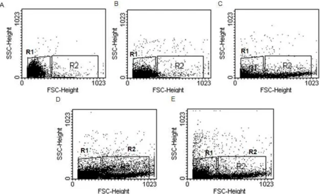
specifically includes B-lymphocytes and small peritoneal macrophages) and R2 (large cells) were created and a shift to R2 was observed following
the antigen stimulation, specially by 1.0 mg/mL Ova and palmitoyl-Ova (Fig 1).
Figure 1 - Representative dot plots showing SSC-Height and FSC-Height for PerC cells by flow cytometry. A: Untreated (control); B: 0.1 mg/mL stimulated; C: 1.0 mg/mL Ova-stimulated; D: 0.1 mg/ml palmitoylOva-stimulated; E: 1.0 mg/mL palmitoyl -Ova-stimulated. One of three representative experiments is shown.
Figures 1–7 summarize the data obtained from the flow cytometry analysis of the cells treated with fluorescent antibodies against CD80 (B7-1), CD86 (B7-2), and CD11b. As seen, PerC cells stimulated with 0.1 mg/mL Ova showed basal levels of costimulatory molecules, suggesting that the mechanisms of oral tolerance driven by 0.1 mg/mL Ova were based on the lack of signaling. T-cell activation in the absence of CD28-B7 signals could lead to a state of anergy. On the other hand, 1.0 mg/mL Ova induced all the costimulatory molecules to the same extent. These findings were in agreement with a study that showed an increase in CD11b-positive cells in the rectal mucosa of rabbits challenged with Ova (Bassan et al. 2005). CD11b deficiency abolished the establishment of oral tolerance to Ova by promoting the Th17 immune deviation (Ehirchiou et al. 2007). Th17 cells and cytokine IL-17 produced a variety of allergic autoimmune diseases, including rheumatoid arthritis, multiple
sclerosis, inflammatory bowel disease and asthma (Fujino et al. 2003; McKenzie; Kastelein; Cua 2006).
Figure 2 - Representative flow cytometry histogram of the immunomarker profiles of unstimulated PerC cells (Ungated). A: PE-conjugated anti-mouse CD80; B: PE-conjugated anti-mouse CD86; C: PE-conjugated anti-mouse CD11b. One of three representative experiments is shown.
Figure 3 – Representative flow cytometry histogram of the immunomarker profiles of 0.1 mg/mL Ova-stimulated PerC cells (Ungated). A: conjugated anti-mouse CD80; B: PE-conjugated anti-mouse CD86; C: PE-PE-conjugated anti-mouse CD11b. One of three representative experiments is shown.
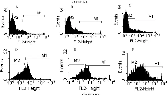
Figure 4 – Representative flow cytometry histogram of the immunomarker profiles of 1.0 mg/mL Ova-stimulated peritoneal macrophages (Gated R1 A-B-C; Gated R2 D-E-F). A-D: conjugated anti-mouse CD80; B-E: conjugated anti-mouse CD86; C-F: PE-conjugated anti-mouse CD11b. One of three representative experiments is shown.
GATED R2 GATED R1
A B C
A B C
A B C
Figure 5 - Representative flow cytometry histogram of the immunomarker profiles of 0,1 mg/mL palmitoyl-Ova-stimulated PerC cells (Gated R1 A-B-C; Gated R2 D-E-F). A-D: conjugated anti-mouse CD80; B-E: conjugated anti-mouse CD86; C-F: PE-conjugated anti-mouse CD11b. One of three representative experiments is shown.
Figure 6 – Representative flow cytometry histogram of the immunomarker profiles of 1.0 mg/mL palmitoyl-Ova-stimulated PerC cell (Gated R1 A-B-C; Gated R2 D-E-F). A-D: conjugated anti-mouse CD80; B-E: conjugated anti-mouse CD86; C-F: PE-conjugated anti-mouse CD11b. One of three representative experiments is shown.
GATED R1
GATED R2
GATED R1
GATED R2
A B C
D E F
A B C
Figure 7 – The CD80, CD86, CD11b immunomarker profiles of PerC cells (Unstimulated; 0,1 mg/mL Ova-stimulated; 1,0 mg/mL Ova-stimulated; 0,1 mg/mL palmitoyl-Ova-stimulated; 1,0 mg/mL palmitoyl-Ova-stimulated): A (Gated R1); B (Gated R2). The median ± SD of three experiments is shown. Statistics: p (a/b/c/d/e/f) < 0.01.
Palmitoyl-Ova, when tested at both
concentrations, induced the expression of CD80, CD86, and CD11b at higher levels. The underlying mechanisms by which lipopeptides, mucosal adjuvants and antigen delivery systems elicit both local and systemic immune responses without an adjuvant are only partially understood. The mechanism of action is assumed to be a complex multistep phenomenon resulting from the physiological conditions and direct interactions of antigens with different cell types at the presumed site of uptake and immune induction (Alves et al. 2005; Oliveira et al. 2007). An elegant study, performed by two-photon excitation, characterized the antigen-specific CD4+ T cells in vivo in real time during the induction of oral priming versus oral tolerance (Zinselmeyer et al. 2005). Dramatic changes in T-cell behavior were simultaneously observed in local (MLN) and peripheral (CLN) lymphoid organs after the oral administration of the antigen, with, or without an adjuvant. The investigators suggested that these changes could be a consequence of variations in the expression of the associated signaling molecules such as chemokines, cytokines, costimulatory molecules, and their receptors. Lipopeptides have been shown to increase the expression of CD11b, CD11c, and costimulatory molecules (BenMohamed et al. 2002) and to stimulate innate immunity by specific interaction with the Toll family receptor (TLR) on APCs. Robinson et al. (1992) have shown that lipopeptides exhibited stronger interactions with specific T-cell clones and APCs than native peptides.
Similar mechanisms were also identified in oral immunization models. Treating mice with the Flt3
ligand, a DC growth factor, and immunization with cholera toxin (CT), was found to convert the tolerant DCs into immunogenic APCs, probably due to the up regulation of CD80 and CD86 (Willianson et al. 1999). Cong et al. (1997) have shown that the role of CT as a mucosal adjuvant involves the selective up regulation of CD86 expression on bone marrow-derived macrophages. An interesting correlation demonstrated that CD86 could be critical for the secretion of TGF-β as well as the production of IgA antibodies (Liu et al. 1999). It has also been shown that CD86 may be more critical for the induction of IL-4-producing cells, and IL-4 is a differentiating factor of TGF-β-producing Th-3 cells. Taken together, these findings could be associated with the present observation that the oral administration of palmitoyl-Ova produced systemic and local immune responses. The present results suggested that the kinetics and the level of expression of the costimulatory molecules on the APCs in turn regulated the oral tolerance, or the systemic and local immune responses. In addition, palmitoyl-Ova-mediated changes in the interaction between APCs and T cells could induce the stimulatory signals and determine the characteristics of the effector response.
Evaluation of Cellular Immune Responses
Since the in vitro stimulation with palmitoyl-Ova strongly induced the B7.1 (CD80), B7.2 (CD86) and CD11b molecules, the effect of this form of the Ag on both in vivo and in vitro T-cell mediated responses was further studied. The response of mice orally treated with saline, Ova, or, palmitoyl-Ova, and immunized with the Ag in
% o f R 1 c a v it y p e ri to n e a l c e ll s CD 80 CD 86 CD 11b 0 20 40 60 80
a a a a a a
b c d b b d e b d
CFA, was assayed by Ag-induced footpad swelling. Footpad swelling did not differ between
the mice pretreated with palmitoyl-Ova
(experimental group), or saline (immune group) but was significantly suppressed in mice pretreated with soluble Ova (data not shown). The effect of the in vitro stimulation of spleen cells obtained from the mice that had been orally
treated with palmitoyl-Ova, before being
immunized with Ova adsorbed in CFA or Al(OH)3 was also studied. The profiles obtained with both the adjuvants were quite similar, although a higher stimulation index was reached when the animals were immunized with CFA. Spleen cells obtained from the mice that had been orally pretreated with palmitoyl-Ova proliferated as much as cells from the immune group (pretreated with saline), but much higher than the cells from the tolerant group (data not shown). The control of T-cell expansion by B7/CD28 signals is both a direct consequence of entry and progression into the cell cycle, and an indirect outcome of an increased production of the T-cell growth factor IL-2 (Appleman et al. 2000). In this way, a strong T-cell proliferative response induced by palmitoyl-Ova may be indirectly related to high CD80 and CD86 expression and production of IL-2. Additionally, ISCOMS-Ova, an antigen entrapped in a lipid structure, also primed
antigen-specific proliferative PLNs responses and
produced a significant amount of IL-2 when administered orally (Maloy et al. 1995).
While palmitoyl-Ova increased B7 expression at both the concentrations, only high dose Ova induced the expression of CD80 and CD86. These results suggested that varying doses of Ova triggered different mechanisms that regulated the induction of oral tolerance by the B7 delivery signals. This postulation was supported by the results of an array examining the B7-CD28 functions. Blocking the B7-CD28 interaction promoted the transplantation tolerance that was initially driven by massive T-cell inhibition (Wells 1999). In addition, T-cell activation in the absence of B7-CD28 signals can lead to a state of anergy (Wells et al. 2003). In agreement with the previous reports, the effector phase of the oral tolerance, subsequent to low dose Ag feeding, was related to a decrease in IL-2 expression in the present work. Furthermore, an ip injection of the anti-CD86 mAb concurrent with a low dose feeding regimen, did not inhibit the production of IL-2 indicating a
possible role in the induction of IL-2 producing cells (Liu et al. 1999).
Thus, these factors appeared to affect the cellular proliferative response against low dose Ova. On the other hand, a high dose of Ova further induced the expression of costimulatory molecules when compared to untreated cells. Importantly, CTLA-4 also binds to the ligands CD80 and CD86, thereby
blocking TCR proximal signaling and
consequently attenuating cell cycle progression, cytokine production, and proliferation (Kumar et al. 2013). Thus, the immune response delivered by APC lymphocytes is a complex interplay of costimulatory signals.
CONCLUSIONS
Taken together, the results suggested a direct effect of palmitoyl-Ova in the increased expression of CD86, CD80 and CD11b in the peritoneal cavity adherent cells through the induction of systemic and local immune responses.
ACKNOWLEDGMENTS
We acknowledge the research supported by CNPq and PRPq-UFMG.
REFERENCES
Alves AC, Gontijo CM, Oliveira MC, Diniz SMF, Oliveira FM, Cardoso VN et al. Biodistribution of Free 99mTc-ovalbumin and 99mTc-ovalbumin encapsulated in liposomes. Braz Arch Biol Tech. 2005; 48: 235-241.
Appleman LJ, Berezovska A, Grass I, Boussiotis VA. CD28 costimulation mediates T cell expansion via IL-2 –independent and IL-2-dependente regulation of cell cycle progression. J Immunol. 2000; 164; 144-151.
Bassan N, Vinuesa M, Roma S. CD11b-positive cells expression in rectal mucosa from ovalbumin sensitized and a challenged rabbits. Acta Gastroenterol Latinoam. 2005; 35(1): 7-12.
BenMohamed L, Krishnan R, Auge C, Primus JF, Diamond DJ. Intranasal administration of a synthetic lipopeptide without adjuvant inducez systemic immune responses. Immunol. 2002; 106(1): 113-121. Bour-Jordan H, Esensten JH, Martinez-Llordella M,
Penaranda C, Stumpf M, Bluestone JA. Intrinsic and extrinsic control of peripheral T-cell tolerance by costimulatory molecules of the CD28/B7 family.
Cong Y, Weaver CT, Elson CO. The mucosal adjuvancity of cholera toxin involves enhancement of costimulatory activity by selective up-regulation of B7.2 expression. J Immunol. 1997; 159:5301.
Curotto de Lafaille MA, Lafaille JJ. Natural and adaptative Foxp3 regulatory T cells: more of the same or a division labor. Immunity. 2009: 30(5): 626-635. Ehirchiou D, Xiong Y, Xu G, Chen W, Shi Y, Zhang L.
CD11b facilitates the development of peripheral tolerance by suppressing Th17 differentiation. J Med Exp Med. 2007: 204(7): 1519-1524.
Friedman A, Weiner HL. Induction of anergy or active suppression following oral tolerance is determined by antigen dosage. Proc Natl Acad Sci. USA. 1994; 91: 6688-6692.
Fujino S, Andoh A, Bamba S, Ogawa A, Hata K, Araki Y, Bamba T, Fujiyama Y. Increased expression of interleukin 17 in inflammatory bowel disease. Gut.
2003; 52:65-70.
Gonella PA, Chen YH, Waldner H, Weiner HL. Induction of oral tolerization in CD86 deficient mice: a role for CD86 and B cells in the up-regulation of TGF-beta. J Autoimmun. 2006; 26(2): 73-81.
Greenwald RJ, Freeman GJ, Sharpe AH. The B7 family revisited. Annu Rev Immunol. 2005: 23:515-548. Kumar S, Verma AK, Das M, Dwivedi PD. A
molecular insight of CTLA-4 in food allergy.
Immunol Letters. 2013; 149(1-2): 101-109.
Lee LF, Xu B, Michie SA, Beilhack GF, Warganich T, Turley S, McDevitt HO. The role of TNF-a in the pathogenesis of type 1 diabetes in the nonobese diabetic mouse: analysis of dendritic cell maturation.
Proc Natl Acad Sci. USA. 2005; 102:15995-16000. Lenschow DJ, Walunas TL, Bluestone JA. CD28/B7
system of T cell costimulation. Annu Rev Immunol. 1996; 14: 233-258.
Lenschow DJ, Zeng Y, Hathcock KS, Zuckerman LA, Freeman G, Thistlethwaite JR et al. Inhibition of transplant rejection following treatment with anti-B7-2 and anti-B7-1 antibodies. Transplantation. 1995; 60(1): 1171-1178.
Liu L, Kuchroo VK, Weiner HL. B7.2 (CD86) but not B7.1 (CD80) costimulation is required for the induction of low dose oral tolerance. J Immunol.
1999; 163(4): 2284-2290.
Maloy KJ, Donachie AM, Mowat AM. Induction of Th1 and Th2 CD4+ T cell responses by oral or parenteral immunization with ISCOMs. Eur J Immunol. 1995; 25: 2835-2841.
McKenzie BS, Kastelein RA, Cua DJ. Understanding the IL-13-IL-17 immune pathway. Trends Immunol. 2006; 27:17-23.
Oliveira FM, Dos Santos EM, Alves AC, Campana-Pereira MA, Ramaldes GA, Cardoso VN et al. Digestion, absorption and tissue distribution of ovalbumin and palmitoyl-ovalbumin: impact on immune responses triggered by orally administered antigens. Scand J Immunol. 2007; 65(2): 139-147.
Oliveira FM, Santos EM, Mota-Santos TA, Ruiz de Souza V, Gontijo CM. Covalent coupling of palmitate to ovalbumin inhibits and blocks the induction of oral tolerance. Scand J Immunol. 2002; 55: 570-576.
Oliveira FM, Silva-Neto AF, Silva CA, Gontijo, CM. Coupling of palmitate residues to ovalbumin affect the induction of oral tolerance against the antigen.
Braz J Med Biol Res. 1998; 31: 1421-1424.
Perez VL, Van Parijs L, Biuckians A, Zheng XX, Strom, TB, Abbas AK. Induction of peripheral T cell tolerance in vivo requires CTLA-4 engagement.
Immunity. 1997; 6: 411-417.
Popov I, Li M, Zheng X, San H, Zhang X, Ichim TE, Suzuki M, Feng B, Vladau C, Zhong R et al. Preventing autoimmune arthritis using antigen-specific immature dendritic cells: a novel tolerogenic vaccine. Arthritis Res Ther. 2006; 8:R141.
Pulendran B, Smith JL, Caspary G, Brasel K, Pettit D, Maraskovsky E, Maliszewski CR. Distinct dendritic cell subsets differentially regulate the class of immune response in vivo. Proc Natl Acad Sci. USA. 1999; 96:1036-1041.
Robinson JH, Case MC, Brooks CG. Palmitic acid conjugation of a protein antigen enhances major complex class II-restricted presentation to T cells.
Immunol. 1992; 76: 593-598.
Sakaguchi S, Yamaguchi T, Nomura T, Ono M. Regulatory T cells and immune tolerance. Cell. 2008; 133:775-787.
Titus RG, Chiller JM. Orally induced tolerance: definition at the cellular level. Int Arch Allergy Appl Immun. 1981; 65: 323-338.
Wells AD, et al. Requirement for T-cell apoptosis in the induction of peripheral transplantation tolerance. Nat Med. 1999; 5:1303-1307.
Wu H, Rodgers JR, Perrard XY, Perrard JL, Prince JE, Abe Y et al. Deficiency of CD11b or CD11d results in reduced staphylococcal enterotoxin-induced T cell response and T cell phenotypic changes. J Immunol.
2004; 173(1): 297-306.
Zhang M, Tang H, Guo Z, An H, Zhu X, Song W, Guo J, Huang X, Chen T, Wang J, Cao X. Splenic stroma drives mature dendritic cells to differentiate into regulatory dendritic cells. Nat Immunol 2004; 5: 1124-1133.
Zhou L, Chong MM, Littman DR. Plasticity of CD4+ T cell lineage differentiation. Immunity. 2009: 30(5): 646-655.
Zinselmeyer BH, Dempster J, Gurney AM, Wokosin D, Miller M, Ho H, et al. In situ characterization of CD4+ T cell behavior in mucosal and systemic lymphoid tissues during the induction of oral priming and tolerance. J Exp Med. 2005; 201(11): 1815-1823.