Vol. 51, n. 5 : pp.1057-1063, September-October 2008
ISSN 1516-8913 Printed in Brazil BRAZILIAN ARCHIVES OF
BIOLOGY AND TECHNOLOGY
A N I N T E R N A T I O N A L J O U R N A L
Gill Histopathological Alterations in Nile Tilapia,
Oreochromis niloticus
Exposed to Treated Sewage Water
1António Fontaínhas-Fernandes*
, Ana Luzio, Sofia Garcia-Santos, João Carrola and Sandra Monteiro
1
Centro de Estudos Tecnológicos do Ambiente e da Vida; Centro de Investigação e de Tecnolgias Agro-Ambientais e Biológicas; Apartado 1013, [email protected]; 5000-911; Vila Real - Portugal
ABSTRACT
Adult Nile tilapia, Oreochromis niloticus, of both sexes were exposed in wastewater from a sewage treatment plant for a period of 4 days. Gill samples were collected after 24, 48, 72 and 96 h and histopathological changes were analyzed by light and scanning electronic microscopy. Gill epithelium of control O. niloticus (freshwater group) was similar to that of other teleosts, while histopathological lesions were observed in exposed fishes. The main histopathological changes were edema, lifting of lamellar and filamentar epithelia and lamellar fusion. Cell proliferation with consequent thickening of the filament epithelium was also found in fishes exposed to the treated sewage water. The severity of the lesions increased with the time of exposure, namely the hyperplasia of the epithelial cells with proliferation of filamentar epithelium and fusion of lamellae observed at 96 h. Additionally, several histopathological results obtained by light microscopy were confirmed through scanning microscopy.
Key words: Gill, tilapia, histopathology, wastewater
*
Author for correspondence
INTRODUCTION
Environmental pollution and its effects on the health of aquatic ecosystems is a great problem that has been studied intensely in the last years. The wastewater treatment plants receive large amounts of compounds from domestic and/or industrial wastes, which are not totally eliminated during the treatment process (Ternes et al., 1999). For this reason, sewage treatment works (STW) effluents represent a complex mixture of substances that include the partially eliminated wastewater molecules and metabolites formed during the treatment process (Carballo et al., 2005). These effluents have been considered as an
important factor contributing to the reduction of species diversity in the world (Schmidt et al., 1999; Amisah and Cowx, 2000).
others have been developed under the experimental conditions (Diniz et al., 2005a). In recent studies, effluents from the STW have been demonstrated to affect fish health, causing ahistological lesions and higher susceptibility to infectious diseases (Escher et al., 1999; Bernet et al., 2001). The analysis of biochemical and histopathological changes is often used to assess the effect of effluents on different tissues in field and experimental studies. However, only a few studies have been carried out to investigate the histopathological effects of STW effluents in freshwater fishes (Billiard and Khan, 2003; Solé et al., 2003; Alberto et al., 2005; Petterson et al., 2006). In Portugal, few studies exist mainly focused on the Lisbon region, showing the presence of estrogenic compounds in sewage and industrial effects in fish (Diniz et al., 2005a, b). The knowledge about the gill histopathological alterations caused by the effluents from STW is scarce.
The gill is the main organ for different functions, such as gas exchange, ion regulation and excretion of metabolic waste products (Wood, 1991). Its complexity and constant contact with the external environment make the gill the first target to waterborne pollutants (Mallatt, 1985; Perry and Laurent, 1993; Fracácio et al., 2003). The pollutants not only enter the organism through the gills, but also exert their primary toxic effects on the branchial epithelium (Playle et al., 1992) which in turn, may influence the general gill functions. The effect of different contaminants on gill biochemical and morphological changes have been analysed in several studies (Monteiro et al., 2005; Garcia-Santos et al., 2006; Romão et al., 2006; Fernandes et al., 2007).
The purpose of this study was to evaluate the impact of a treated effluent from a STW (Vila Real, Portugal) on gill morphology in Nile tilapia,
Oreochromis niloticus. To our knowledge, it is the first time that this kind of study has been assessed using fishes exposed to a sewage effluent in Portugal.
MATERIALS AND METHODS
Animals
Nile tilapia (Bouaké strain) was originally obtained from the Institute Nationale de Recherche
Agronomique (Rennes, France) and raised in the Aquaculture Station of the University of Trás-os-Montes and Alto Douro (UTAD, Vila Real, Portugal) for three generations. The fishes used for this experiment were maintained in 600 L recirculating tanks, filled with dechlorinated tap water (pH 6.5-7.5; alkalinity 60 mg L-1 as HCO3-; conductivity 63 µS cm-1; Na+, 14 mg L-1; K+, 2.3 mg L-1; Ca2+, 4.1 mg L-1; Mg2+, 6.5 mg L-1; Cl-, 19.5 mg L-1; NO3-, 27 mg L-1; NO2-, 0.5 mg L-1). Fishes were fed daily to satiation with a previous tested diet (Fontaínhas-Fernandes et al., 1999), kept at a constant temperature of 25 ± 1 ºC and controlled photoperiod (12D: 12L). Supplemental aeration was provided to maintain the dissolved oxygen near saturation.
Experimental Design
An urban sewage treatment work was selected to study the effects of the treated effluents on Nile tilapia, O. niloticus. This STW is located in Vila Real (Portugal), a densely populated area it was chosen because it receives large and variable quantities of domestic wastewater that is treated before being discharged into the river.
Adult male and female tilapia (97.6 ± 0.5 g of mean body weight and 17.5 ± 0.7 cm fork lenght) were used as the experimental animals. Fishes were transferred from the stock tank to six experimental 200 L tanks. There were 10 fishes per tank in triplicate for each treatment. Fish from three tanks were exposed to treated sewage effluent at a nominal concentration of 100% during a period of 4 days. The remaining tanks containing tap water were the control group. Each
tank was supplied with a constant flow rate of 5 L
min-1. The tanks were aerated and the temperature
(25 ± 1 ºC) and photoperiod (12D: 12L) were controlled. The physical and chemical parameters of the treated water were as follows: pH 7.9; DOC 44.4 mg L-1; NH4-N 30.1 mg L-1; NO3 34 mg L-1; NO2 32.9 mg L-1; Ca 11.1 mg L-1; Mg 4 mg L-1; Na 79.6 mg L-1; K 18 mg L-1; Cd 2.75 µg L-1; Pb 0.38 µg L-1; Zn 0.4 mg L-1; Cu 25.3 µg L-1; Cr 2.5 µg L-1 and Ni 4 µg L-1. The experiments described complied with the Guidelines of the European Union Council (86/609/EU).
Histopathological analysis
A gill arch of the right side of each fish was collected and fixed in Bouin’s fluid for 24 h, dehydrated in graded ethanol concentrations and embedded in paraffin wax. Sagittal sections (5 µm of thickness) were cut and mounted on glass slides. Sections were deparaffinized in xylene, hydrated in ethanol and stained with hematoxylin/eosin. Changes induced by treatment in the gill tissue were photographed and analyzed by light microscopy (Nikon® Labophot). Furthermore, a representative sample of gills were fixed at 4 ºC in 4% glutaraldheyde buffered with 0.1 M cacodylate pH 7.4 during 2 h for scanning electron microscopy (SEM). After proper rinses and dehydration in ethanol, the tissues were then critical point dried with CO2 and mounted onto aluminum stub and coated with gold palladium. The preparations were then examined under a Fey Quanta 400 EFEM.
RESULTS AND DISCUSSION
Gill epithelium of control group was similar to that of other teleostean fishes (Fig. 1A and 2A). However, tilapia exposed to the treated water from the STW showed several gill histopathological alterations.
Epithelial cell lifting, epithelial hypertrophy and hyperplasia, slight deformations of the lamellae, and fusion of adjacent lamellae were more prevalent and more pronounced in exposed tilapia than in the control group (Fig. 1A and 2A). The gill lesions included edema that was more evident at 24 h of exposition, with lifting of lamellar and filamentary epithelium (Fig. 1C). The severity of the lesions observed in this study increased with the time of exposure, namely the hyperplasia of the epithelial cells with proliferation of filamentar epithelium and fusion of lamellae at 96 h (Fig. 1B e 2B, C). The vasodilatation of lamellae was severe and extended to the entire lamellar axis (Fig. 1C). Necrosis was also observed in the
filamentar epithelium (Fig. 1C). Aneurisms were found in lamellae, probably induced by the loss of support capacity of the pillar cells (Fig. 1D). The types of histopathological lesions did not follow a consistent pattern of variation with exposition time in each treatment.
The scanning electron micrographs of the gill epithelium also revealed that tilapia of untreated group showed a normal architecture. In contrast, several histological lesions observed in the present study were similar to those observed in trout submitted to residual water, namely lifting of the lamellar epithelium, hypertrophy and hyperplasia of epithelial cells, as well as fusion of adjacent lamellae (Escher et al., 1999; Bernet et al., 2004). Bernet et al. (2004) developed two monitoring approaches (active and passive) to assess the effects of wastewater disposal on histological alterations in brown trout (Salmo trutta) and data were not completely consistent. The gills were the most sensitive organ to the effects of treated wastewater in the active monitoring, but were not affected in fishes collected in the river (passive program). However, in an experiment extending over 3 months, brown trout (S. trutta) were exposed to dilutions of treated wastewater from a large sewage plant and no effects were detectable in the gills (Bucher and Hofer, 1993).
On the other hand, gill sections from S. trutta
exposed to STW effluent in cages showed moderate histopathological changes (Escher et al., 1999). This study concluded that the most prevalent lesions were distortion of lamellae and clubbing and fusion of the secondary lamellae due to a hypertrophy and a moderate hyperplasia of the respiratory epithelium and cells at the base of the secondary lamellae.
In contrast, a higher percentage of gill lesions, especially hyperplasia were observed in cunner,
Tautogolabrus adspersus, exposed to municipal and industrial effluents (Billiard and Khan, 2003). Cellular hyperplasia was also the most common gill lesion observed in Winter flounder taken from similar site (Khan et al., 1996).
Figure 1 - Photomicrographs of the gill epithelium of Nile tilapia, Oreochromis niloticus. (A) Gill of untreated fish under control conditions showing the central venous sinus (SVC) covered by filamentar epithelium (EF) which is perpendicularly intersected by lamellae (L). (B, C, D) Fish exposed to the sewage treated water. (B) At 96 h tilapia showed an intensive filamentar epithelium (EF) proliferation, which led to almost completely lamellar fusion (FL). Sometimes, thickness of lamellar epithelium is also detected (arrowhead). The inset shows focal lamellar fusion observed in fish exposed for 72 hours. (C) Lifting of lamellar and, sometimes, filamentar epithelium is observed (arrows). Intensive vasodilatation (V) of the lamellar vascular axis, necrosis (n) and edema (**) are found at 96 h. (D) Aneurisms (A) in the apical region of the lamellar vascular axis and proliferation of filamentar epithelium (EF) is seen at 96 h. CC, chloride cell; M, mucous cell; CP, pavement cell; CPi, pillar cell. Bar = 30µm.
In conclusion, this is the first Portuguese study that has tried to assess the gill morphological alterations caused by wastewater treatment effluents. This study showed that treated domestic effluent from a STW located in Vila Real (Portugal) caused gill histological lesions. However, further studies that could have
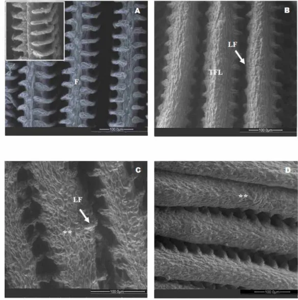
Figure 2 - Scanning electron micrographs of the gill epithelium of Nile tilapia, Oreochromis niloticus. (A) Untreated fish under control conditions; (B, C, D) Exposed fish. B) Lamellar fusion (LF) and thickening of filamentar epithelium (TFE). C, D) Lamellar fusion (LF) and thickening of filamentar epithelium showing high severity (**) at 72 h (C) and 96 h (D).
RESUMO
Tilápias adultas, Oreochromis niloticus, de ambos os sexos foram expostas em águas residuais de uma estação de tratamento de esgoto durante 4 dias. Amostras de brânquia foram recolhidas após 24, 48, 72 e 96 h e as alterações histopatológicas foram analisadas por microscopia óptica e eletrônica de varredura. O epitélio da brânquia do grupo controle apresentou uma morfologia similar à de outros peixes teleosteos, enquanto foram observadas lesões nos peixes expostos. As principais alterações histopatológicas foram
REFERENCES
Alberto, A.; Camargo, A. F. M.; Verani, J. R.; Costa, O. F. T. and Fernandes, M.N. (2005), Health variables and gill morphology in the tropical fish Astyanax
fasciatus from a sewage-contaminated river.
Ecotoxicol. Environ. Saf.,61, 247-255.
Amisah, S. and Cowx, I. G. (2000), Response of the fish populations of the river Don in South Yorkshire to water quality and habitat improvements. Environ. Pollut.,108, 191-199.
Bernet, D.; Schmidt, H.; Wahli, T. and Burkhardt-Holm, P. (2001), Effluent from a sewage treatment works causes changes in serum chemistry of brown trout (Salmo trutta). Ecotoxicol. Environ. Saf., 48, 140-147.
Bernet, D.; Schmidt-Posthaus, H.; Wahli, T. and Burkhardt Holm, P. (2004), Evaluation of two monitoring approaches to assess effects of waste water disposal on histological alterations in fish. Hydrobiologia, 524, 53-66.
Billiard, S.M. and Khan, R.A. (2003), Chronic stress in cunner, Tautogolabrus adspersus, exposed to municipal and industrial effluents. Ecotoxicol. Environ. Saf.,55, 9-18.
Bucher, F. and Hofer, R. (1993), The effects of treated domestic sewage on three organs (gills, kidney, liver) of brown trout (Salmo trutta). Water Res.,27(2), 255-261.
Carballo, M.; Aguayo, S.; de la Torre, A. and Muñoz, M. J. (2005), Plasma vitellogenin levels and gonad morphology of wild carp (Cyprinus carpio L.) in a receiving rivers downstream of sewage treatment plants. Sci. Total Environ.341, 71-79.
Diniz, M. S.; Peres, I. and Pihan, J. C. (2005a), Comparative study of the estrogenic responses of mirror carp (Cyprinus carpio) exposed to treated municipal sewage effluent (Lisbon) during two periods in different seasons. Sci. Total Environ.349, 129-139.
Diniz, M. S.; Peres, I.; Magalhães-Antoine, I.; Falla, J. and Pihan, J.C. (2005b), Estrogenic effects in crucian carp (Carassius carassius) exposed to treated sewage effluent. Ecotoxicol. Environ. Saf.,62, 427-435. Escher, M.; Wahli, T.; Büttner, S.; Meier, W. and
Burkhardt-Holm, P. (1999), The effect of sewage plant effluent on brown trout (Salmo trutta fario). Aquat. Sci., 61, 93-110.
Fernandes, C.; Fontaínhas-Fernandes, A.; Monteiro, S. M. and Salgado,, M.A. (2007), Changes in plasma electrolytes and gill histopathology in wild Liza saliens from the Esmoriz-Paramos coastal lagoon. Bull. Environ. Contam Toxicol., 79, 301-305.
Fontaínhas-Fernandes, A.; Gomes, E.; Reis-Henriques, M. A. and Coimbra, J. (1999), Replacement of fish meal by plant proteins in the diet of Nile tilapia: digestibility and growth performance. Aquac. Intern., 7, 57-67.
Fracácio, R.; Verani, N. F.; Espíndola, E. L.; Rocha, O.; Rigolin-Sá, O. and Andrade, C. A. (2003), Alterations on growth and gill morphology of Danio rerio (Pisces, Ciprinidae) exposed to the toxic sediments. Braz. Arch. Biol. Technol., 46, 685-695. Garcia-Santos, S.; Fontaínhas-Fernandes, A. and
Wilson, J. M. (2006), Cadmium tolerance in the Nile tilapia (Oreochromis niloticus) following acute exposure: Assessment of some ionoregulatory parameters. Environ. Toxicol., 21 (6), 33-46.
Garric, J.; Vollat, B.; Nguyen, D.K.; Bray, M.; Migeon, B. and Kosmala, A. (1996), Ecotoxicological and chemical characterization of municipal wastewater treatment plant effluents. Water Sci. Technol.,33, 83-91.
Grizzle, J. M.; Hurowitz, S. A. and Strenght, D. R. (1988), caged fish as monitors of pollution: Effects of chlorinated effluent from a wastewater treatment plant. Water Res. Bullet.,24, 951-959.
Khan, R. A.; Barker, D. E.; Ryan, K.; Murphy, B. and Hooper, R.G. (1996), Abnormalities in winter flounder (Pleuronectes americanus) living near a paper mill n the Humber Arm, Newfoundland. In: Servos, M. R.; Munkittrick, K. R.; Carey, J. H. and Van der Kraak G. J. (eds.), Environmental Fate and Effects of Pulp and Paper Mill Effluents. St Lucie Press, FL, pp. 511-523.
Kosmala, A.; Charvet, S.; Roger, M.C. and Faessel, B. (1999), Impact assessment of a wastewater treatment plant effluents using in stream invertebrates and the Ceriodaphnia dubia chronic toxicity test. Water Res., 33(1), 266-278.
Mallatt, J. (1985), Fish gill structural changes induced by toxicants and other irritants: a statistical review. Can. J. Fish. Aquat. Sci.,42, 630-648.
Monteiro, S. M.; Mancera, J. M.; Fontaínhas-Fernandes, A. and Sousa, M. (2005), Copper induced alterations of biochemical parameters in the gill and plasma of Oreochromis niloticus. Comp. Biochem. Physiol., 141, 375-383.
Perry, S.F. and Laurent, P. (1993), Environmental effects on fish gill structure and function. In: Rankin, J.C., Jensen, F.B. (Eds.), Fish Ecophysiology. Chapman and Hall, London, pp. 231-264.
Petterson, M.; Adolfsson-Erici, M.; Parkkonen, J.; Förlin, L. and Asplund, L. (2006), Fish bile used to detect estrogenic substances in treated sewage water. Sci. Total Environ., 366, 174-186.
Playle, R.C.; Gensemer, R. W. and Dixon, D. G. (1992), Copper accumulation on gills of fathead minnows: influence of water hardness, complexation and pH of the gill micro-environment. Environ. Toxicol Chem., 11, 381-391.
Schmidt, H. D.; Bernet, D.; Wahli, T.; Meier, W. and Burkhardt-Holm, P. (1999), Active bio monitoring with brown trout and rainbow trout in diluted sewage plant effluents. J. Fish Biol., 54, 585-596.
Solé, M.; Raldua, D.; Barceló, D. and Porte, C. (2003), Long-termed exposure effects in vitellogenin, sex hormones, and biotransformation enzymes in female carp in relation to a sewage treatment works. Ecotoxicol. Environ. Saf.,56, 373-380.
Ternes, T. A.; Stumpf, M.; Mueller, J.; Haberer, K.; Wilken, R. D. and Servos, M. (1999), Behaviour and occurrence of estrogens in municipal sewage treatment plant: I Investigations in Germany, Canada and Brazil. Sci. Total Environ.,225, 81-90.
Wood, C. M. (1991), Branchial ion and acid-base transfer in freshwater teleost fish: environmental hyperoxia as a probe. Physiol. Zool., 64, 68-102. Wright, J. A.; Chessman, B. C.; Fairweather, P. G. and
Benson, L. J. (1995), Measuring the impact of sewage effluent on the macro invertebrate community of an upland stream: The effect of different levels of taxonomic resolution and quantification. Austral. J. Ecol., 20, 142-149.