(Annals of the Brazilian Academy of Sciences)
Printed version ISSN 0001-3765 / Online version ISSN 1678-2690 www.scielo.br/aabc
http://dx.doi.org/10.1590/0001-3765201520150008
Bone turnover markers for early detection of fracture healing disturbances: A review of the scientific literature
CRISTINA P. SOUSA1,2,3, ISABEL R. DIAS1,2,3, MÓNICA LOPEZ-PEÑA4, JOSÉ A. CAMASSA1, PAULO J. LOURENÇO5,6, FERNANDO M. JUDAS5,6, MANUELA E. GOMES2,3 and RUI L. REIS2,3
1
Departamento de Ciências Veterinárias, Escola das Ciências Agrárias e Veterinárias, Universidade de Trás-os-Montes e Alto Douro, Quinta de Prados, Apartado 1013, 5000-801 Vila Real, Portugal
2Grupo de Investigação 3B’s, Departamento de Engenharia de Polímeros, Universidade do Minho,
Avepark - Parque de Ciência e Tecnologia, Zona Industrial da Gandra, 4805-017 Barco GMR, Portugal
3
Instituto de Investigação em Ciências da Vida e Saúde (ICVS), Laboratório Associado ICVS/3B’s, Universidade do Minho, Campus de Gualtar, 4710-057 Braga, Portugal
4
Department of Veterinary Clinics Sciences, Faculty of Veterinary Medicine, University of Santiago de Compostela, University Campus, Av. Carballo Calero, 27002 Lugo, Spain
5Serviço de Ortopedia, Centro Hospitalar e Universitário de Coimbra, EPE, Av. Bissaya Barreto
Praceta Mota Pinto, Hospitais Universitários de Coimbra, Rua Fonseca Pinto, 3000-075 Coimbra, Portugal
6
Faculdade de Medicina, Universidade de Coimbra, Pólo III – Pólo das Ciências da Saúde, Azinhaga de Santa Comba, Celas, 3000-548 Coimbra, Portugal
Manuscript received on January 12, 2015; accepted for publication on January 30, 2015
ABSTRACT
Imaging techniques are the standard method for assessment of fracture healing processes. However, these methods are perhaps not entirely reliable for early detection of complications, the most frequent of these being delayed union and non-union. A prompt diagnosis of such disorders could prevent prolonged patient distress and disability. Efforts should be directed towards the development of new technologies for improving accuracy in diagnosing complications following bone fractures. The variation in the levels of bone turnover markers (BTMs) have been assessed with regard to there ability to predict impaired fracture healing at an early stage, nevertheless the conclusions of some studies are not consensual. In this article the authors have revised the potential of BTMs as early predictors of prognosis in adult patients presenting traumatic bone fractures but who did not suffer from osteopenia or postmenopausal osteoporosis. The available information from the different studies performed in this field was systematized in order to highlight the most promising BTMs for the assessment of fracture healing outcome.
Key words: bone formation markers, bone resorption markers, delayed union, fracture healing, osteoclast regulatory proteins,non-union process.
Correspondence to: Isabel R. Dias E-mail: [email protected]
INTRODUCTION
The expected outcome of a fracture is the bone healing, defined as the functional stage of bone
factors, such as the degree of injury of the adjacent soft tissue, the displacement of the fracture ends and the degree of comminution, the selected method for the stabilization of the fracture in addition to other patient related factors, namely the age of the patient and the presence of co-morbidities (diabetes, tobacco use, malnutrition and prolonged use of non-steroidal anti-inflammatory drugs) (Giannoudis et al. 2000, Buchwalter et al. 2010). It is estimated that 5-10% of all patients with long bone fractures should develop impaired fracture healing processes, especially delayed union and non-union processes. Delayed union corresponds to the pathological fracture healing process that takes almost twice the necessary length of time for the formation of the fracture callus, due to inadequate fracture stabilization and lack of patient rest as the most frequent causes of this complication. The development of a non-union process, the most commonly diagnosed fracture healing complication, corresponds to a failure of the fracture repair process (Marsh and Li 1999), generally due to extensive lesions of adjacent soft tissues and inadequate vascularisation of the trauma site (Court-Brown and McQueen 1987, Calori et al. 2007). The tibial fractures, due to the particularities of this bone, namely its blood supply and lack of muscle tissue on its anterior surface, present themselves as those most often affected by the phenomena of disturbances of the bone healing process, in which non-union occur in up to 10-20% of patients with tibial shaft fractures. In clinical practice, the fracture healing process is normally evaluated by physical and radiographic examinations. However, there is a lack of consensus in the assessment of this, rendering an estimation of the incidence of fracture healing complications difficult (Hernandez et al. 2012). The interpretation of radiographic signs of an impaired fracture healing process is subjective and the expected time span for their appearance is variable, so that the early diagnosis of fracture healing complications can prove difficult (Bishop et al. 2012). However, fracture healing complications are associated with
prolonged pain and functional impairment, making the early diagnosis of such complications mandatory (Zimmermann et al. 2007, Buchwalter et al. 2010).
(Khosla 2001, Theoleyre et al. 2004). The balance between the amount of OPG and RANKL regulates osteoclastic activity.
More recently, serum levels of growth factors, including transforming growth factor beta (TGF-β1) and bone morphogenetic proteins (BMPs) produced at the fracture site, have also been studied as non-invasive tools in order to assess the fracture healing process (Zimmermann et al. 2005). Growth factors regulate the different steps of the cellular events that lead to normal bone union, from the initial haematoma to the final remodelling stage (Zimmermann et al. 2005). Therefore, abnormal growth factor expression and release in the systemic circulation might be associated with impaired fracture healing processes.
The measurement of BTMs during the fracture healing process could enhance the accuracy of the bone healing stage assessment, allowing early detection in patients at risk of the development of fracture complications (Cox et al. 2010), and enabling precocious treatment of these complications and prevention of prolonged patient distress and disability (Coulibaly et al. 2010). This study has aimed to investigate the clinical effectiveness of BTMs in monitoring the fracture healing process and identifying patients at risk of developing impaired fracture healing processes.
MATERIALS AND METHODS
Literature searches were undertaken on PubMed, ISI Web of Knowledge, Ebsco, Scopus and the US National Library of Medicine databases, by using a specific query. There was supplemented by a search of reference lists of the studies included and relevant reviews and by Internet searching in order to minimize publication bias. The base search strategy included the following components: (1) biological marker terms and (2) fracture healing terms. The paper selection process included a two-step approach in which the screening of title and abstract was followed by the application of inclusion criteria to the full paper of the selected
studies. The inclusion criteria required that the studies: (1) should be conducted among adult patients who had undergone a traumatic fracture, (2) should be based on the application to BTMs in the follow-up of the fracture healing process, (3) should evaluate the fracture healing outcome, (4) should compare serum BTM levels in patients with a normal versus impaired fracture healing process. Inclusion was not restricted by date of publication or type of study. Studies conducted among elderly patients or patients suffering from osteopenia or postmenopausal osteoporosis were excluded from this review in order to minimize a confounding bias, since reduced bone mineral density could influence the bone fracture healing process. Studies were then analyzed according to the type of study and main issue covered and was classified in two groups of interest: (i) observational studies conducted in post-traumatized human and animal patients, and (ii) experimental studies.
RESULTS
As a result of the search, 2,528 papers were identified for initial screening. Of these, 43 were retrieved as full papers. Twenty one studies were later excluded because they were conducted amongst in osteoporotic or post-menopausal women with the aim of assessing the healing process after fragility fractures, manly hip, vertebral and femoral neck fractures. An additional seven studies were excluded because they didn´t compare serum BTM levels between a normal fracture healing process and an impaired fracture healing process. We included one abstract because no additional information was available and there was sufficient outcome data to extract (Fig. 1).
TABLE II
Significance of bone formation biomarkers as early
indicators of prognosis of bone healing process.
Authors Results
ALP
Oni et al. (1989) No significant differences were observed between normal and impaired healing groups.
Komnenou et al. (2005)
In dogs with non-union process, the serum activity did not exceed the upper limit of reference during the study period. In dogs with normal fracture healing process, the serum activity was higher on 10th day after surgery than the normal reference
interval (p<0.05).
Sousa et al. (2011) In normal healing group, the mean values of serum ALP were significantly higher than that of impaired healing group (p<0.05).
Singh Ajai et al. (2013)
In normal healing group, the mean values of serum ALP were significantly higher
than that of impaired healing group. In non-union group, the mean serum ALP levels remained within normal limits throughout the entire follow-up.
BALP
Emami et al. (1999)
Patients with delayed healing process presented lower serum activity between 4th and 7th weeks after intramedullary nailing of tibial fractures than patients with normal fracture healing (p<0.05).
Herrmann et al. (2002) No significant differences between the two groups.
Southwood et al. (2003) Rabbits with osteomyelitis showed lower serum activity at 4
th post-operative week
than rabbits with no infected fractures (p<0.05).
Klein et al. (2004) No significant differences between the two groups.
Marchelli et al. (2009) No significant differences between the two groups.
Sousa et al. (2011)
In dogs with normal fracture healing process, the serum activity was always higher during the follow-up after surgery than the normal reference interval measured in normal healthy dogs (p<0.05).
In the non-union group, the serum levels didn´t exceed the reference limits during the study period.
Moghaddam et al. (2011) No significant differences between the two groups.
OC
Oni et al. (1989) Patients with normal fracture healing presented higher serum levels compared with patients with delayed healing (p<0.05).
Emami et al. (1999) No significant differences between the two groups.
Herrmann et al. (2002)
Patients with normal fracture healing process presented an increase exceeding baseline levels at the 42nd day after traumatic fracture (p=0.034). In patients with
delayed healing, serum levels began to increase 1 month later than patients with normal fracture healing process (28th day versus 60th day).
Southwood et al. (2003)
Rabbits with osteomyelitis showed lower serum levels at 4th post-operative week
and higher serum levels at 16th week post-operative week than rabbits with no
infected fractures (p<0.05).
Marchelli et al. (2009) No significant differences between the two groups.
PINP Moghaddam et al. (2011) No significant differences between the two groups.
PICP Klein et al. (2004) No significant differences between the two groups.
PIIINP Kurdy(2000)
PIIINP levels were significantly higher in delayed fracture healing at 10th week
after fracture than in normal fracture healing (p<0.05).
Klein et al. (2004) No significant differences between the two groups.
Authors Results
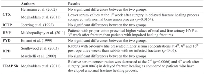
CTX
Herrmann et al. (2002) No significant differences between the two groups. Moghaddam et al. (2011) Lower serum values at the 1
st
week after surgery in delayed fracture healing process compared with normal bone union process (p=0.0164).
ICTP Joerring et al. (1992) No significant differences between the two groups.
HYP Mukhopadhyay et al. (2011) Patients with proper union presented higher values of total and free urinary HYP at 3rd week after fracture than patients with impaired healing fractures.
PYD Emami et al. (1999) No significant differences between the two groups.
DPD Southwood et al. (2003)
Rabbits with osteomyelitis presented higher serum concentrations at 4th, 8th and 16th
post-operative weeks than rabbits with no infected fractures (p<0.05). Marchelli et al. (2009) No significant differences between the two groups.
TRAP 5b Moghaddam et al. (2011)
Relative serum concentration was decreased at the 2nd (p=0.0066) and 4th week after
surgery (p=0.0043) in delayed fracture healing as compared to patients who have developed a normal fracture healing process.
CTX: Cross-linked C-terminal telopeptides of type I collagen; ICTP: Carboxy-terminal telopeptide of type I collagen; HYP: Hydroxyproline; DPD: Deoxypyridinoline; PYD: Pyridinoline; TRAP5b: Tartrate-resistant acid phosphatase isoenzyme 5b.
TABLE III
Significance of bone resorption biomarkers as early
indicators of prognosis of bone healing process.
Authors Result
OPG Marchelli et al. (2009)
Patients with atrophic nonunion process of diaphyseal long bone fractures presented higher serum levels than control groups: subjects that have already healed from the same type of fracture (p<0.001) and subjects that were healing from the same type of fracture (p<0.001).
RANK
Ligand Marchelli et al. (2009) No significant differences between the groups.
TGF-β1 Zimmermann et al. (2005) Four weeks after surgery, patients with delayed union process presented a significantly lower TGF-β1 concentration (p=0.002). Sarahrudi et al. (2011) No significant differences between the groups.
BMP
2,4,6,7,9 Baardewijk et al. (2013) No significant differences between the groups.
OPG: Osteoprotegerin; RANK: Receptor activator of nuclear factor NF-κB; TGF-β1: Transforming growth factor beta 1;
BMP: Bone morphogenetic protein.
TABLE IV
Significance of osteoclasts regulatory proteins and growth factors
as early indicators of prognosis of bone healing process.
DISCUSSION
In several studies it was demonstrated that serum and urinary BTMs are capable of reflecting the healing process with their levels dependent on the localization, type and size of the fracture (Laurer et al. 2000, Stoffel et al. 2007). Joerring et al. (1994) and Veitch et al. (2006) demonstrated that in patients with tibial shaft fractures the type of
treatment, whether cast immobilization or surgical osteosynthesis methods, doesn’t produce significant differences in BTM levels.
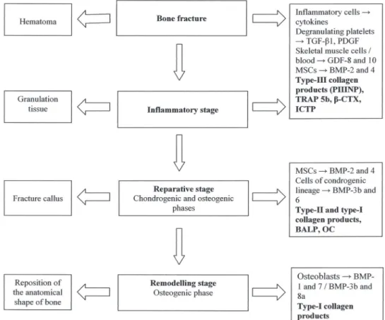
and osteoclast derived enzymes has been shown to occur (Joerring et al. 1994, Stoffel et al. 2007), probably due to the osteoclastic removal of the necrotic tissues at the fracture margins, a small segment of cortices at each side of the fracture (Fig. 2). This event is associated with an equivalent
decrease in bone-formation markers (Bowles et al. 1996, Laurer et al. 2000, Stoffel et al. 2007), associated with an inhibition of osteoblastic synthesis activity which seems to be related to the release of cytokines by inflammatory cells (Laurer et al. 2000, Cox et al. 2010) (Fig. 2).
Figure 1 - Flow of studies through the review.
Type-III collagen products, such as the amino-terminal propeptide of type III procollagen (PIIINP), increase at this early stage during the endochondral fracture healing process and reach their maximal concentration in tibial shaft fractures by the 2nd
week (Joerring et al. 1994) (Fig. 2). After this stage, a significant increase in bone formation markers has been demonstrated - that of the enzyme BALP and OC, since they were derived from osteoblast synthesis, produced during bone ECM mineralization and osteoblast maturation respectively (Obrant et al. 1990, Bowles et al. 1996, Laurer et al. 2000), of type-I collagen products (Veitch et al. 2006, Stoffel et al. 2007), and the persistence of high PIIINP levels, which are released during the formation as well as during the degradation of the fibrous tissue (Joerring et al. 1992, 1994) (Fig. 2). Stoffel et al. (2007) and Veitch et al. (2006) verified that in patients with tibial fractures, after bone union is achieved, the bone resorption markers and bone formation markers remain augmented, corresponding to the remodelling stage of the bone healing process. Stoffel et al. (2007) also demonstrated that the normalization of PIIINP levels precedes radiographic evidence of bone union. Despite serum BTM levels possibly being associated with the different stages of the bone fracture healing process, their clinical effectiveness in predicting impaired fracture healing processes at an early stage isn’t clear.
In a study performed by Komnenou et al. (2005) on dogs, and in one performed by Singh Ajai et al. (2013) on human patients, the serum alkaline phosphatase (ALP) activity was determined throughout the healing process of long bone diaphyseal fractures. In both studies it was observed that serum ALP activity remained within the reference limits during the entire postoperative period in patients that had developed a non-union process, probably indicating a suppression of osteoblastic activity. Singh Ajai et al. (2013) also observed significantly higher levels of serum ALP activity in the group of patients that presented a normal bone union as compared with the delayed healing group. The serum levels of ALP
are the sum of four isoenzymes: intestinal, placental, liver and bone. The bone (BALP) and liver isoforms represent the most relevant fraction of total ALP activity, with an almost equal contribution to about 95% of this enzyme. In the absence of pregnancy and liver or intestinal disorder, ALP activity could be an inexpensive marker for monitoring the bone fracture healing process.
As regards the bone specific isoform-BALP, Emami et al. (1999) found lower levels in patients with delayed union at an early time point during the fracture healing process (by the 4th week after the fracture occurrence) than patients with a normal bone union. Similar results were found from a small study conducted on animals (Sousa et al. 2011). Nevertheless, these results are not in accordance with those obtained from further research conducted by Herrmann et al. (2002), Marchelli et al. (2009) and Moghaddam et al. (2011), BALP activity being shown to be incapable of predicting bone fracture healing outcome, because in these latter studies no significant differences in serum BALP activity were found during the healing process in either patient groups. In these latter studies serum BALP activity, rather than its concentration, was found, this being a more reliable indicator of osteoblastic activity.
entire study period, while in the second study there was a lag in the rise of OC concentration during the first 2 months amongst the group of patients that underwent a delayed union. Nevertheless, Emami et al. (1999) and Marchelli et al. (2009) found that serum OC concentration was unable to differentiate between a delayed union and a normal bone union. These studies present some limitations that could call into question the comparability of the results from the different study groups, results which are mainly related to the small number of patients included in each group. Additionally, the work conducted by Marchelli et al. (2009) is a cross-sectional study, rather than a prospective study like all the others studies, in which the levels of BTMs from three groups of patients at different time points in the fracture healing process were compared.
An evaluation of the serum PINP levels derived from a collagen molecule revealed no differences between patients who, after undergoing a traumatic fracture, progressed into a non-union or a normal bone union (Moghaddam et al. 2011), whereas PICP serum levels were higher in patients with delayed fracture healing by the 2nd week after fracture than in patients undergoing a normal fracture healing process (p=0.07) (Joerring et al. 1994). However, extrapolating the conclusions of this work should be done with some caution due to the small number of patients that the study groups comprised.
Kurdy (2000) found significantly higher serum PIIINP concentrations in patients with delayed union than in patients who were undergoing an adequate fracture healing process. This correlates well with previous histological findings in relation to non-union fractures (Lawton et al. 1997). In the normal fracture healing process it was observed that the expression of collagen type-III is restricted to an early stage during bone regeneration and produced by MSCs and fibroblastic cells, while impaired healing fractures showed prolonged high amounts of collagen type-III also produced by mature osteoblasts which were located on woven bone surfaces (Lawton et al. 1997).
Emami et al. (1999) observed that patients with delayed healing did not differ in cross-linked telopeptides, namely serum PYD levels, when compared with patients with normal fracture healing. In a study, Moghaddam et al. (2011) observed that patients with delayed fracture healing presented lower serum CTX values early in the postoperative period than patients with normal bone union. Analogous results were found for total and free urinary HYP, with initially lower levels after the fracture in patients with impaired bone healing (Mukhopadhyay et al. 2011). The overall role of TRAP 5b as a prognostic indicator of fracture healing was also assessed, and the authors observed lower serum TRAP 5b activity at early stages in patients with delayed fracture healing than in patients who had presented normal fracture healing, probably reflecting disturbances in bone resorption during the normal process of bone regeneration (Moghaddam et al. 2011). This is the only research available that addresses the role of TRAP 5b in the early detection of fracture healing disturbances, the results presented justifying undertaking further research based on a larger population. As regards other issues, TRAP 5b proved to be one of the most promising markers of bone resorption, being very sensitive and specific and capable of detecting early bone metastases (Halleen 2003), predicting the occurrence of fractures (Gerdhem et al. 2004) and diagnosing aseptic loosening of hip arthroplasty (Landgraeber et al. 2010).
during the period of time under consideration (Colombini et al. 2011).
Zimmermann et al. (2005) and Sarahrudi et al. (2011) compared the serological levels of TGF-β1 after bone fracture in patients with normal and impaired fracture healing, but obtained different results. Zimmermann et al. (2005) verified that the
serological variation of TGF-β1 could contribute to an early detection of an impaired fracture healing process. However, in a more recent research conducted by Sarahrudi et al. (2011) no differences were found between these groups. Similar results were obtained by Baardewijk et al. (2013), when the differences in the serum levels of BMP-2, 4, 6, 7 and 9 in patients with impaired and normal fracture healing process were compared.
Southwood et al. (2003) developed a non-union model in rabbits by injecting Staphylococcus aureus
into femoral fracture defects, and compared serum BTM levels in this group with levels obtained in a group of animals undergoing a normal fracture healing process. The measurement of serum BALP activity and OC and DPD levels was demonstrated to be effective for the assessment of the fracture healing process and for the early diagnosis of osteomyelitis, since the authors confirmed lower serum levels of BALP and OC and higher serum levels of DPD in the non-union group by the 4th postoperative week than in the non-infected control group.
In an experimental research conducted on sheep, a standardized midshaft osteotomy of the tibia was performed, the tibia being stabilized with either a unilateral external fixator or an unreamed tibial nail locked with mediolateral inserted bolts (Klein et al. 2004). During the healing process, interfragmentary movements and axial torsion was significantly higher in the group stabilized by means of the tibial nail than in the group with external fixators. Therefore, since the degree of stability at the bone ends determines the progression of fracture healing, the groups presented different healing evolutions, with the first group showing a delayed union
compared to the second group. Despite differences in the level of fracture callus consolidation, the BTMs were not sufficiently sensitive to detect changes in the healing process and there were no significant differences observed in the biomarkers studied, that is serum BALP activity and serum PICP and PIIINP levels (Klein et al. 2004). Nevertheless, regarding the limitations of experimental studies and in comparison to clinical situations, the observed delayed fracture healing could not entirely demonstrate the pathophysiological mechanisms underlying an impaired healing process.
The main limitation for the clinical application of BTMs in patients is related to a remarkable biological variability, rendering it difficult to establish normal limits of serum and urinary BTM levels which are required for clinical application (Souberbielle et al. 1999). Many biological factors cannot be controlled, such as age, gender and ethnicity, the comparison of the results with reference intervals created for these factors being mandatory (Seibel 2005). Besides these, most of the biochemical markers also show a circadian rhythm with a coefficient of variation that can reach 60%. The circadian rhythm is particularly marked in BTMs measured in urine, presenting higher values in the morning and lower values in the evening and at night. The only known factor which significantly influences this circadian variation is fasting, which can reduce the range of variation by 25%, if samples are collected in the morning before feeding and after 12 hours of fasting (Schlemmer and Hassager 1999, Clowes et al. 2002, Qvist et al. 2002). Hence, controlling the timing of sampling is crucial in order to obtain results with clinical significance (Seibel 2006).
temperature of -70°C for serum samples and -30°C for urinary samples is recommended for storage (Seibel 2006). Additionally, BTMs are affected by freeze-thaw cycles, which should be avoided by separating the samples in different vials, when it is necessary to perform several assays. The reproducibility of the results thus depends on the standardization of sample processing and preservation in order to ensure their stability (Seibel 2000). Another obstacle to ensuring reproducibility of the results is related to a high inter-laboratory variability. It was demonstrated that results obtained from identical samples and determined by means of the same analytical method may have a coefficient of variation exceeding 48%, thereby hampering the comparison of the results between different laboratories (Seibel et al. 2001).
CONCLUSIONS
The need for an accurate outcome measurement in fracture healing has been emphasized. The changes in BTM levels and their capacity to predict impaired fracture healing processes were discussed by several authors, but it is difficult to achieve consensus. The evidence that is available was heterogeneous, thus making it difficult to draw conclusions as to whether or not BTMs were able to identify patients at risk of developing impaired fracture healing processes.
One of the major limitations of some of these studies is the loss of statistical power due to the inclusion of only a small number of participants, which comprises the external validity of the studies undertaken up to the present. Moreover, even if we control the listed issues that contribute to biological and analytical variability, in order to understand the value of BTMs as prognostic indicators in the bone healing process it would be necessary to carry out more observational studies involving the elimination of additional factors that may influence the serum levels in patients with bone fractures. Thus, since the amount of fracture callus formation could differ depending on the type[s] of fracture, on
osteosynthesis methods and fracture localization, in order to exclude the influence of these possibly confusing factors, such issues should be used to match patients of the different study groups, those with normal and impaired fracture healing processes, during the study design and statistical analysis of the results. However, it is worth noting that the exact match is difficult to achieve in observational studies.
At the present time, there is evidence that supports the undertaking of further studies using an adequate number of patients and appropriate study design, since in several studies the BTMs were suitable for discriminating between normal and impaired fracture healing processes at an early point in time. Impaired healing fractures are associated with abnormally high amounts of collagen type-III expressed by osteoblasts on woven bone surfaces, which are correlated with elevated serum PIIINP levels observed in patients at an early stage. These findings suggest that serum PIIINP levels can be a promising marker for the detection of fracture healing disturbances.
The importance of osteoclast regulatory proteins, namely OPG, RANK and RANKL, as prognostic indi-cators in fracture healing, is relatively unrecognized. Additional studies are needed to verify the potential of these markers for predicting the evolution of traumatic bone fractures after their treatment.
The authors declare themselves not to have any conflict of interest.
ACKNOWLEDGMENTS
This study was supported by the PhD grant - SFRH/ BD/45018/2008, attributed to Cristina Sousa by the Portuguese Foundation for Science and Technology.
RESUMO
frequentes destas sendo o atraso da união e a não-união. Um diagnóstico eficaz destas desordens poderia prevenir a dor e a incapacidade prolongada do paciente. Esforços devem ser dirigidos no sentido do desenvolvimento de novas tecnologias para melhorar a exatidão no diagnóstico de complicações após fraturas ósseas. A variação nos níveis dos marcadores do turnover ósseo (BTMs) têm sido avaliados com vista à sua capacidade para prever o comprometimento da cicatrização das fraturas numa fase inicial, no entanto, as conclusões de alguns estudos não são consensuais. Neste artigo os autores fizeram uma revisão do potencial dos BTMs como fatores de previsibilidade precoce do prognóstico em doentes adultos que apresentavam fraturas ósseas traumáticas mas que não sofriam de osteopenia ou osteoporose pós-menopausa. A informação disponível nos diferentes estudos realizados neste campo foi sistematizada com vista a evidenciar-se os BTMs mais promissores para a avaliação da evolução da cicatrização das fraturas.
Palavras-chave: marcadores de formação óssea, marcadores de reabsorção óssea, atraso da união, cicatrização das fraturas, proteínas reguladoras de osteoclastos, processo de não-união.
REFERENCES
ASAGIRI M AND TAKAYANAGI H. 2007. The molecular under-standing of osteoclast differentiation. Bone 40: 251-264.
AUBIN JE. 2008. Mesenchymal stem cells and osteoblast
differentiation. In: Bilezikian JP, Raisz LG and Martin TJ (Eds), Principles of Bone Biology. 3rd ed., San Diego: Academic Press, p. 85-107.
BAARDEWIJK L, ENDE J, LISSENBERG-THUNNISSEN S, ROMIJN
LM, HAWINKELS LJ, SIER CF AND SCHIPPER IB. 2013.
Circulating bone morphogenetic protein levels and delayed fracture healing. Int Orthop 37: 523-527.
BISHOP JA, PALANCA AA, BELLINO MJ AND LOWENBERG DW.
2012. Assessment of compromised fracture healing. J Am Acad Orthop Surg 20: 273-282.
BOWLES SA, KURDY N, DAVIS AM, FRANCE MW AND MARSH
DR. 1996. Serum osteocalcin, total and bone-specific
alkaline phosphatase following isolated tibial shaft fracture. Ann Clin Biochem 33(Pt 3): 196-200.
BUCHWALTER JA, EINHORN TA, MARSH LJ, GULOTTA L,
RANAWAT A AND LANE J. 2010. Bone and joint healing. In: Buchholz RW, Court-Brown C, Heckman JD and Tornetta III P (Eds), Rockwood and Green’s Fractures in Adults. 7th ed., Philadelphia Lippincott: Williams & Wilkins, p. 85-103.
CALORI GM, ALBISETTI W, AGUS A, IORI S AND TAGLIABUE
L. 2007. Risk factors contributing to fracture non-unions. Injury 38(Suppl. 2): S11-18.
CLOWES JA, HANNON RA, YAP TS, HOYLE NR, BLUMSOHN
A AND EASTELL R. 2002. Effect of feeding on bone
turnover markers and its impact on biological variability of measurements. Bone 30: 886-890.
COLOMBINI A, LOMBARDI G, GALLIERA E, DOGLIOTTI G, RANDELLI P, MEERSSEMANN A, MINEO G, CABITZA P
AND CORSI MM. 2011. Plasma and drainage fluid levels of soluble receptor activator of nuclear factor-kB (sRANK),
soluble receptor activator of nuclear factor-kB ligand
(sRANKL) and osteoprotegerin (OPG) during proximal
humerus fracture healing. Int Orthop 35: 777-782.
COULIBALY MO, SIETSEMA DL, BURGERS TA, MASON J,
WILLIAMS BO AND JONES CB. 2010. Recent advances
in the use of serological bone formation markers to monitor callus development and fracture healing. Crit Rev Eukaryot Gene Expr 20: 105-127.
COURT-BROWN C AND MCQUEEN M. 1987. Compartment
syndrome delays tibial union. Acta Orthop Scand 58: 249-252.
COX G, EINHORN TA, TZIOUPIS C AND GIANNOUDIS PV. 2010.
Bone-turnover markers in fracture healing. J Bone Joint Surg Br 92: 329-334.
CREMERS S, GARNERO P AND SEIBEL MJ. 2008. Biochemical Markers of Bone Metabolism. In: Bilezikian JP, Raisz LG and Martin TJ (Eds), Principles of Bone Biology. 3rd ed.,
San Diego: Academic Press, p. 1857-1881.
EMAMI A, LARSSON A, PETREN-MALLMIN M AND LARSSON
S. 1999. Serum bone markers after intramedullary fixed
tibial fractures. Clin Orthop Relat Res 368: 220-229.
GERDHEM P ET AL. 2004. Biochemical markers of bone
metabolism and prediction of fracture in elderly women. J Bone Miner Res 19: 386-393.
GIANNOUDIS PV, MACDONALD DA, MATTHEWS SJ, SMITH
RM, FURLONG AJ AND DE BOER P. 2000. Nonunion of
the femoral diaphysis. The influence of reaming and non-steroidal anti-inflammatory drugs. J Bone Joint Surg Br
82: 655-658.
HALLEEN JM. 2003. Tartrate-resistant acid phosphatase 5B
is a specific and sensitive marker of bone resorption.
Anticancer Res 23: 1027-1029.
HERNANDEZ RK, DO TP, CRITCHLOW CW, DENT RE AND JICK
SS. 2012. Patient-related risk factors for fracture-healing
complications in the United Kingdom General Practice
Research Database. Acta Orthop 83: 653-660.
HERRMANN M, KLITSCHER D, GEORG T, FRANK J, MARZI I AND
HERRMANN W. 2002. Different kinetics of bone markers in normal and delayed fracture healing of long bones. Clin Chem 48: 2263-2266.
KHOSLA S. 2001. Minireview: The OPG/RANKL/RANK
system. Endocrinology 142: 5050-5055.
KLEIN P, BAIL HJ, SCHELL H, MICHEL R, AMTHAUER H,
BRAGULLA H AND DUDA GN. 2004. Are bone turnover
KOMNENOU A, KARAYANNOPOULOU M, POLIZOPOULOU ZS,
CONSTANTINIDIS TC AND DESSIRIS A. 2005. Correlation of serum alkaline phosphatase activity with the healing process of long bone fractures in dogs. Vet Clin Pathol 34: 35-38.
KOMORI T. 2013. Functions of the osteocyte network in the regulation of bone mass. Cell Tissue Res 352: 191-198.
KURDY NM. 2000. Serology of abnormal fracture healing: the role of PIIINP, PICP, and BsALP. J Orthop Trauma 14: 48-53. JOERRING S, JENSEN LT, ANDERSEN GR AND JOHANSEN JS.
1992. Types I and III procollagen extension peptides in serum respond to fracture in humans. Arch Orthop Trauma Surg 111: 265-267.
JOERRING S, KROGSGAARD M, WILBEK H AND JENSEN LT.
1994. Collagen turnover after tibial fractures. Arch Orthop Trauma Surg 113: 334-336.
LANDGRAEBER S, LOER F, HEEP H, CLASSEN T, GRABELLUS F,
TOTSCH M AND VON KNOCH M. 2010. Tartrate-resistant acid phosphatase 5b and C-terminal telopeptides of type I collagen as markers for diagnosis of aseptic loosening after total hip replacement. Arch Orthop Trauma Surg 130: 441-445.
LAURER HL, HAGENBOURGER O, QUAST S, HERRMANN W AND
MARZI I. 2000. Sequential changes and pattern of
bone-specific alkaline phosphatase after trauma. Eur J Trauma
1: 33-38.
LAWTON DM, ANDREW JG, MARSH DR, HOYLAND JA AND
FREEMONT AJ. 1997. Mature osteoblasts in human
non-union fractures express collagen type III. Mol Pathol 50: 194-197.
LEEMING DJ, ALEXANDERSEN P, KARSDAL MA, QVIST
P, SCHALLER S AND TANKO LB. 2006. An update on biomarkers of bone turnover and their utility in biomedical research and clinical practice. Eur J Clin Pharmacol 62: 781-792.
LI J ET AL. 2000. RANK is the intrinsic hematopoietic cell
surface receptor that controls osteoclastogenesis and regulation of bone mass and calcium metabolism. Proc Natl Acad Sci USA 97: 1566-1571.
MARCHELLI D, PIODI LP, CORRADINI C, PARRAVICINI L, VERDOIA
C AND ULIVIERI FM. 2009. Increased serum OPG in atrophic nonunion shaft fractures. J Orthop Traumatol 10: 55-58. MARSH DR AND LI G. 1999. The biology of fracture healing:
Optimising outcome. Br Med Bull 55: 856-889.
MOGHADDAM A, MULLER U, ROTH HJ, WENTZENSEN A,
GRUTZNER PA AND ZIMMERMANN G. 2011. TRACP 5b
and CTX as osteological markers of delayed fracture healing. Injury 42: 758-764.
MUKHOPADHYAY M, SINHA R, PAL M, BHATTACHARYYA S, DAN A AND ROY MM. 2011. Role of common biochemical markers for the assessment of fracture union. Indian J Clin Biochem 26: 274-248.
OBRANT KJ, MERLE B, BEJUI J AND DELMAS PD. 1990. Serum bone-gla protein after fracture. Clin Orthop Relat Res (258): 300-303.
ONI OO, MAHABIR JP, IQBAL SJ AND GREGG PJ. 1989. Serum osteocalcin and total alkaline phosphatase levels as prognostic indicators in tibial shaft fractures. Injury 20: 37-38.
QVIST P, CHRISTGAU S, PEDERSEN BJ, SCHLEMMER A AND
CHRISTIANSEN C. 2002. Circadian variation in the serum concentration of C-terminal telopeptide of type I collagen (serum CTx): Effects of gender, age, menopausal status, posture, daylight, serum cortisol, and fasting. Bone 31: 57-61.
SARAHRUDI K, THOMAS A, MOUSAVI M, KAISER G,
KÖTTSTORFER J, KECHT M, HAJDU S AND AHARINEJAD
S. 2011. Elevated transforming growth factor-beta 1
(TGF-β1) levels in human fracture healing. Injury 42:
833-837.
SCHLEMMER A AND HASSAGER C. 1999. Acute fasting
diminishes the circadian rhythm of biochemical markers of bone resorption. Eur J Endocrinol 140: 332-337.
SEIBEL MJ. 2000. Molecular markers of bone turnover:
biochemical, technical and analytical aspects. Osteoporos Int 11(Suppl. 6): S18-29.
SEIBEL MJ. 2005. Biochemical markers of bone turnover: Part I: Biochemistry and variability. Clin Biochem Rev 26: 97-122. SEIBEL MJ. 2006. Clinical application of biochemical markers of
bone turnover. Arq Bras Endocrinol Metabol 50: 603-620.
SEIBEL MJ, LANG M AND GEILENKEUSER WJ. 2001.
Interlaboratory variation of biochemical markers of bone turnover. Clin Chem 47: 1443-1440.
SINGH AJAI AS, MAHDI AA AND SRIVASTAVA RN. 2013.
Evaluation of serum alkaline phosphatase as a biomarker of healing process progression of simple diaphyseal fractures in adult patients. Int Res J Biol Sci 2: 40-43. SOUBERBIELLE JC, CORMIER C AND KINDERMANS C. 1999.
Bone markers in clinical practice. Curr Opin Rheumatol 11: 312-219.
SOUSA C, ABREU H, AZEVEDO JT, VIEGAS CA, REIS RL, GOMES
ME AND DIAS IR. 2011. Serum total and bone alkaline phosphatase and tartrate-resistant acid phosphatase activities for the assessment of bone fracture healing in dogs. Arq Bras Med Vet Zoot 63: 1007-1011.
SOUTHWOOD LL, FRISBIE DD, KAWCAK CE AND MCILWRAITH
CW. 2003. Evaluation of serum biochemical markers of bone metabolism for early diagnosis of nonunion and infected nonunion fractures in rabbits. Am J Vet Res 64: 727-735.
STOFFEL K, ENGLER H, KUSTER M AND RIESEN W. 2007.
Changes in biochemical markers after lower limb fractures. Clin Chem 53: 131-134.
TEITELBAUM SL AND ROSS FP. 2003. Genetic regulation of osteoclast development and function. Nat Rev Genet 4: 638-649.
THEOLEYRE S, WITTRANT Y, TAT SK, FORTUN Y, REDINI
F AND HEYMANN D. 2004. The molecular triad OPG/
RANK/RANKL: Involvement in the orchestration of
pathophysiological bone remodeling. Cytokine Growth Factor Rev 15: 457-475.
VEITCH SW, FINDLAY SC, HAMER AJ, BLUMSOHN A, EASTELL
ZIMMERMANN G, HENLE P, KÜSSWETTER M, MOGHADDAM A, WENTZENSEN A, RICHTER W AND WEISS S. 2005. TGF-β1
as a marker of delayed fracture healing. Bone 36: 779-785. ZIMMERMANN G, MUELLER U AND WENTZENSEN A. 2007. The
value of laboratory and imaging studies in the evaluation of long-bone non-unions. Injury 38: S33-S37.
SUPPLEMENTARY MATERIAL
TABLE SI - Characteristics of the studies that assess the role of BTMs in predicting the prognosis of bone fracture
healing. ALP: Alkaline phosphatase; BALP: Bone specific
alkaline phosphatase; OC: Osteocalcin; PINP: Amino-terminal procollagen propep tides of collagen type I; PICP:
Carboxy-terminal procollagen propeptides of collagen type I; PIIINP: Amino-terminal procollagen propeptides of collagen type III; CTX: Cross-linked C-terminal telopeptides of type I collagen; ICTP: Carboxy-terminal telopeptide of type I collagen; HYP: Hydroxyproline; DPD: Deoxypyridinoline; PYD: Pyridinoline; ICTP: Carboxy-terminal telopeptide of type I collagen; TRAP5b: Tartrate-resistant acid phosphatase isoenzyme 5b;
OPG: Osteoprotegerin; RANK: Receptor activator of nuclear factor NF-κB; TGF-β1: Transforming growth factor beta 1;