Alternatively spliced
MEFV
transcript lacking exon 2 and its protein isoform
pyrin-2d implies an epigenetic regulation of the gene in inflammatory cell
culture models
Gokce Celikyapi Erdem
1, Sule Erdemir
1, Irem Abaci
1, Asli K. Kirectepe Aydin
1, Elif Everest
1and
Eda Tahir Turanli
1,21
Department of Molecular Biology Genetics and Biotechnology, Dr. Orhan Ocalgiray Molecular Biology
and Genetics Research Centre, Graduate School of Science, Engineering and Technology,
Istanbul Technical University, Istanbul, Turkey.
2Molecular Biology and Genetics Department, Faculty of Science and Letters, Istanbul Technical University,
Istanbul, Turkey.
Abstract
The function of gene body DNA methylation in alternative splicing, and its relation to disease pathogenesis is not fully elucidated. The gene for familial Mediterranean fever (MEFV) encodes the pyrin protein and contains a 998 bp CpG island, covering the second exon, which is differentially methylated in FMF patients compared to healthy controls. Our further observation of increased exon 2-splicedMEFV transcript in leukocytes of FMF patients provoked us to test the role of exon methylation in alternative splicing using inflammatory cell culture models. First,in vitro exon methylation triggered an increased level of exon 2 exclusion using a splicing cassette in a promyelocytic leukemia cell line (HL-60). HL-60 cells subjected to methylating and demethylating agents, as well as cells differentiated to neutrophil-like cells, exhibited different levels of spliced/unspliced transcripts. We observed increased levels of spliced transcripts in neutrophil-like (p = 0.0005), activated (p = 0.0034) and methylated cells (p < 0.0001), whereas decreased levels in demethylated cells (p = 0.0126) compared to control untreated HL-60 cells. We also showed that the protein isoform of pyrin lacking the exon 2 has an adverse subcellular localization in neutrophil-like cells. There-fore, it remains in the cytoplasm rather than the nucleus. This may point to an epigenetic involvement in an important inflammatory gene.
Keywords: Exon methylation, MEFV, pyrin, alternative splicing, inflammation.
Received: September 7, 2016; Accepted: March 2, 2017.
Introduction
DNA methylation reduces gene expression by either blocking transcription start sites or interacting with nucleo-somes that leads to heterochromatinization. Studies also show that promoter silencing precedes DNA methylation, which acts as a “lock” rather than a “silencer” (Locket al., 1987; Ohmet al., 2007; Schlesingeret al., 2007; Widsch-wendteret al., 2007; Gal-Yamet al., 2008). DNA methy-lation can also enhance gene expression through alternative promoters or by blocking insulators (reviewed in Jones, 2012). More recently, multiple sources of evidence posi-tively correlate exon methylation with active gene tran-scription, which is also conserved across species (Jones, 1999; Nguyen et al., 2001; Hellman and Chess, 2007;
Cokuset al., 2008; Hodgeset al., 2009; Fenget al., 2010; Gonzalezet al., 2016).
Furthermore, the link between exonic DNA methy-lation and alternative mRNA splicing through chromatin regulation (reviewed in Lev Maoret al., 2015) has become better understood. Various studies performed with diverse cells and/or tissues from diverse sources show that alternate exons exhibit differential methylation patterns, being either abundantly or weakly methylated (Gelfman et al., 2013; Wanet al., 2013; Gutierrez-Arcelus et al., 2015; Vujicet al., 2015). Moreover, studies have demonstrated that the in-terruption of DNA methylation like DNA methyltrans-ferase 3 (DNMT3) blockage/deletion (Li-Byarlay et al., 2013; Vujicet al., 2015) or 5-aza-2’-deoxycytidine treat-ment (Yanget al., 2014) altered the splicing pattern.
Two possible mechanisms were proposed for the ex-planation of the effect of exonic DNA methylation on alter-native splicing: a) methylation interferes with RNA Pol II elongation (Shuklaet al., 2011; Maunakeaet al., 2013), and
DOI: http://dx.doi.org/10.1590/1678-4685-GMB-2016-0234
Send correspondence to Eda Tahir Turanli. Molecular Biology and Genetics Department, Faculty of Science and Letters, Istanbul Technical University, Ayazaga Campus, 34469, Maslak, Istanbul, Turkey. E-mail: [email protected].
b) recruitment of splicing factors to methylated sites (Saint-Andréet al., 2011; Yearimet al., 2015). Argonaute proteins (1 and 2), generally known for their role in trans-criptional silencing, combine these two mechanisms by re-cruiting splice factors as well as slowing down the RNA Pol II elongation rate in a study which used CD44 gene as a model (Ameyar-Zazouaet al., 2012).
However, a more specific indication of methylation in alternative splicing and chromatin dynamics, and its rela-tion to disease pathogenesis is limited. MEFV contains a 998 bp CpG island (NC_000016.10 from 3254057 to 3255054) encompassing its whole exon 2. We have previ-ously observed a negative correlation between methylation and expression levels of MEVF gene transcripts in familial Mediterranean fever (FMF) patients and control groups, in which patients were showing slightly higher exon 2 methy-lation levels (p = 0.049) (Kirectepeet al., 2011a). Interest-ingly, exon 2 spliced transcript (MEFV-d2) levels were significantly higher in leukocytes of FMF patients com-pared to healthy controls (p = 0.026) (Kirectepe et al., 2011b). This finding is compatible with the general notion that gene body methylation is positively correlated with ex-pression (Yanget al., 2014).
MEFV(MEditerranean FeVer) is the gene responsi-ble for FMF, which is an autoinflammatory disease charac-terized by acute episodes of inflammation, with a high inci-dence in Mediterranean populations. It is suggested that pathogenic variants onMEFVgene result in defective pyrin production, which in turn affects FMF pathology (The In-ternational FMF Consortium, 1997). However, there are certain percentages of FMF patients (5–15%), depending on the ethnic background, who do not carryMEFV patho-genic variants but still present a full FMF phenotype (Lidar and Livneh, 2007).MEFVprotein product pyrin is known to have a regulatory role in inflammation as part of the inflammasome complex. MEFV is mainly expressed in neutrophils, eosinophils, monocytes, dendritic cells and synovial fibroblasts (Centolaet al., 2000) and its expres-sion is increased by proinflammatory agents such as inter-feron(IFN-), tumor necrosis factor(TNF-), lipopoly-saccharide (LPS) and interleukin 1 (IL-1) (Matzner et al., 2000).
AlthoughMEFVis generally transcribed into a major full-length transcript, 14 alternatively spliced transcripts are known, and among those only six get translated into protein isoforms; d2, d2/8ext, d2/9ext, 8ext, 2a, 2a/4a (Grandemangeet al., 2009, Medlej-Hashimet al., 2010). Pyrin-d2, which is the first described pyrin isoform, is gen-erated by in-frame alternative splicing of exon 2 and ex-pressed in peripheral blood leukocytes (PBLs) (Papinet al., 2000). The subcellular localization of full-length pyrin and its d2 isoform were investigated in several cell lines. Full-length pyrin (pyrin-fl) is cytoplasmic and d2 isoform is mainly nuclear (Papin et al., 2000; Tidow et al.; 2000, Cazeneuveet al., 2003). As an exception, one study
indi-cated that myc-tagged d2 was not exclusively nuclear and was regularly cytoplasmic in synovial fibroblasts, suggest-ing that it may shuttle between cytoplasm and nucleus (Diazet al., 2004). Nonetheless, native pyrin, which con-sists predominantly of pyrin-fl, was nuclear in synovial fibroblasts, neutrophils, and dendritic cells, but was cyto-plasmic in monocytes. Moreover, the localization of pyrin-fl and pyrin-d2 was not affected by the most frequent
MEFVpathogenic variants (Cazeneuveet al., 2003). Here, we aimed to analyze the possible relationship between splicing ofMEFVexon 2 and its methylation using
in vitrocell culture model systems to further investigate our
in vivo results from FMF patients (Kirectepe et al., 2011a,b). HL-60 promyelotic cells were first transfected with methylated and non-methylated splicing constructs using a splicing cassette, as a preliminaryin vitrostudy to assess the possible role of methylation on the alternative splicing ofMEFVsecond exon. Later, expression levels of the exon 2 lacking transcripts were analyzed in cell culture models, using methanol as methylating and 5-aza-2’deoxy-cytidine as demethylating agents, DMSO for differentia-tion to neutrophil-like cells, and LPS as an activating agent. Methylation status analysis of cell culture systems was also performed using real-time quantitative PCR analysis, which allowed us to explore the methylation level ofMEFV
CpG island. We have shown thatin vitromethylation of the splicing cassette containing the second exon of MEFV
leads to its splicing. We also observed thatMEFV-d2 tran-script levels were increased when cells were subjected to methylation, differentiated to neutrophil-like cells or acti-vated, and decreased when the cells were demethylated.
Because abnormal localization of proteins partici-pates in the pathogenesis of many human diseases (Hung and Link, 2011; Agostinhoet al., 2015; Liu and Hu, 2016), we also studied localization differences of pyrin-fl and pyrin-d2 exploring the localization of recombinant con-structs via confocal microscopy. Our results also confirmed that pyrin full-length form was localized in cytoplasm and exon 2 spliced form in nucleus of HL-60 cell-line. On the other hand, unlike previous findings, both forms were found to be localized in the cytoplasm of neutrophil-like cells. Our results showed for the first time that methylation causes splicing of the second exon, which leads to the in-ability of pyrin-d2 form to localize into nucleus in neutro-phil-like cells. These findings strengthen our hypothesis of
MEFV-d2 transcripts having a role in inflammatory condi-tions through epigenetic modificacondi-tions.
Material and Methods
Splicing reporter assay using pSpliceExpress cassette
nucleo-tides from intron 1 and 83 nucleonucleo-tides from intron 2 was amplified from peripheral blood genomic DNAs of healthy control samples without any pathogenic or benignMEFV
variations via PCR, using the primers given in Supplemen-tary Table S1.
A second PCR reaction was performed to add the ap-propriate recombination sites (attb 1 and 2) with primers given in supplementary Table S2. Amplicons were cloned to pSpliceExpress (Kishore et al., 2008) vector using amplicon Gateway® BP Clonase® II Enzyme (Invitrogen, Waltham, Massachusetts, USA), and the recombination product was transformed toE. coliTop 10 cells via heat-shock. After overnight incubation, plasmid isolation was performed from colonies using High Pure Plasmid Isola-tion Kit (Roche Diagnostics, Mannheim, Germany), fol-lowed by measurement of the plasmid concentrations using a Nanodrop (Thermo Fisher Scientific Inc., Waltham, MA USA) spectrophotometer. Later, half of the amount was methylated with CpG Methylase (M.SssI) (Zymo Re-search, Irvine, CA, USA) overnight at 30 °C, and the other half was left unmethylated. The methylation of the insert was confirmed with digestion using SmaI enzyme, which cuts at non-methylated CCC/GGG sites.
HL-60 cells were cultured in RPMI 1640 medium containing 10% FBS and 300L penicillin/streptomycin. Then the cells (2 x 10⁶) were transfected with methylated and unmethylated pSpliceExpress cassettes containing the CpG island DNA element (2g), together with the empty pSpliceExpress vector as a negative control, by nucleo-fection using Amaxa® Cell Line Nucleofector® Kit V (Amaxa, Cologne, Germany). Transfected cells were incu-bated for 24 h at 37 °C in a humidified atmosphere contain-ing 5% CO2, and RNA isolation was performed using High
Pure RNA Isolation Kit (Roche Diagnostics, Mannheim, Germany). cDNA synthesis was done using High Capacity cDNA Reverse Transcription Kit (Applied Biosystems, Inc. Foster City, California). PCR reaction was setup with rat insulin primers (given in supplementary Table S3), which are specific to the rat insulin exons present within the pSpliceExpress vector, known to be concurrently spliced.
Cell culture models
HL-60 promyelotic cells were cultured in liquid sus-pension in RPMI 1640 medium supplemented with 10% fe-tal bovine serum, 2 mM L-glutamine, 100 U/mL penicillin and 100 pg/mL streptomycin (each from Lonza, Amaxa, Cologne, Germany). The cells were cultured at 37 °C in a humidified atmosphere containing 5% CO2. Different cell
culture models were generated to mimic differentin vivo
conditions as explained below.
a) Neutrophil-like cells
Cells were induced to differentiate into neutrophils with 1.75% (vol/vol) DMSO for 6 days. 2 x 10⁵cells/mL, 1.5 x 10⁵cells/mL, 1 x 10⁵cells/mL and 0.5 x 10⁵cells/mL
were used as initial cell numbers. Cell cultures were diluted with a fresh medium every 48 h, and DMSO concentrations were adjusted accordingly. Differentiation into neutro-phil-like cells was assessed via confocal imaging subse-quent to 4’,6-diamidino-2-phenylindole (DAPI) staining as well as by flow cytometry assay using PE-CD44 antibody (Mollet al., 1998; Springet al., 1988). Granularity was also assessed via side-scattered light (SSC).
b) LPS-activated cells
HL-60 cells (8 x 10⁶) were transferred into 6-well plates and incubated overnight for LPS activation. The me-dia was switched to 0.5% serum containing meme-dia and incu-bated for an additional 24 h. HL-60 cells were stimulated by 100 g/mL LPS. Additionally 10 ng/L phorbol-12-myristate-13-acetate (PMA) was added 5 h before LPS in-duction to enhance inflammation (Galarceet al., 2008).
c) Globally methylated and demethylated cells
HL-60 (1 x 10⁶) cells were incubated with 5% (v/v) methanol for 2 days to increase methylationin vitro(Huang
et al., 2001). Also, 20 M 5-aza-2’deoxycytidine (deazacytidine) was added into cell culture media for 72 h to reduce global methylation.
Quantitative real-time PCR for exon 2 transcription analysis
Total RNA isolation from the established cell cultures (5 x 10⁶cells approximately) was done by High Pure RNA Isolation Kit (Roche Diagnostics, Mannheim, Germany). High Capacity Reverse Transcription Kit (Applied Bio-systems, Inc. Foster City, California) was used for cDNA synthesis.
Expression analyses were done using Power SYBR® Green Master Mix (Thermo Fisher Scientific Inc., Wal-tham, MA USA) and performed at StepOnePlus Real-Time PCR System (Applied Biosystems®). Primers listed in sup-plementary Table S4 were used for detecting different lev-els ofMEFVtranscripts of the cell culture models: The 1–3 primer, encompassing the junction of exons 1 and 3, ampli-fies MEFV transcripts without exon 2. The 2–3 primer, binding to the junction of exons 2 and 3, amplifies all other forms containing exon 2. Through these amplifications, we could acquire all possibleMEFVtranscripts with these two primers. GAPDH was used as a house-keeping gene.
The relative expression level was calculated using the
CT method. All reactions were done in duplicates (techni-cal replicates) and were repeated three times (biologi(techni-cal replicates). To compare the MEFV-d2 transcript ratio in cell culture models, and considering that the relative ex-pression of both transcripts varies greatly in these cells, we normalized our data as follows:
Ratio of d2 transcript =
CTd CTd CTfl
Ratio of fl transcript =
CTfl CTd2 CTfl
Region-specific methylation analysis of the cell culture models
DNA was isolated from the established cell cultures (5 x 10⁶cells approximately) using DNA Isolation Kit for Cells and Tissues (Roche Diagnostics, Mannheim, Ger-many). Methylation analyses were performed with One-Step qMethyl Kit (Zymo Research, Irvine, CA, USA) using primers amplifying theMEFV CpG island, analyzed via qRT-PCR. Two reactions were setup as Test and Reference reactions: The Test reaction includes Methylation Sensitive Restriction Enzymes (MSREs) to cut at the methylated nu-cleotides, whereas the Reference reaction does not contain these enzymes. Therefore, the Test reaction samples are cut if methylated, creating smaller fragments, which result in lower Ct values.
The data was analyzed with qMethyl Calculator, which calculates the methylation ratio as follows: Percent methylation = 100 x 2-Ct.
whereCt is the average Ct value from the Test tion minus the average Ct value from the Reference reac-tion.
Pyrin localization analysis
Plasmid constructs containing CMV promoter and GFP tagged full-length cDNA sequence (pCMV6-AC-GFP-MEFV-fl) (Figure S1) and the cDNA sequence with-out the second exon ofMEFV(pCMV6-AC-GFP-MEFV -d2) (Figure S2) were manufactured by Origene Technol-ogies, Inc. (Rockville, Maryland, ABD). These constructs (2 g) were transfected to 2 x 10⁶ HL-60 cells, neutro-phil-like cells and LPS+PMA induced cells using Lonza Nucleofection Kit V (Amaxa, Cologne, Germany) with ap-propriate Nucleofector Program X-001 (X-01) for Nucleo-fector® I Device. For fluorescent staining and imaging, 24 hours after transfection, cells were fixed with methanol, and DAPI was used for cell nucleus imaging. Cells were vi-sualized using laser confocal microscopy (Leica TCS SP2 SE, Wetzlar, Germany). Localization studies were per-formed with at least three transfections and the images were obtained with a 63 oil objective.
Statistical analysis
Expression variations ofMEFVtranscripts, as well as methylation ratios were analyzed by using two-tailed un-paired t-test in Graphpad Prism (v. 6.0) software (GraphPad Software Inc, La Jolla, CA USA) and were re-ported as means and two-sided 95% confidence intervals.
Results
Analysis of splicing reporter assay of methylated MEFV-exon2
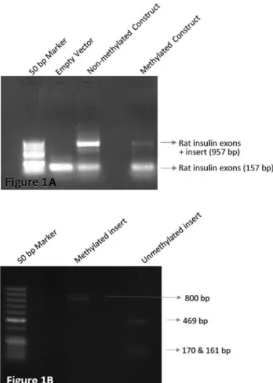
We first tested whether methylation affects splicing by transfecting HL-60 cells with methylated and unmethylated constructs containing the 633 bpMEFVexon 2 plus the intron sequences, 800 bp in total, cloned in pSpliceExpress splicing vector. This vector allows tracing splicing events by means of its rat insulin exons splicing concurrently. We have shown thatMEFVexon 2 is spliced when methylated (Figure 1), resulting in a smaller amplicon that lacks the second exon ofMEFV.
Splicing analysis of the cells’MEFVtranscripts
Different cell culture inflammation models were es-tablished to analyze the effect of methylation on the exon 2 splicing of the cells’ endogenousMEFVtranscripts. To this end, HL-60 promyelotic cells were used as control groups and were induced with different agents: DMSO for
neutrophil-like transformation, PMA and LPS for activa-tion, methanol for methylation and deazacytidine for demethylation.
Differentiation of HL-60 cells into neutrophil-like cells was achieved as shown in DAPI staining (Figure S3), flow cytometry analysis using PE-CD44 antibody (Figure S4) as well as SSC results (Figure S5), given in supplemen-tary data. CD44 is a cell surface glycoprotein involved in cell–cell interactions, cell adhesion and migration, and is expressed in different cell types including hematopoietic cells. DMSO-induced differentiation towards neutrophils causes downregulation of CD44 from the surface of cells compatible with a similar reduction in CD44 expression during normal granulopoiesis process (Mollet al., 1998; Springet al., 1988).
Methanol is known as a toxic and mutagenic sub-stance, generally used as a fixing agent in cell imaging. It has also been shown to increase genomic methylation by incorporating methyl residues into DNA (Huang et al., 2001). In our methylation cell model, we also used 5% (vol/vol) methanol to increase global methylation levels in HL-60 cells.
Expression studies were performed via two sets of primers amplifying cells endogenous MEFV exon 2-lacking and exon 2-containing transcripts and the data was normalized by dividing individual CTs to the total tran-scripts’. We observed that neutrophil-like cells (p = 0.0005, 2-fold increase), activated cells (p = 0.0034; 2.5-fold in-crease) and methylated cells (p < 0.0001; 2.5-fold inin-crease) exhibit an increased MEFV-d2 transcript expression whereas demethylated cells (p = 0.0126; 1.7-fold decrease) exhibit a decreased level, compared to untreated HL-60 cells (Figure 2).
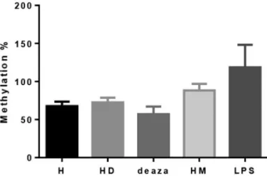
Analysis of induced and repressed global methylation studies in cell culture models confirmed that DMSO, PMA
& LPS and methanol treatments increase, while deaza-cytidine decreasesMEFV second exon methylation (Fig-ure 3). The differences in methylation level were not statistically significant, with a positive trend in HL-60 cells treated with DMSO (p = 0.4) and HL-60 cells treated with PMA & LPS (p = 0.053).
Cellular localization of pyrin and its isoform pyrin-d2
The localization of full-length pyrin and exon 2-lacking pyrin isoform (pyrin-d2) were investigated in HL-60 cells along with neutrophil-like cells via confocal mi-croscopy, since our findings imply thatMEFV-d2 transcript is increased in these cells.
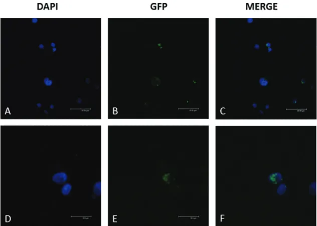
In untreated HL-60 controls, theMEFV-fl-GFP pro-tein was localized in the cytoplasm andMEFV-d2-GFP was localized in the nucleus (Figure 4). In contrast, in neutro-phil-like cells, bothMEFV-fl-GFP andMEFV-d2-GFP pro-ducts were localized in the cytoplasm (Figure 5).
Discussion
Alternative splicing causes the production of protein isoforms with differential subcellular localization, which may lead to altered functions (Stammet al., 2005; Hughes, 2006). More than 90% of human genes are subjected to al-ternative splicing, which is an evolutionarily conserved mechanism, ensuring proteomic diversity (Ast, 2004; Pan
et al., 2008). Co-transcriptional splicing enables the forma-tion of alternative transcripts through epigenetic regulaforma-tion of the chromatin (Iannone and Valcárcel, 2013). Epige-nomic studies have illustrated that CG dinucleotides were more abundant on exonic sites than intronic sites, and 3’ and 5’ splice sites were mostly methylated (nearly 100%) compared to surrounding CG rich regions (Gelfmanet al., 2013).
Figure 2- Relative expression results of different cell culture models. The 1-3 are exon 2 spliced transcripts’ expressions and 2-3 exon 2 containing transcripts’ expressions. H: untreated HL-60 cells; HD: HL-60 cells dif-ferentiated to neutrophil-like cells via DMSO; deaza: demethylated HL-60 cells using deazacytidine; HM: methylated HL-60 cells with methanol; LPS: HL-60 cells activated through PMA & LPS. The expression compar-isons were significant with p < 0.0001 for Hvs.HM, p = 0.0005 for Hvs. HD, p = 0.0034 for Hvs.LPS and p = 0.0126 for Hvs.deaza (p < 0.05 was considered statistically significant, N = 4 for each cell model).
Figure 4- Localization of recombinantMEFVproteins in untreated HL-60 cells.MEFV-fl-GFP andMEFV-d2-GFP constructs were transfected via Nucleofection and the localization of their products was analyzed using confocal microscopy. The transfection efficiency was 67% forMEFV-fl and 58% forMEFV-2d. A) DAPI staining of HL-60 cells transfected withMEFV-fl-GFP; B) GFP visualization ofMEFV-fl-GFP; C) Merged image of A and B; D) DAPI staining of HL-60 cell transfected withMEFV-d2-GFP; E) GFP visualization ofMEFV-d2-GFP; F) Merged image of D and E.
Previously, we showed thatMEFV exon 2 methy-lation levels of patients were slightly but significantly higher than the levels of controls (p = 0.049) (Kirectepeet al., 2011b). Furthermore, we observed a negative correla-tion between methylacorrela-tion and expression in all groups (r=–0.29, p = 0.041), which was more accentuated in the patient group (r=–0.36, p = 0.035). Here, we proposed a model where the methylation ofMEFVexon 2 is related to its alternative splicing and expression. To test that hypothe-sis,MEFVexon 2 including approximately 80 nucleotides of its flanking introns was cloned into a splicing reporter vector, which proved that transfected MEFV exon 2 is spliced when methylated. Then, different cell culture mod-els were constructed to confirm this finding with endoge-nousMEFVexpression: HL-60 cells were used as controls and DMSO induced cells, activated cells trough PMA and LPS, methylated cells with methanol, demethylated cells via deazacytidine constituted the induced group where the expression level of MEFV exon 2-containing and lack-ing-transcripts were analyzed. The results indicated that methylation increased the expression of MEFV-d2 tran-script, along with neutrophil transformation and activation with PMA & LPS, contrary to demethylated cells in which it was decreased. These findings suggest that methylation might play a role in the splicing of the second exon. The methylation levels of the CpG island of these cell culture models were further analyzed (Figure 3). Although the dif-ferences were not statistically significant, they exhibited similar increased methylation patterns, implying that methylation increases in inflammatory-like conditions.
An increasing number of studies are pointing to the involvement of DNA methylation in the regulation of alter-native splicing. A genome wide study performed in mouse retina and brain showed that differently methylated regions regulate alternative splicing in a tissue-specific manner (Wanet al., 2013). Another genome-wide study on honey bees, in which the authors inhibited the expression of Dnmt3 (DNA (cytosine-5)-methyltransferase 3), changed the alternative splicing pattern due to the decrease in methylation levels (Li-Byarlayet al., 2013). A recent study proposed that alternative splicing of sarcomeric geneMyh7
may be linked to the cardiac epigenome, which may lead to disease formation (Vujic et al., 2015). Absence of the
MEFVexon 2-deleted form in mice and rats, which do not contain a CpG island on the MEFV gene (Papin et al., 2000), also points towards the possible role of DNA metlation in alternative splicing, and also strengthens our hy-pothesis.
Several mechanisms have been proposed in attempt-ing to explain the role of epigenetic modifications in alter-native splicing. Some studies have shown that methylation leads to the inclusion of alternative exons. For example, MeCP2 (methyl-CpG binding protein 2) and HP1 (hetero-chromatin protein 1) are proteins found to participate in exon retention in the presence of DNA methylation
(Yea-rimet al., 2015). Adversely, another protein, CTCF, was found to play a role in the recognition of a weak exon signal in the CD45 gene in the absence of DNA methylation (Shukla et al., 2011). Although our research on the CTCFBSDB database, a database for CTCF binding sites, has shown the absence of CTCF in the MEFV gene in HL-60 cells, the CTCF prediction tool on the same site pre-dicts a binding site 35 bp from the exon recognition consen-sus sequence. As alternative splicing is regulated via splice site strength, and, thus, stronger sites increase the inclusion of alternative exons (Schwartzet al., 2009), recognition of weak splice sites via reducing RNA PolII elongation rate is crucial for the retention of subjected exons. Methylation may also simply blockcis-acting sequences where RNA-binding proteins bind to enhance the inclusion level. We found many putative exonic splicing enhancer sites con-taining a CG dinucleotide, which could be blocked by methylation (Human Splicing Finder). Therefore, differen-tial methylation of specific sites within a CpG island may be responsible for exon inclusion levels, rather than methy-lation of a specific region. It would be very informative to perform an analysis of a potential protein, like CTCF, slow-ing down the elongation rate of the RNA PolII and thus en-abling the recognition of theMEFVexon 2 signal, leading to its inclusion. This would also suggest methylation of its specific binding site, which will be further analyzed.
Point mutations/variations are also known to cause al-ternative splicing. Rittoreet al.(2014) showed three varia-tions [rs4149570(c.-610G > T), rs767455(c.36A > G,pPro12Pro), rs1800692(c.473-33C > T)] in the pro-moter, exon 1 and intron 2, respectively, of TNFRSF1A gene enhance the splicing of exon 2in vitro. These varia-tions appeared to have a role in the pathogenesis of TRAPS disease (Rittore et al., 2014). Furthermore, in another study, Toneet al.(2011) suggested that c.910G > A variant (rs75977701) leads to the skipping of MEFV exon 2. This rare variant, which has a Minor Allele Frequency (MAF) of T = 0.0074/37 in the Thousands Genomes Project, was ab-sent in our FMF cohort. In Japan, where this variant is more frequently encountered, its frequency in FMF patients is 0.9% (Kishidaet al., 2014). This is in contradiction with our finding of higher MEFV-d2 transcript levels in FMF patients compared to healthy controls (Kirectepe et al., 2011a), suggesting a different mechanism for the splicing of exon 2, at least for the Turkish population.
Since many diseases are shown to be caused by ab-normal localization of proteins, the localization differences of GFP tagged full-length pyrin (pyrin-fl) and exon 2 spliced forms (pyrin-d2) were also analyzed in inflamma-tion related cell culture models. Both MEFV-fl-GFP and
(Figure 5). This type of experiment requires a western blot confirmation in cytoplasmic vs. nuclear fractions. How-ever, only a maximum of 40% of HL-60 cells differentiated into neutrophil-like cells, and the transfection efficiency was not constant among the experiments. Thus western blot studies had inconsistencies in thesein vitromodels. There-fore, we increased the repeats of our confocal experiments to obtain reliable results. The 14.3.3 proteins, potent anti-apoptotic factors that control intracellular signaling, cell cycle and apoptosis, interact with pyrin-fl but not pyrin-d2 through three serine residues located in exon 2. Jéruet al.
(2005) showed that this interaction caused full-length pyrin to be retained in the cytoplasm. Furthermore, the lack of in-teraction with pyrin-d2 isoform resulted in protein translo-cation to the nucleus. Thus, cell-specific post-translational processing and/or protein–protein interactions, may ulti-mately determine subcellular localization relating to patho-logical functions in different cell types. Although alterna-tive splicing of exon 2 creates a nuclear localization signal, it does not correspond to known NLS motifs. Exon 1-3 junction encodes a domain, which is necessary but not suf-ficient to target the spliced form to nucleus. Thus, this do-main may be required for the activity of another NLS-like motif located elsewhere in the spliced form. We and other authors previously suggested that pyrin-d2 may have a role as a transcription factor, and its function may be impaired during inflammation (Kirectepeet al., 2011a). Thus, under-standing the proteins that interact with the exon 2-deleted but not the full-length pyrin protein and their functions may lead to identification of a mechanism involved in nuclear import and inflammatory diseases.
Several other studies analyzed similar features: Tidowet al.(2000) studied the localization of full-length pyrin in COS-1 and found it to be cytoplasmic. They also found that DMSO induction increases the full-length pression in HL-60 cells but they did not investigate the ex-pression of exon 2 spliced transcript. Diaz et al. (2004) induced the expression of exon 2 spliced form by LPS in synovial fibroblasts and monocyte-enriched PBLs from pa-tients; however, they found it to be still two times less abun-dant than the full-length form. We had opposite results with HL-60 promyelotic cells, which are known to be neutro-philic precursors, suggesting that the expression of spliced transcript may change with harmful stimuli, since neutro-phils are the first responders in cases of inflammation. Cazeneuveet al.(2003) reported pyrin-d2 to be in the nu-cleus of HeLa cells; nevertheless, they also found that it in-teracts with ASC in the cytoplasm. In a recent study, pyrin was shown to co-localize with actin in HL-60 cells (Akka-ya-Ulumet al., 2015). Knowing that the overexpression of recombinant products transfected to cells in a transient manner could disrupt physiological pathways for protein transport (Kremmidiotiset al., 1999), it could be of interest to generate an antibody specific to exon 2 spliced form to
detect the localization of native protein in different cells via confocal microscopy and western blot analyses.
Despite the fact that FMF is reported to be a reces-sively inherited disease, 15 to 25% of patients from loca-tions where FMF is less prevalent present no pathogenic variations in the MEFVgene. A modifier gene has been suggested as a primary alternative, and studies were con-ducted to identify another gene or locus having an epistatic interaction with MEFV. Touitou et al. (2001) proposed
MICAas a modifier gene in FMF; however, they could not find a significant result for affected patients from different ethnic origins. Hence, any disruption leading to truncated pyrin formation may have a role in the pathology of FMF. Our findings clearly show the differences between undif-ferentiated and difundif-ferentiated cells. Research should be conducted with patients in active inflammation and relapse periods and healthy controls to further understand the exact mechanism. However, methylation leading to the splicing ofMEFVsecond exon, and consequently to a protein that has an aberrant localization, may explain the pathogenesis of FMF without theMEFVpathogenic variants.
Acknowledgments
This study was supported by the Scientific Research Projects Department of Istanbul Technical University (Grant No: 33931, 35076, 35077). GCE was supported by TUBITAK BIDEB-2211, National PhD Grant Program. We would also like to express our gratitude to Koray Kirimtay and Ayse Erozenci for their contributions, as well as to Gul Yazici Kaya for linguistic advice.
References
Agostinho P, Pliássova A, Oliveira CR and Cunha RA (2015) Lo-calization and trafficking of amyloid- protein precursor and secretases: Impact on Alzheimer’s disease. J Alzhei-mer’s Dis 45:329-347.
Akkaya-Ulum YZ, Balci-Peynircioglu B, Purali N and Yilmaz E (2015) Pyrin-PSTPIP1 colocalises at the leading edge dur-ing cell migration. Cell Biol Int 39:1384-1394.
Ameyar-Zazoua M, Rachez C, Souidi M, Robin P, Fritsch L, Young R, Morozova N, Fenouil R, Descostes N, Andrau J,et al.(2012) Argonaute proteins couple chromatin silencing to alternative splicing. Nat Struct Mol Biol 19:998-1004. Ast G (2004) How did alternative splicing evolve? Nat Rev Genet
5:773-782.
Cazeneuve C, Papin S, Jeru I, Duquesnoy P and Amselem S (2003) Subcellular localisation of marenostrin/pyrin iso-forms carrying the most common mutations involved in fa-milial Mediterranean fever in the presence or absence of its binding partner ASC. J Med Genet 41:e24.
Centola M, Wood G, Frucht DM, Galon J, Aringer M, Farrell C, Kingma DW, Horwitz ME, Mansfield E, Holland SM,et al. (2000) The gene for familial Mediterranean fever, MEFV, is expressed in early leukocyte development and is regulated in response to inflammatory mediators. Blood 95:3223-3231.
(2008) Shotgun bisulphite sequencing of the Arabidopsis genome reveals DNA methylation patterning. Nature 452:215-219.
Diaz A, Hu C, Kastner DL, Schaner P, Reginato AM, Richards N and Gumucio DL (2004) Lipopolysaccharide-induced ex-pression of multiple alternatively spliced MEFV transcripts in human synovial fibroblasts: A prominent splice isoform lacks the C-terminal domain that is highly mutated in famil-ial Mediterranean fever. Arthritis Rheum 50:3679-3689. Feng S, Cokus SJ, Zhang X, Chen PY, Bostick M, Goll MG,
Hetzel J, Jain J, Strauss SH, Halpern ME,et al.(2010) Con-servation and divergence of methylation patterning in plants and animals. Proc Natl Acad Sci U S A 107:8689-8694. Galarce GD, Foncea RE, Edwards AM, Pessoa-Mahana H,
Pes-soa-Mahana CD and Ebensperger RA (2008) Biological evaluation of novel 6-Arylbenzimidazo[1,2-c]quinazoline derivatives as inhibitors of LPS-induced TNF-alpha secre-tion. Biol Res 41:43-50.
Gal-Yam EN, Egger G, Iniguez L, Holster H, Einarsson S, Zhang X, Lin JC, Liang G, Jones PA and Tanay A (2008) Frequent switching of Polycomb repressive marks and DNA hyper-methylation in the PC3 prostate cancer cell line. Proc Natl Acad Sci U S A 105:12979-12984.
Gelfman S, Cohen N, Yearim A and Ast G (2013) DNA-methy-lation effect on cotranscriptional splicing is dependent on GC architecture of the exon-intron structure. Genome Res 23:789-799.
González C, Salces-Ortiz J, Calvo JH and Serrano MM (2016)In silico analysis of regulatory and structural motifs of the ovine HSP90AA1 gene. Cell Stress Chaperones 21:415-427.
Grandemange S, Soler S and Touitou I (2009) Expression of the familial Mediterranean fever gene is regulated by non-sense-mediated decay. Hum Mol Genet 18:4746-4755. Gutierrez-Arcelus M, Ongen H, Lappalainen T, Montgomery SB,
Buil A, Yurovsky A, Bryois J, Padioleau I, Romano L, Planchon A,et al.(2015) Tissue-Specific Effects of Genetic and Epigenetic Variation on Gene Regulation and Splicing. PLoS Genet 11:e1004958.
Hellman A and Chess A (2007) Gene body-specific methylation on the active X chromosome. Science 315:1141-1143. Hodges E, Smith AD, Kendall J, Xuan Z, Ravi K, Rooks M,
Zhang MQ, Ye K, Bhattacharjee A, Brizuela L,et al.(2009) High definition profiling of mammalian DNA methylation by array capture and single molecule bisulfite sequencing. Genome Res 19:1593-1605.
Huang YS, Held GA, Andrews JE and Rogers YM (2001)
14
C-methanol incorporation into DNA and proteins of orga-nogenesis stage mouse embryosin vitro. Reprod Toxicol 15:429-435.
Hughes TA (2006) Regulation of gene expression by alternative untranslated regions. Trends Genet 22:119-122.
Hung MC and Link W (2011) Protein localization in disease and therapy. J Cell Sci 124:3381-3392.
Iannone C and Valcárcel J (2013) Chromatin’s thread to alterna-tive splicing regulation. Chromosoma 122:465-474. Jéru I, Papin S, L’hoste S, Duquesnoy P, Cazeneuve C, Camonis J
and Amselem S (2005) Interaction of pyrin with 14.3.3 in an isoform-specific and phosphorylation-dependent manner re-gulates its translocation to the nucleus. Arthritis Rheum 52:1848-1857.
Jones PA (1999) The DNA methylation paradox. Trends Genet 15:34-37.
Jones PA (2012) Functions of DNA methylation: Islands, start sites, gene bodies and beyond. Nat Rev Genet 13:484-492. Kirectepe AK, Celikyapi Erdem G, Senturk N, Arisoy N, Hatemi
G, Ozdogan H, Kasapcopur O and Tahir Turanli E (2011a) Increased expression of exon 2 deleted MEFV transcript in familial Mediterranean fever patients. Int J Immunogenet 38:327-329.
Kirectepe AK, Kasapcopur O, Arisoy N, Celikyapi Erdem G, Hatemi G, Ozdogan H and Tahir Turanli E (2011b) Analysis of MEFV exon methylation and expression patterns in fa-milial Mediterranean fever. BMC Med Genet 12:105. Kishida D, Nakamura A, Yazaki M, Tsuchiya-Suzuki A, Matsuda
M and Ikeda S (2014) Genotype-phenotype correlation in Japanese patients with familial Mediterranean fever: Differ-ences in genotype and clinical features between Japanese and Mediterranean populations. Arthritis Res Ther 16:439. Kishore S, Khanna A and Stamm S (2008) Rapid generation of
splicing reporters with pSpliceExpress. Gene 427:104-110. Kremmidiotis G, Lensink IL, Bilton RL, Woollatt E, Chataway
TK, Sutherland GR and Callen DF (1999) The Batten dis-ease gene product (CLN3p) is a Golgi integral membrane protein. Hum Mol Genet 8:523-531.
Lev Maor G, Yearim A and Ast G (2015) The alternative role of DNA methylation in splicing regulation. Trends Genet 31:274-280.
Li-Byarlay H, Li Y, Stroud H, Feng S, Newman TC, Kaneda M, Hou KK, Worley K, Elsik CG, Wickline SA,et al.(2013) RNA interference knockdown of DNA methyl-transferase 3 affects gene alternative splicing in the honey bee. Proc Natl Acad Sci U S A 110:12750-12755.
Lidar M and Livneh A (2007) Familial Mediterranean fever: Clin-ical, molecular and management advancements. Neth J Med. 65:318-324.
Liu Z and Hu J (2016) Mislocalization-related disease gene dis-covery using gene expression based computational protein localization prediction. Methods 93:119-127.
Lock LF, Takagi N and Martin GR (1987) Methylation of theHprt gene on the inactive X occurs after chromosome inactiva-tion. Cell 48:39-46.
Matzner Y, Abedat S, Shapiro E, Eisenberg S, Bar-Gil-Shitrit A, Stepensky P, Calco S, Azar Y and UrieliShoval S (2000) Ex-pression of the familial Mediterranean fever gene and activ-ity of the C5a inhibitor in human primary fibroblast cultures. Blood 96:727-731.
Maunakea AK, Chepelev I, Cui K and Zhao K (2013) Intragenic DNA methylation modulates alternative splicing by recruit-ing MeCP2 to promote exon recognition. Cell Res 23:1256-1269.
Medlej-Hashim M, Nehme N, Chouery E, Jalkh N and Megarbane A (2010) Novel MEFV transcripts in familial Mediterranean fever patients and controls. BMC Med Genet 11:87. Moll J, Khaldoyanidi S, Sleeman JP, Achtnich M, Preuss I, Ponta
H and Herrlich P (1998) Two different functions for CD44 proteins in human myelopoiesis. J Clin Invest 102:1024-1034.
Nguyen CT, Gonzales FA and Jones PA (2001) Altered chromatin structure associated with methylation-induced gene silenc-ing in cancer cells: Correlation of accessibility, methylation, MeCP2 binding and acetylation. Nucleic Acids Res 29:4598-4606.
sup-pressor genes to DNA hypermethylation and heritable si-lencing. Nat Genet 39:237-242.
Pan Q, Shai O, Lee LJ, Frey BJ and Blencowe BJ (2008) Deep sur-veying of alternative splicing complexity in the human transcriptome by high-throughput sequencing. Nat Genet 40:1413-1415.
Papin S, Duquesnoy P, Cazeneuve C, Pantel J, Coppey-Moisan M, Dargemont C and Amselem S (2000) Alternative splic-ing at theMEFVlocus involved in familial Mediterranean fever regulates translocation of the marenostrin/pyrin pro-tein to the nucleus. Hum Mol Genet 9:3001-3009.
Rittore C, Sanchez E, Soler S, Barat-Houari M, Albers M, Obici L, McDermott MF, Touitou I and Grandemange S (2014) Identification of a new exon 2-skipped TNFR1 transcript: Regulation by three functional polymorphisms of the TNFR-associated periodic syndrome (TRAPS) gene. Ann Rheum Dis 73:290-297.
Saint-André V, Batsche E, Rachez C and Muchardt C (2011) Histone H3 lysine 9 trimethylation and HP1g favor inclu-sion of alternative exons. Nat Struct Mol Biol 18:337-344. Schlesinger Y, Straussman R, Keshet I, Farkash S, Hecht M,
Zimmerman J, Eden E, Yakhini Z, Ben-Shushan E, Reubi-noff BE,et al.(2007) Polycomb-mediated methylation on Lys27 of histone H3 pre-marks genes forde novo methy-lation in cancer. Nat Genet 39:232-236.
Schwartz S, Meshorer E and Ast G (2009) Chromatin organiza-tion marks exon-intron structure. Nat Struct Mol Biol 16:990-995.
Shukla S, Kavak E, Gregory M, Imashimizu M, Shutinoski B, Kashlev M, Oberdoerffer P, Sandberg R and Oberdoerffer S (2011) CTCF-promoted RNA polymerase II pausing links DNA methylation to splicing. Nature 479:74-79.
Stamm S, Ben-Ari S, Rafalska I, Tang Y, Zhang Z, Toiber D, Thanaraj TA and Soreq H (2005) Function of alternative splicing. Gene 344:1-20.
Spring FA, Dalchau R, Daniels GL, Mallinson G, Judson PA, Par-sons SF, Fabre JW and Anstee DJ (1988) The Ina and Inb blood group antigens are located on a glycoprotein of 80,000 MW (the CDw44 glycoprotein) whose expression is influ-enced by theIn(Lu)gene. Immunology 64:37-43.
The International FMF Consortium (1997) Ancient missense mu-tations in a new member of the roret gene family are likely to cause familial Mediterranean fever. Cell 90:797-807. Tidow N, Chen X, Müller C, Kawano S, Gombart AF,
Fischel-Ghodsian N and Koeffler HP (2000) Hematopoietic-specific expression of MEFV, the gene mutated in familial Mediter-ranean fever, and subcellular localization of its correspond-ing protein, pyrin. Blood 95:1451-1455.
Tone Y, Toma T, Toga A, Sakakibara Y, Wada T, Yabe M, Kusafuka H and Yachie A (2011) Enhanced exon 2 skipping caused by c.910G > A variant and alternative splicing of MEFV genes in two independent cases of familial Mediter-ranean fever. Mod Rheumatol 22:45-51.
Touitou I, Picot M, Domingo C, Notarnicola C, Cattan D, De-maille J and Koné-Paut I (2001) The MICA region deter-mines the first modifier locus in familial Mediterranean fe-ver. Arthritis Rheum 44:163-169.
Vujic A, Robinson EL, Ito M, Haider S, Ackers-Johnson M, See K, Methner C, Figg N, Brien P, Roderick HL,et al.(2015) Experimental heart failure modelled by the cardiomyocyte-specific loss of an epigenome modifier, DNMT3B. J Mol Cell Cardiol 82:174-183.
Wan J, Oliver VF, Zhu H, Zack DJ, Qian J and Merbs SL (2013) Integrative analysis of tissue-specific methylation and alter-native splicing identifies conserved transcription factor binding motifs. Nucleic Acids Res 41:8503-8514.
Widschwendter M, Fiegl H, Egle D, Mueller-Holzner E, Spizzo G, Marth C, Weisenberger DJ, Campan M, Young J, Jacobs I,et al.(2007) Epigenetic stem cell signature in cancer. Nat Genet 39:157-158.
Yang X, Han H, De Carvalho DD, Lay FD, Jones PA and Liang G (2014) Gene body methylation can alter gene expression and is a therapeutic target in cancer. Cancer Cell 26:577-590. Yearim A, Gelfman S, Shayevitch R, Melcer S, Glaich O, Mallm
JP, Nissim-Rafinia M, Cohen AH, Rippe K, Meshorer E,et al.(2015) HP1 is involved in regulating the global impact of DNA methylation on alternative splicing. Cell Rep 10:1122-1134.
Internet Resources
CTCFBSDB 2.0: A database for CTCF binding sites and genome organization, http://insulatordb.uthsc.edu (February 10, 2016).
Human Splicing Finder, http://www.umd.be/HSF3/ index.html (February 10, 2016).
qMethyl Calculator - for determination of % methylation from
du-plicate samples,
http://www.zymoresearch.com/tools/qmethyl-calculator/du plicate-samples (December 3, 2015).
Supplementary material
The following online material is available for this article: Table S1 – Primers used for the amplification of pSpliceExpress insert sequence.
Table S2 – Primers used for adding attb 1&2 recombination sites.
Table S3 – Rat insulin primers.
Table S4 – Primers for quantitative real-time PCR. Figure S1 – DMSO induction of HL-60 cells.
Figure S2 – CD44 expression analysis of undifferentiated and differentiated HL-60 cells.
Figure S3 – Granularity analysis of undifferentiated and differentiated HL-60 cells via side-scattered light (SSC). Figure S4 – pCMV6-AC-GFP-MEFV-FL (OriGene) con-struct.
Figure S5 - Granularity analysis of undifferentiated and dif-ferentiated HL-60 cells via side-scattered light (SSC).
Associate Editor: Juan Lucas Argueso Almeida