A Deep Insight into the Sialome of Rhodnius
neglectus, a Vector of Chagas Disease
Paula Beatriz Santiago
1, Teresa C. F. Assumpç
ão
1,2, Carla Nunes de Araújo
1,3, Izabela
Marques Dourado Bastos
1, David Neves
1, Ionizete Garcia da Silva
4, Sébastien Charneau
1,
Rayner Myr L. Queiroz
1, Tainá Raiol
1,5, Jo
ão Victor de Araújo Oliveira
6, Marcelo Valle
de Sousa
1, Eric Calvo
2, José M. C. Ribeiro
2, Jaime M. Santana
1*
1 Department of Cell Biology, The University of Brasília, Brasília, Brazil, 2 Vector Biology Section, Laboratory of Malaria and Vector Research, National Institute of Allergy and Infectious Disease, Rockville, Maryland, United States of America, 3 Ceilândia Faculty, The University of Brasília, Brasília, Brazil, 4 Department of Parasitology, The University of Goiás, Jataí, Brazil, 5 Instituto Leônidas e Maria Deane -Fiocruz Amazônia, Manaus, Brazil, 6 Department of Computer Science, The University of Brasília, Brasília, Brazil
Abstract
Background
Triatomines are hematophagous insects that act as vectors of Chagas disease. Rhodnius
neglectus
is one of these kissing bugs found, contributing to the transmission of this
Ameri-can trypanosomiasis. The saliva of hematophagous arthropods contains bioactive
mole-cules responsible for counteracting host haemostatic, inflammatory, and immune
responses.
Methods/Principal Findings
Next generation sequencing and mass spectrometry-based protein identification were
per-formed to investigate the content of triatomine R. neglectus saliva. We deposited 4,230
cod-ing DNA sequences (CDS) in GenBank. A set of 636 CDS of proteins of putative secretory
nature was extracted from the assembled reads, 73 of them confirmed by proteomic
analy-sis. The sialome of R. neglectus was characterized and serine protease transcripts
detected. The presence of ubiquitous protein families was revealed, including lipocalins,
serine protease inhibitors, and antigen-5. Metalloproteases, disintegrins, and odorant
bind-ing protein families were less abundant.
Conclusions/Significance
The data presented improve our understanding of hematophagous arthropod sialomes, and
aid in understanding hematophagy and the complex interplay among vectors and their
ver-tebrate hosts.
a11111
OPEN ACCESS
Citation: Santiago PB, Assumpção TCF, Araújo CNd, Bastos IMD, Neves D, Silva IGd, et al. (2016) A Deep Insight into the Sialome of Rhodnius neglectus, a Vector of Chagas Disease. PLoS Negl Trop Dis 10 (4): e0004581. doi:10.1371/journal.pntd.0004581 Editor: Paulo Filemon Pimenta, Fundaçao Oswaldo Cruz, BRAZIL
Received: October 22, 2015 Accepted: March 7, 2016 Published: April 29, 2016
Copyright: This is an open access article, free of all copyright, and may be freely reproduced, distributed, transmitted, modified, built upon, or otherwise used by anyone for any lawful purpose. The work is made available under theCreative Commons CC0public domain dedication.
Data Availability Statement: All Invertebrate sample from Rhodnius neglectus salivary glands files are available from the BioSample database (accession number SAMN03975952).http://www.ncbi.nlm.nih. gov/biosample/3975952
Funding: This work was supported by grants 476252/2012-1 (http://www.cnpq.br) from the Brazilian National Council for Scientific and Technological Development (CNPq), PROAP-2013 (http://www.capes.gov.br) from the Coordenação de Aperfeiçoamento de Pessoal de Nível Superior (CAPES), CT-Infra 2011 (http://www.finep.gov.br) from the Financiadora de Estudos e Projetos (Finep),
Author Summary
Chagas disease is caused by the Trypanosoma cruzi protozoan, which is transmitted to
ver-tebrates through the feces of infected triatomines during blood sucking. The vascular
injury caused by the bite triggers mechanisms capable of preventing the association with
hosts, such as immune response, inflammation and haemostasis. However,
hematopha-gous insects are able to counteract these defenses through a complex repertoire of salivary
molecules that have specific targets in the host. Our results show that R. neglectus salivary
glands express different protein gene families, possessing multi-functional features directly
related to different anti-haemostatic activities. For instance, lipocalins are proteins
pos-sessing anti-coagulant and vasodilator functions. Saliva contents have evolved to adapt to
blood-feeding habit, ensuring the maintenance of blood flow, the success of the meal, and
transmission of diseases.
Introduction
Blood-sucking triatomines (Hemiptera: Reduviidae) feed exclusively on blood in all life stages.
They obtain their blood meal from venules or arterioles of their vertebrate hosts. The steps
dur-ing feeddur-ing include piercdur-ing of the host skin, followed by a probdur-ing period, and finally
engorge-ment [
1
]. In support of this habit, these arthropods have evolved effective mechanisms to
counteract host responses, such as haemostasis, inflammation and immunological reactions.
While biting, their salivary glands (SG) release potent pharmacological substances, including
vasodilator, anti-inflammatory, antiplatelet, anticlotting and immunomodulatory molecules, to
enable the arthropod to obtain a successful blood-meal [
2
,
3
]. These bioactive salivary
compo-nents represent a promising source of molecules with therapeutic potential for treating
circula-tory disorders [
4
,
5
].
In the 1990s, multinational control programs against Chagas disease led to a significant
reduction of acute cases in many endemic regions of Latin America, mainly through a
reduc-tion of domestic vectors [
6
]. However, factors such as the wide geographical distribution of
triatomine species and the availability of different infection reservoirs remain multifactorial
obstacles in the control of the disease. Nowadays, there is constant concern regarding the
spo-radically or progressive (re)invasion and (re)colonization of human dwellings by wild
second-ary vectors [
7
,
8
]. Rhodnius neglectus is found in the Brazilian Savanna (Cerrado) in association
with different wild palms, playing an important role in the sylvatic maintenance of T. cruzi and
Trypanosoma rangeli [
9
–
11
]. In nature, R. neglectus feeds mainly on birds and much less on
rodents, and rarely on opossum [
12
]. This species is able to act as a secondary vector, being
observed in both intra and peridomestic environments in five Brazilian states [
13
–
17
], a
possi-ble result of deforestation and wild ecotope invasion. These anthropogenic environmental
changes favor vector dispersion, bridging sylvatic/domestic cycles of the disease.
Sialome studies (from the Greek sialo = saliva) have been developed for many species of
bloodsucking insects, which are frequently vectors of human and animal diseases. Sanger
auto-mated sequencing technology has been used to investigate the salivary transcriptome for
almost two decades. However, Next Generation Sequencing (NGS) is capable of providing
much more sequence data in a single run, with a higher resolution than that from the Sanger
technique, allowing for deeper analysis of the transcripts. One important application of NGS is
RNA sequencing (RNA-seq), used to describe transcriptomes of cells and tissues. Deep
sequencing increases the possibilities of finding new biological molecules in the saliva of
and Pronex 476252/2012-5 (http://www.fap.df.gov.br) from the Fundação de Apoio à Pesquisa do Distrito Federal (FAPDF) to JMS; and Z01 AI000810-18 (http://www.niaid.nih.gov/Pages/default.aspx) to Vector-Borne Diseases: Biology of Vector Host Relationship, from the National Institute of Allergy and Infectious Diseases (NIAID) to JMCR. The funders had no role in study design, data collection and analysis, decision to publish, or preparation of the manuscript.
Competing Interests: The authors have declared that no competing interests exist.
bloodsucking insects, offering a new array of substances to be further investigated and
func-tionally characterized.
The aim of this report is to catalog the transcripts of R. neglectus SGs with probable function
in hematophagy using RNAseq and mass spectrometry. This strategy was used to describe the
bioactive molecules in triatomine saliva and improve our understanding on the dynamics of
the blood-feeding process, vector-host interaction and disease transmission. The data is
avail-able at the National Center for Biotechnology Information (NCBI) and can be used in different
scientific research projects.
Methods
Insects and Transcriptome Salivary Gland Preparation
R. neglectus triatomines originating from insects collected in 1982 at Itambaracá, in Paraná
State, Brazil, were reared in the insectarium at the University of Brasília (Brazil). They were
kept at 27±1°C, a relative humidity of 70–75%, under a 12 h/12 h light/dark cycle. The blood
source of these insects was Gallus gallus domesticus. The SGs of 5
thinstar nymphs and adults
were dissected at 5, 12, and 24 days post blood meal in cold Trizol reagent (Invitrogen,
Carls-bad, CA, USA). A pool of thirty SG pairs was stored at -80°C prior to RNA extraction.
Salivary Gland RNA Isolation, Library Preparation and Sequencing
Total RNA was extracted following the Trizol manufacturer’s instructions. RNA integrity and
concentration were checked by lab-on-chip analysis using an Agilent 2100 Bioanalyzer (Agilent
Technologies, USA). A RNA sample was sent to the Federal District High-Performance
Genome Center (DF, Brazil) for Illumina cDNA library construction and next generation
sequencing. A Library was prepared with standard protocols using TruSeq RNA kit, v2
(Illu-mina, San Diego, CA). To generate paired-end reads of 300 nucleotides in length, the
sequenc-ing of cDNA libraries was performed on an Illumina MiSeq sequencer (Illumina, USA). One
lane of the MiSeq machine was used for sequencing this and another library, distinguished by
bar coding. The RNA-seq sequencing generated a total of 12,049,305 reads. The nominal length
of the sequences was 301 nt. Following trimming of low quality bases (quality 20 or lower), the
average length was 248.07, the median was 301 and L50 was 296 nt. Sequences smaller than 25
nt or with average quality
< 20 were rejected.
Bioinformatic Analysis
Bioinformatic analyses were conducted as previously described [
18
]. As there was no reference
genome to map, the strategy was to perform a de novo assembly with Abyss [
19
] and
Soapde-novo Trans [
20
] assemblers using different kmer (k) values (from 20 to 90). The resulting
assemblies were joined by an iterative BLAST and cap3 assembler [
21
]. Sequence
contamina-tion between bar-coded libraries were identified and removed when their sequence identities
were over 98%. Coding sequences (CDS) were extracted based on the existence of a signal
pep-tide and on similarities to other known proteins [
22
]. Coding and protein sequences were
mapped into a hyperlinked Excel spreadsheet. Reads were mapped into contigs using blastn
[
23
] with a word size of 25, masking homonucleotide decamers and allowing mapping to up to
five different CDS if the BLAST results had the same scores. Mapping of the reads was also
included in the Excel spreadsheet. CDS were automatically annotated a program written by
JMCR that searched a vocabulary of nearly 250 words for matches various databases, including
Swissprot, Gene Ontology, KOG, PFAM, and SMART, and a subset of the non-redundant
pro-tein database containing propro-teins from vertebrates (NCBI). Further manual annotation was
done as required. Alignment analysis were done with Bioedit software [
24
] after sequence
alignment performed using ClustalW [
25
]. Phylogenetic analysis and statistical
neighbor-join-ing bootstrap tests of the phylogenies were done with Mega package [
26
]. The sequences used
in alignments with R. neglectus CDS were obtained from the non-redundant protein database
of the NCBI and are represented by six letters followed by the NCBI GI number. The letters
derive from the first three letters of the genus and the first three letters of the species name.
Data Availability
The raw reads were deposited at the Sequence Read Archive (SRA) in NCBI under bioproject
PRJNA292130. A total of 4,230 coding sequences were deposited in DDBJ/EMBL/GenBank
through the Transcriptome Shotgun Annotation portal under the accession GDKW00000000.
LC-MS/MS Protein Identification
The SGs were dissected from 5
thinstar nymphs and adults at 5, 12 and 24 days post blood meal
and carefully punctured at 4°C. Following centrifugation (16.000 × g, 15 min, 4°C), the soluble
protein fraction from fifteen pairs of SG homogenates was ethanol/acetone precipitated.
Resus-pended proteins were consecutively alkylated, reduced, digested by trypsin, and subjected to
LC-MS/MS analysis as previously described [
27
]. Briefly, the tryptic peptides were loaded onto
a 2 cm fused silica trap column (150
μm inner diameter) packed in-house with reverse phase
capillary column ReproSil-Pur C18-AQ 5
μm resin (Dr. Maisch GmbH, Germany) and
sepa-rated using a DIONEX 3000 nanoUPLC system coupled to an LTQ-Orbitrap Elite mass
spec-trometer (Thermo Scientific, Waltham, USA). MS1 spectra were recorded in the Orbitrap mass
analyzer with 120,000 resolution. After ion fragmentation, MS/MS spectra of the 15 most
intense ions were acquired. Raw files were generated and used for protein identification using
Proteome Discoverer v.1.3 (Thermo Scientific, Waltham, USA) with in-house SequestHT
algo-rithm for R. neglectus SG transcriptome and human keratins, BSA and porcin trypsin. The false
discovery rate was less than 1%, with peptide rank of 1 and at least 2 peptides per protein.
Results and Discussion
General Description of the Sialome of R. neglectus
The assembly of R. neglectus SG transcriptome enabled the extraction of 5,705 CDS. These
CDS mapped a total of over 11 million reads. Following automated and manual annotation,
the CDS were classified into putative secreted, housekeeping, unknown, transposable element,
and viral product. The CDS of the housekeeping class comprised the largest class (
Table 1
).
They were further characterized into 24 subclasses, according to their possible function,
sum-marized in
Table 2
.
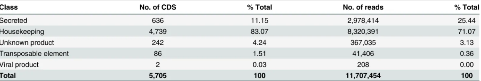
Table 1. Classification and abundance of coding sequences extracted from the salivary gland transcriptome of R. neglectus.
Class No. of CDS % Total No. of reads % Total
Secreted 636 11.15 2,978,414 25.44 Housekeeping 4,739 83.07 8,320,391 71.07 Unknown product 242 4.24 367,035 3.13 Transposable element 86 1.51 41,406 0.36 Viral product 2 0.03 208 0.00 Total 5,705 100 11,707,454 100 doi:10.1371/journal.pntd.0004581.t001
Putative Secreted Proteins
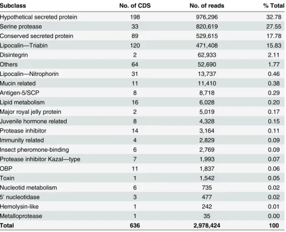
The secreted class was organized in subclasses that include previously known gene families
present in hematophagous saliva, such as lipocalin, nitrophorin, antigen-5, as well as gene
fam-ilies not commonly reported in triatomine saliva, such as serine protease and disintegrin
(
Table 3
). The following section describes the putative secreted proteins present in R. neglectus
sialome, highlighting the remarkable finding of serine proteases in this group.
Lipocalins
Lipocalins comprised one of the most abundant groups of transcripts, with 16.29% of
puta-tively secreted reads. These include a large group of extracellular proteins that usually bind to
small hydrophobic molecules, cell surface receptors or other proteins. The members of this
family have little similarity in peptide sequence, however share a conserved three-dimensional
structure, comprised of a single eight-stranded antiparallel
β-barrel [
28
]. In blood-sucking
insect and tick saliva the lipocalins are abundantly expressed, but not in Diptera or fleas. In
ticks, their function is associated with binding to histamine and serotonin [
29
]. Triabin and
nitrophorin, the two major groups found here, are discussed below.
Lipocalins of the triabin family.
First isolated from the saliva of the Triatoma pallidipennis
kissing bug [
30
], triabin is a lipocalin-like thrombin inhibitor, which inhibits thrombin-induced
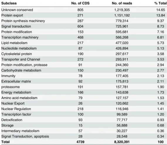
Table 2. Classification and abundance of coding sequences of putative housekeeping function extracted from the sialotranscriptome of R. neglectus.
Subclass No. of CDS No. of reads % Total
Unknown conserved 805 1,219,305 14.65
Protein export 271 1,151,192 13.84
Protein synthesis machinery 287 779,314 9.37
Signal transduction 604 725,961 8.73
Protein modification 153 595,681 7.16
Transcription machinery 468 566,268 6.81
Lipid metabolism 217 477,020 5.73
Nucleotide metabolism 87 426,894 5.13
Cytoskeletal protein 190 297,617 3.58
Transporter and Channel 272 293,911 3.53
Protein modification, protease 91 244,360 2.94
Carbohydrate metabolism 150 230,497 2.77
Immunity 78 177,405 2.13
Extracellular matrix 92 175,813 2.11
proteasome 191 157,781 1.90
Energy metabolism 166 143,638 1.73
Amino acid metabolism 79 127,157 1.53
Nuclear Export 26 120,662 1.45 Nuclear Regulation 218 116,946 1.41 Transcription factor 100 99,589 1.20 Detoxification 93 77,717 0.93 Storage 15 56,888 0.68 Intermediary metabolism 57 30,227 0.36
Signal Transduction, apoptosis 28 28,548 0.34
Total 4739 8,320,391 100
platelet aggregation, and prolongs thrombin clotting time through the formation of a
noncova-lent complex with thrombin at a 1:1 molar ratio. Previous analysis revealed that triabin is a
com-pact one-domain molecule essentially consisting of an eight-stranded
β-barrel and inhibits
thrombin exclusively via its fibrinogen-recognition exosite [
31
]. Thrombin is the ultimate serine
protease formed during activation of the blood coagulation cascade, which catalyzes the
poly-merization of fibrinogen to fibrin, the solid fibrillar component of the blood clot, thereby being
a fundamental promoter of blood clotting. Thus, the triabin-like lipocalins may function as
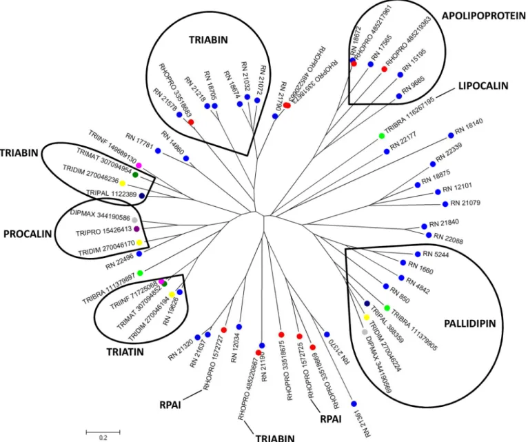
thrombin inhibitors in R. neglectus saliva. The library analysis shows 120 different CDS from
lipocalin family containing the triabin conserved domain, such as triabin, pallidipin,
apolipo-protein, procalin and triatin. The alignment of these members with lipocalins already described
in triatomines resulted in a phylogram containing different clades (
Fig 1
). In addition, it is
possi-ble to note two divergent clades containing only R. neglectus and Rhodnius prolixus sequences
(RPAI and Apolipoprotein), which may represent additional gene members present in Rhodnius
spp. The presence of different clades indicates the expansion of this gene family by gene
duplica-tion events, suggesting that, for R. neglectus, lipocalins exert a crucial role in success feeding.
Lipocalins of the nitrophorin family.
Rhodnius spp. show a characteristic red coloration
in their saliva due to the presence of haemoproteins called nitrophorins (NPs). These molecules
form a stable complex with nitric oxide (NO), which is sensitive to pH variation, being stabilized
by low pH in the lumen of the SGs (pH ~5), and released at neutral pH in the host (pH ~7.5)
[
32
]. The secretion of NO is an efficient way to counteract haemostasis, acting both as a potent
vasodilator and as an antagonist of platelet activation. NPs 1–4 can additionally sequester
Table 3. Classification and abundance of coding sequences of putative secretory function extracted from the sialotranscriptome of R. neglectus.
Subclass No. of CDS No. of reads % Total
Hypothetical secreted protein 198 976,296 32.78
Serine protease 33 820,619 27.55
Conserved secreted protein 89 529,615 17.78
Lipocalin—Triabin 120 471,408 15.83 Disintegrin 2 62,933 2.11 Others 64 52,690 1.77 Lipocalin—Nitrophorin 31 13,737 0.46 Mucin related 11 11,410 0.38 Antigen-5/SCP 8 8,718 0.29 Lipid metabolism 16 6,028 0.20
Major royal jelly protein 2 5,019 0.17
Juvenile hormone related 8 4,328 0.15
Protease inhibitor 14 3,164 0.11
Immunity related 4 2,829 0.09
Insect pheromone-binding 6 2,769 0.09
Protease inhibitor Kazal—type 7 1,993 0.07
OBP 11 1,837 0.06 Toxin 1 1,542 0.05 Nucleotid metabolism 6 735 0.02 5’ nucleotidase 3 477 0.02 Hemolysin-like 1 242 0.01 Metalloprotease 1 35 0.00 Total 636 2,978,424 100 doi:10.1371/journal.pntd.0004581.t003
histamine released by host mast cells, reducing inflammation and immune response [
33
,
34
].
NP 2 inhibits clotting in a mechanism independent of NO or histamine binding, acting as a
spe-cific inhibitor of the intrinsic factor X-(FX)-activating complex [
35
]. As well as reversibly
bind-ing to NO or histamine, NP 7 also inhibits prothrombin activation by blockbind-ing phospholipid
binding sites for the prothrombinase complex on the surfaces of vesicles and activated platelets
through binding to phosphatidylserine [
36
]. The current sialotranscriptome identified 13,737
reads related to the diversity of NPs. The NPs of R. neglectus also appear to be a gene family that
expanded during evolutionary processes, as inferred by the phylogenetic tree (
Fig 2
). Notice that
there are several sequences homolog to NP1-4 and 7, NPs described in R. prolixus saliva.
Fig 1. Phylogram of lipocalin containing triabin domain from R. neglectus SG transcriptome. Phylogenetic tree derived from the alignment of R. neglectusCDS and other triatomine lipocalin sequences as described in Methods section. The bar at the bottom represents 20% amino acid substitution. The colored circles indicate each species whose sequences were used: blue, R. neglectus sequences from SG transcriptome; red, R. prolixus; yellow, Triatoma dimidiata; green, Triatoma brasiliensis; dark green, Triatoma matogrossensis; dark blue, T. pallidipennis; purple, Triatoma protacta; magenta, Triatoma infestans; gray, Dipetalogaster maxima.
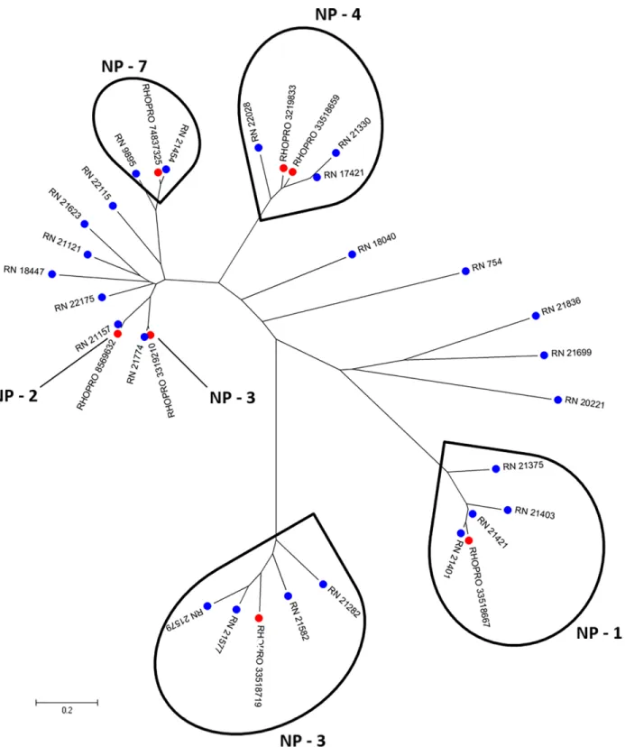
Fig 2. Phylogram of lipocalin containing nitrophorin domain from R. neglectus SG transcriptome. Phylogenetic tree derived from the alignment of R. neglectusCDS and R. prolixus nitrophorin sequences as described in Methods section. The bar at the bottom represents 20% amino acid substitution. The colored circles indicate each species whose sequences were used: blue, R. neglectus sequences from SG transcriptome and red, R. prolixus sequences from NCBI.
The mean number of nitrophorins in salivary electrophoretic profiles varies among
Rhod-nius species, with R. neglectus showing the fewest. The high polymorphism of NPs may help in
the identification of Rhodnius species [
37
]. The lower proportion of nitrophorin content in the
saliva compared to those found in the saliva of other Rhodnius spp. might not, by itself, explain
the reduced feeding performance of R. neglectus on mammals. For instance, although R.
neglectus shows lower amounts of nitrophorins, it feeds more efficiently than R. robustus [
37
].
It is important to note that the exact contribution of each class of saliva molecules on the
feed-ing process is unknown.
Antigen-5 Family
The CAP superfamily members [Cysteine-Rich Secretory Proteins (CRISPS), Antigen 5 (Ag5),
and Pathogenesis-Related 1 (Pr-1)] are found in a wide range of organisms, most often as
secreted proteins [
38
]. Ag5, present in the venom of wasps and ants, are considered potent
aller-gens to mammals [
39
,
40
]. This superfamily can also block smooth muscle contraction when
present in snake venom [
41
] and act in the defense response in plants [
42
]. They have been
described in the saliva of some hematophagous, including mosquitoes [
43
,
44
] and sand flies
[
45
]. Among triatomines, Ag5 genes have been reported in the sialotranscriptomes of R. prolixus
[
46
], T. infestans [
47
], D. maxima [
48
], T. matogrossensis [
49
] and Triatoma rubida [
50
]. Their
functions in blood-feeder saliva remained unexplored for a long time, but a recent report
revealed salivary Ag5 of D. maxima and T. infestans as Cu
+2-dependent antioxidant enzymes
that inhibit neutrophil oxidative burst and platelet aggregation induced by collagen [
51
].
The sialotranscriptome analysis revealed eight CDS related to the Ag5 family. The
align-ment of R. neglectus Ag5 with other triatomine Ag5 sequences showed some conserved motifs
(
S1 Fig
). Phylogenetic analysis offers support for the formation of clades I and II comprising
triatomine and Diptera sequences, respectively (
Fig 3
).
Serine Protease Inhibitors
For blood-feeders, targeting components of the coagulation cascade is essential to attenuate the
haemostatic response of their hosts. All enzymes participating in this cascade are serine
prote-ases associated with complement activation [
52
,
53
]. The R. neglectus sialotranscriptome
exhib-ited a variety of transcripts coding for proteins with serine protease inhibitory function,
comprising 14 CDS and 3,164 reads. Based on their Pfam signature, kazal, pacifastin and serpin
families were extracted.
Kazal family.
Kazal-type domain-containing proteins are serine protease inhibitors
play-ing important functions in invertebrates, mainly havplay-ing vasodilation, antimicrobial, and
thrombin inhibition effects. These protease inhibitors are single or multidomain proteins that
share a conserved sequence motif, a distinctive cysteine distribution pattern and highly similar
three-dimensional structure [
54
]. Rhodniin is a kazal-type thrombin inhibitor isolated from R.
prolixus [
55
,
56
]. Dipetalogastin from D. maxima [
57
], infestin from T. infestans [
58
] and
bra-siliensin from T. brasiliensis [
59
] are thrombin inhibitors located in the intestines. From the
horse fly Hybomitra bimaculata (Diptera, Tabanidae) SGs, a vasodilator named vasotab was
identified as a member of Kazal-type protease inhibitor family acting through ion channel
inhi-bition and vasodilation [
60
].
Seven CDS in R. neglectus sialotranscriptome possessed the typical sequence of nonclassical
Kazal domains characterized by a shorter distance between the first and second cysteine
resi-due, unlike the seven or eight spacer residues found in the classical configuration [
55
,
57
]. The
alignment showed a low degree of conserved amino acids, but confirmed the presence of the
six cysteine residues responsible for the formation of disulfide bridges (
Fig 4
). The relative
posi-tions of cysteine residues were the same in the compared sequences.
Additionally, one contig was identified as dipetalogastin due to the cysteine residues
distri-bution and the presence of the conserved motif CGXDXXTYXNXC, a distinguishing repeat of
Kazal-type inhibitors [
57
]. This transcript is full length and possesses the signal peptide
indica-tive of secretion. The alignment with other protein sequences with the same features revealed a
high degree of conserved amino acids (
S2 Fig
).
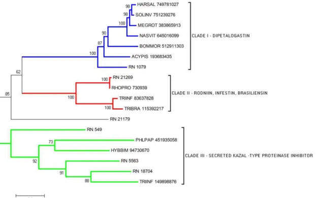
The phylogram of serine protease inhibitor members clearly shows the formation of three
clades, with a good bootstrap support, each one representing a different family of serine
prote-ase inhibitor discussed above (
Fig 5
). The CDS RN_21179 is notably distinct from the clades,
suggesting the presence of a divergent gene. The different clades may represent sequences
dif-ferentially expressed sharing the same function regarding haemostasis inhibition.
Fig 3. Phylogram of Antigen-5 proteins from R. neglectus SG transcriptome. Phylogenetic tree derived from the alignment of R. neglectus CDS and other insect antigen-5 sequences as described in Methods section. The bar represents 10% amino acid substitution.
doi:10.1371/journal.pntd.0004581.g003
Fig 4. Kazal-type members from R. neglectus SG transcriptome. ClustalW alignment of Kazal-type domain-containing members from R. neglectus salivary transcriptome (RN_5563 and RN_549) and other insect kazal-type sequences, identified as described in Methods section. The alignment indicates conserved residues in black and similar residues in gray background, the six conserved cysteines (boxes) and the blue bar indicates the signal peptide indicative of secretion.
Pacifastin family.
Pacifastin is a family of serine protease inhibitors, mostly multi-domain
proteins, first isolated from the plasma of the crayfish Pacifastacus leniusculus. The protein is
heterodimeric, comprising both a transferrin chain (heavy chain, PHC) and a protease
inhibi-tor chain (light chain, PLC) [
61
,
62
]. Insect pacifastins may have multiple functions, acting as
regulators of a wide variety of serine peptidase-dependent processes such as immunity and
reproduction [
63
]. In Hemiptera, two pacifastin-like protease inhibitors from T. infestans eggs
were functionally characterized, suggesting a role in insect immune response [
64
]. Here, two
CDS are related to pacifastin, RN_17301 and RN_20047, and their alignment with other
mem-bers of the pacifastin family reveals four conserved domains, containing the cysteine-rich
inhibitory pattern of PLC comprised of a triple-stranded antiparallel beta-sheet connected by
three disulfide bridges (
S3 Fig
). This is the first time pacifastin members are identified in
triato-mine SGs, their function in this organ is still unknown but it might be related to insect
immunity.
Serpin family.
Serpins are a large family of structurally related proteins found across taxa,
showing diverse activities not limited to inhibition of serine proteases [
65
]. In vertebrates,
ser-pins play crucial control in blood coagulation, fibrinolysis and inflammation. Dysfunction,
deficiencies or over-expression of serpins can cause either abnormal bleeding or thrombosis
[
66
]. The function of this protein in saliva of mosquitoes is related to host haemostasis
regula-tion, seeming to act as a potent reversible inhibitor of the host factor Xa [
67
]. In Ixodes ricinus
ticks, the molecule was also associated with inhibition of blood coagulation and fibrinolysis of
the vertebrate host [
68
–
70
]. The consensus three-dimensional fold of serpins is comprised of a
bundle of 8
–9 α-helices and a β-sandwich composed of three β-sheets [
71
].
Here, four CDS from R. neglectus sialotranscriptome were classified as serpins. The
phylo-gram showed four clades with a good bootstrap support (
Fig 6
). Transcripts RN_9905,
Fig 5. Phylogram of Serine Protease inhibitors from R. neglectus SG transcriptome. Phylogenetic tree derived from the alignment of R. neglectus CDS and other insect sequences as described in Methods section. The bar at the bottom represents 20% amino acid substitution.
RN_20002 and RN_10079 grouped each one in a separate clade while the fourth CDS,
RN_2083, seemed to be a distant divergent gene. In R. neglectus saliva this inhibitor might also
function in the modulation of coagulation cascade.
Proteases
Metalloprotease.
One CDS found in the R. neglectus sialotranscriptome is related to the
zinc-dependent metalloproteases from the astacin-like metalloproteases, a family of the
met-zincins superfamily. There are three conserved regions in proteins from this family. The first
one is the distinguishing family signature sequence HEXXHXXGXXHE, which is the
zinc-binding active site. The second region, RXDRD, is a hydrophilic region, and the third highly
conserved region, MXY, is the methionine-containing turn (the Met-turn) [
72
–
74
].
This gene family comprises many proteins from diverse species. In the venom of different
spider species, there is a common toxin with the ability to hydrolyze fibrinogen and fibronectin
[
75
–
77
], suggesting a relationship between this proteolytic activity with local hemorrhage,
since fibronectin plays a role in platelet aggregation, blood vessel stability and wound healing
[
78
]. Therefore, the biologic function of astacin-like proteases in triatomine saliva could be
related to the maintenance of blood flow at the bite site. Astacin domain metalloproteases were
already reported in T. matogrossensis sialotranscriptome [
49
]. The CDS RN_21266 is a
full-Fig 6. Phylogram of serpin proteins from R. neglectus SG transcriptome. Phylogenetic tree derived from the alignment of R. neglectus CDS and other insect sequences as described in Methods section. The bar represents 20% amino acid substitution.
length sequence containing a signal peptide indicative of secretion. Its alignment with other
metalloproteases revealed the three conserved motifs of the family (
Fig 7A
). The phylogenetic
tree suggests, with a good bootstrap support, that the secreted metalloproteases are closely
related proteins (
Fig 7B
).
ADAMTS (ADAM with thrombospondin motifs)/Disintegrins.
Two further members
of the metzincin metalloprotease superfamily were identified in R. neglectus transcriptome and
are related to the adamalysin/reprolysin family, which includes ADAM (A Disintegrin And
Metalloproteinase domain) and ADAMTS (A Disintegrin And Metalloproteinase with
Throm-bospondin motifs). ADAMTS is a group of secreted, extracellular and multidomain proteases
that have diverse roles in both mammals and invertebrates [
79
,
80
]. They are cysteine-rich
mol-ecules that selectively block the function of integrin receptors on the cell membrane surface
[
81
,
82
], exhibiting a thrombospondin-like (TS) repeat and a cysteine-rich domain typical of
disintegrins [
79
]. In this family, the third histidine in the family signature sequence containing
three zinc ligands is followed by a conserved aspartic acid, HEXXHXXGXXHD. Moreover, it
lacks the fifth zinc ligand and the methionine residue of the consensus Met-turn is placed
within the sequence V/I-M-A/S [
74
,
79
].
Together with snake venom metalloproteinases (SVMP), ADAM and ADAMTS are a group
of versatile molecules in viper venom that affects different elements in haemostasis [
83
]. The
disintegrins can bind to platelets and act as potent inhibitors of platelet aggregation [
84
–
86
].
The molecule can also bind to endothelial cells [
87
,
88
], as well as neutrophils and phagocytes
[
89
]. Rhodostomin is a disintegrin that inhibits activity of LPS-treated monocytes via
αvβ3
integrin affecting haemostasis, cell-cell interaction and suppresses tumor growth [
90
]. In
hematophagous organisms, the disintegrins have been described in tick and leech saliva [
91
].
Here, one CDS related to disintegrins was identified in the R. neglectus SG transcriptome. The
alignment exhibited a high degree of homology between R. neglectus disintegrin sequence and
others of the same family (
Fig 8
). The identification of ADAMTS is one of the main findings
from the sialotranscriptome of R. neglectus.
Serine proteases and trypsin-like proteins.
The R. neglectus SG transcriptome revealed
serine proteases reads as the second most abundant group in the secreted class, comprising
820,619 reads. The majority of the sequences identified as serine proteases exhibited the trypsin
domain (Tryp-SPc) of the CDD and Smart databases. RN_1189 was assembled from 768,048.
Its alignment with serine proteases from other organisms revealed conserved residues located
around the cleavage and active sites (
Fig 9A
), and the presence of a CUB (complement C1r/
C1s, Uegf, Bmp1) domain, a structural motif of approximately 110 residues found almost
exclusively in extracellular and plasma membrane-associated proteins. This domain is also
present in honeybee allergens Api SI and Api SII, which are probably components of the
hon-eybee defense system [
92
,
93
].
Some serine proteases can function as regulators of coagulation. Thrombin can participate
in this regulation by binding to thrombomodulin, a membrane protein present in host
endo-thelial cells. This complex is able to activate Protein C (a serine protease), which acts as a potent
anticoagulant enzyme by inactivating factors V and VIII, impairing thrombus progression [
94
,
95
]. In snake venom, blockage of thrombus formation by serine proteases has also been
reported. SPSV (Serine Protease Snake Venom) releases a unique fibrinopeptide that produces
only instable monomers of fibrin, leading to clots that are rapidly dispersed [
96
]. Although the
specific role in hematophagous saliva is still unknown, an active serine protease was described
in T. infestans [
97
], as well as in horse fly Tabanus yao saliva, which functions as a
fibrinogen-olytic enzyme [
98
]. RN_22226, RN_21634, RN_19989, RN_17969, and RN_10652 were
matched by blastp to serine proteases of T. infestans, T. braziliensis, Panstrongylus megistus,
and R. prolixus. Serine proteases also play important roles in fertilization, embryonic
Fig 7. The secreted metalloprotease from R. neglectus SG transcriptome. (A) ClustalW alignment of the secreted metalloprotease from R. neglectus salivary transcriptome (RN_21266) and other metalloproteases sequences, identified as described in Methods section. The alignment indicates conserved residues in black and similar residues in gray background. The blue bar indicates the signal peptide indicative of secretion. The boxes limit the family signature sequences showing the determinant residues (black asterisk). (B) Phylogenetic tree derived from the alignment of R. neglectus CDS and other metalloproteases sequences as described in Methods section. The bar represents 10% amino acid substitution.
development, and in the processes of molting and metamorphosis of insects [
99
,
100
]. In our
sample, the triatomines did not show any sign of larval molting at SG dissection.
Sequences containing CLIP, LDLa and SUSHI domains, which are cysteine-stabilized
struc-tures for molecular recognition, were also identified. The CLIP domain is restricted to the
Arthropoda and was found N-terminally to the Tryp-SPc domain of RN_16275, RN_18155,
Fig 8. The ADAMTS sequence from R. neglectus SG transcriptome. ClustalW alignment of the ADAMTS sequence from R. neglectus salivary
transcriptome (RN_11351) and other insect ADAMTS sequences, identified as described in Methods section. The alignment indicates conserved residues in black and similar residues in gray background. The bar indicates the signal peptide indicative of secretion. The boxes limit the family signature sequences showing the determinant residues (black asterisk). The symbols (black hash) above indicate the conserved cysteines.
Fig 9. Serine proteases from R. neglectus SG transcriptome. (A) ClustalW alignment of a serine protease from R. neglectus SG transcriptome (RN_1189) and other serine proteases members, identified as described in Methods section. The alignment indicates conserved residues in black and similar residues in gray background. The blue bar indicates the signal peptide indicative of secretion. The symbols above the residues indicate (black circle) cleavage site and (black triangle) active site showing the HDS triad. (B) Phylogenetic tree was built from the alignment of R. neglectus CDS and other insect sequences as described
and RN_7118. Both domains belong to the serine proteases of the trypsin-like S1 family, that
are typically secreted enzymes associated with extracellular proteolysis [
101
]. CLIP domain has
been suggested to be important for dimerization, mediating specific protein-protein
interac-tions involved in the regulation of serine protease activities. The LDLa domain was identified
in RN_12992, RN_12776, RN_12432, and RN_21634. The last two sequences also presented
the SUSHI motif of smart database which is known as CCP (Complement Control Protein)
module, containing approximately 60 amino acid residues identified in several proteins of the
complement system. These R. neglectus putative secreted serine proteases may play critical
roles in many key biological processes as blood coagulation and immunity. In the vertebrate
hosts, allergenicity may reinforce the toxic effect of serine proteases, independently of their
cat-alytic activity, as proposed by Georgieva and colleagues [
93
].
The phylogram clearly showed four different groups (
Fig 9B
), indicating the expression of
at least four genes related to serine proteases in the saliva of R. neglectus. In regard to the large
amount of reads, the results observed here suggests that, to R. neglectus, the serine proteases
arise as an important salivary secreted gene family, a probably evolutionary adaptation where
the protein could gain a new function as a result of selective pressure for the blood-feeding
behavior success. These proteases could act in the vertebrate host, as well as in the insect, on
pathogens that were ingested with blood. Further experiments are necessary to address the
pos-sible roles of those genes on the biology of R. neglectus.
OBP
The odorant-binding protein (OBP) family is a chemosensory protein ubiquitous in insects
commonly associated with solubilizers and carriers of odorants and pheromones. Although
associated with chemosensory organs, in recent times this family has been related to other roles
such as hydrophobic chemical transportation [
102
]. The OBPs are characterized by a variable
amino acid sequence, but conserve a pattern of six conserved cysteines residues paired to form
three disulfide bridges [
103
]. The folding is a typical six
α-helices assembled in a compact and
stable structure [
104
,
105
]. Eleven CDS containing protein sequences related to OBPs were
rec-ognized in our transcriptome analysis, all possessing signal peptide prediction. The conserved
cysteine residues of R. neglectus CDS (
Fig 10
) were seen during alignment. Phylogenetic
analy-sis with good bootstrap support shows Clade I containing most R. neglectus sequences grouped
with R. prolixus OBP. However, RN_3440 was grouped in Clade III, suggesting this is a more
distant OBP (
S4 Fig
).
The Proteome of R. neglectus Saliva
R. neglectus saliva content was tryptic digested and subjected to mass spectrometry to validate
the analysis of the transcripts possibly associated with secreted products. Among the 73
identi-fied secreted proteins groups, 48 were from the lipocalin family, including triabin, pallidipin
and nitrophorin proteins, reaffirming their abundance (
Table 4
). Other soluble proteins,
pre-dicted as being secreted by these arthropods, were: secreted metalloprotease, antigen-5, serpin
and trypsin-like protease, each with at least one observation. It is intriguing that only one serine
protease was detected by proteomic analysis, regardless the high number of transcripts reads
assigned to this subclass of putative secreted proteins (
Table 3
). There are several possible
explanations for this observation. First, the proteins are expressed in a such a small amount not
in Methods section The bar at the bottom represents 20% amino acid substitution. The colored circles identify the sequences used: blue, R. neglectus sequences from SG transcriptome; red, Hemiptera order; green, Hymenoptera order; magenta, Dictyoptera order.
detectable by our proteomic approach; second, the proteins are not secreted; third and most
likely, these proteins present in SGs are expressed upon specific physiological conditions, such
as during stimulation of salivation (feeding). In addition, it is also possible that those enzymes
have both intracellular and extracellular functions as many other proteases do.
Comparison of Protein Contents between R. neglectus and R. prolixus
A comparative blastp analysis was employed to address the similarity of the SGs proteins from
R. neglectus compared to R. prolixus. The two species do not show high evolutionary
diver-gence, presenting at least 80% identity in analyzed sequences (
Table 5
), suggesting both species
share a common ancestral lineage. As described before, this high degree of protein similarity
was also seen with R. brethesi and R. robustus in the Amazon rainforest [
106
].
Fig 10. The secreted OBP family from R. neglectus SG transcriptome. ClustalW alignment of secreted OBPs from R. neglectus SG transcriptome (RN_5213, RN_3440 and RN_18954) and other members from the OBP family, identified as described in Methods section. The alignment indicates conserved residues in black and similar residues in gray background. The blue bar indicates the signal peptide indicative of secretion and the boxes, the six conserved cysteines.
doi:10.1371/journal.pntd.0004581.g010
Table 4. Classification and abundance of proteins from the salivary proteome of R. neglectus based on LC-MS/MS.
Class No. of protein groups % Total
Lipocalin–Triabin 34 46.58
Lipocalin–Nitrophorin 17 23.29
Others 13 17.81
Conserved secreted protein 2 2.74
Antigen-5/SCP 1 1.37
Inositol polyphosphate phosphatase 1 1.37
Metalloprotease 1 1.37
Protease inhibitor–Serpin 1 1.37
Chitinase-like lectin 1 1.37
Trypsin-like protease 1 1.37
Juvenile hormone related 1 1.37
Total 73 100
Identity among sequences was greater in housekeeping class members, showing that these
proteins have a lower evolutionary rate than those of the secreted class. This indicates that
anti-haemostatic proteins evolve faster after divergence. Different molecular mechanisms may be
responsible for the variation between these closely related Rhodnius species, expanding their
biological diversity patterns. The particularity of each species could be related to their different
habitats, including different prey and abiotic factors.
Final Considerations
Hematophagy evolved independently at least six times in approximately 15,000 species
allow-ing for adaptation to an existallow-ing complex host haemostatic system [
5
,
107
]. Thus, many
sali-vary molecules target different pathways for the insect to achieve a successful blood meal. Here,
we described R. neglectus sialome in all its complexity to expand our knowledge of the salivary
proteins from hematophagous triatomine bugs.
R. neglectus is considered of secondary importance in the transmission of T. cruzi, causative
agent of Chagas disease. The analysis of salivary secretory products of R. neglectus that might
be involved in vector-host interactions share similarity with other triatomine species, which
can also be infected by and transmit the protozoan.
It is possible that the expression of putative trypsin-like serine proteases in the SGs of R.
neglectus correlates with blood sources of this species of triatomine. Their role and that of other
secreted class, hypothetical and conserved secreted proteins, in hematophagy should be
ana-lyzed in future works, and we accentuate that sialome study is still an open field for new
discoveries.
Table 5. Identities of R. neglectus proteins compared to R. prolixus (v 3.0) proteins by blastp.
Class Average identity SE* N
Secreted 86.46 1.47 200
Immunity 91.17 3.76 35
Transporters and channels 92.33 1.22 114
Extracelular matrix 94.07 1.81 43 Transposable elements 94.64 2.50 22 Protein export 95.09 0.84 166 Signal transduction 95.39 0.54 285 Storage 95.50 1.44 8 Cytoskeletal 95.64 0.96 88 Unknown conserved 95.99 0.44 369
Protein modification 95.99 0.78 144
Metabolism 96.28 0.45 405 Transcription machinery 96.50 0.50 250 Proteasome machinery 96.59 0.80 122 Nuclear regulation 96.65 0.75 95 Nuclear export 96.67 1.05 12 Unknown 97.04 1.47 24 Detoxification 97.15 1.16 48 Transcription factor 97.43 0.63 53 Protein synthesis 97.95 0.42 180 Total 2,663
*SE: standard error
Supporting Information
S1 Fig. Antigen-5 proteins from R. neglectus SG transcriptome.
ClustalW alignment of
anti-gen-5 members from R. neglectus salivary transcriptome (RN_21331, RN_21434 and
RN_21812) and other hemiptera sequences, identified as described in Methods section. The
alignment indicates conserved domains in black and similar domains in gray background.
(TIF)
S2 Fig. The dipetalogastin from R. neglectus SG transcriptome.
ClustalW alignment of a
dipetalogastin member from R. neglectus salivary transcriptome (RN_1079) and other
sequences from dipetalogastin family, identified as described in Methods section. The
align-ment indicates conserved residues in black and similar residues in gray background. The boxes
indicate conserved motifs, and the blue bar indicates the signal peptide indicative of secretion.
(TIF)
S3 Fig. Pacifastin members from R. neglectus SG transcriptome.
ClustalW alignment of
pacifastin members from R. neglectus salivary transcriptome (RN_17301 and RN_20047) and
other sequences from the pacifastin family of proteins, identified as described in Methods
sec-tion. The alignment indicates conserved residues in black and similar residues in gray
back-ground. The bars indicate the four conserved pacifastin motifs.
(TIF)
S4 Fig. Phylogram of the secreted OBP family from R. neglectus SG transcriptome.
Phyloge-netic tree was built from the alignment of R. neglectus CDS and other OBP sequences as
described in Methods section. The bar represents 20% amino acid substitution.
(TIF)
Acknowledgments
We thank Ana Cristina Gomes and Fabiano Bastos for technical assistance.
Author Contributions
Conceived and designed the experiments: PBS CNdA JMCR JMS. Performed the experiments:
PBS RMLQ TR JVdAO EC JMCR. Analyzed the data: PBS TCFA CNdA JMCR JMS.
Contrib-uted reagents/materials/analysis tools: IMDB DN IGdS SC MVdS JMCR JMS. Wrote the
paper: PBS TCFA CNdA IMDB DN IGdS SC RMLQ TR JVdAO MVdS EC JMCR JMS.
References
1. LAVOIPIERRE MM, DICKERSON G, GORDON RM. Studies on the methods of feeding of blood-sucking arthropods. I. The manner in which triatomine bugs obtain their blood-meal, as observed in the tissues of the living rodent, with some remarks on the effects of the bite on human volunteers. Ann Trop Med Parasitol. 1959; 53:235–50. PMID:14414675.
2. Fontaine A, Diouf I, Bakkali N, Misse D, Pages F, Fusai T, et al. Implication of haematophagous arthropod salivary proteins in host-vector interactions. Parasit Vectors. 2011; 4:187. doi:10.1186/ 1756-3305-4-187PMID:21951834; PubMed Central PMCID: PMC3197560.
3. Champagne DE. Antihemostatic strategies of blood-feeding arthropods. Current drug targets. 2004; 4 (4):375–96. PMID:15578959.
4. Champagne DE. Antihemostatic molecules from saliva of blood-feeding arthropods. Pathophysiology of haemostasis and thrombosis. 2005; 34(4–5):221–7. doi:10.1159/000092428PMID:16707932. 5. Ribeiro JM. Blood-feeding arthropods: live syringes or invertebrate pharmacologists? Infect Agents
Dis. 1995; 4(3):143–52. PMID:8548192.
6. Steverding D. The history of Chagas disease. Parasit Vectors. 2014; 7:317. doi: 10.1186/1756-3305-7-317PMID:25011546; PubMed Central PMCID: PMCPMC4105117.
7. Guhl F, Pinto N, Aguilera G. Sylvatic triatominae: a new challenge in vector control transmission. Mem Inst Oswaldo Cruz. 2009; 104 Suppl 1:71–5. PMID:19753461.
8. Fitzpatrick S, Feliciangeli MD, Sanchez-Martin MJ, Monteiro FA, Miles MA. Molecular genetics reveal that silvatic Rhodnius prolixus do colonise rural houses. PLoS Negl Trop Dis. 2008; 2(4):e210. doi:
10.1371/journal.pntd.0000210PMID:18382605; PubMed Central PMCID: PMC2270345.
9. Abad-Franch F, Monteiro FA, Jaramillo O N, Gurgel-Gonçalves R, Dias FB, Diotaiuti L. Ecology, evo-lution, and the long-term surveillance of vector-borne Chagas disease: a multi-scale appraisal of the tribe Rhodniini (Triatominae). Acta Trop. 2009; 110(2–3):159–77. doi:10.1016/j.actatropica.2008.06. 005PMID:18619938.
10. Gurgel-Goncalves R, Galvao C, Costa J, Peterson AT. Geographic distribution of chagas disease vectors in Brazil based on ecological niche modeling. J Trop Med. 2012; 2012:705326. doi:10.1155/ 2012/705326PMID:22523500; PubMed Central PMCID: PMC3317230.
11. Gurgel-Goncalves R, Cura C, Schijman AG, Cuba CA. Infestation of Mauritia flexuosa palms by triato-mines (Hemiptera: Reduviidae), vectors of Trypanosoma cruzi and Trypanosoma rangeli in the Brazilian savanna. Acta Trop. 2012; 121(2):105–11. doi:10.1016/j.actatropica.2011.10.010PMID:22037200. 12. Rodrigues VL, Pauliquevis C Junior, da Silva RA, Wanderley DM, Guirardo MM, Rodas LA, et al.
Col-onization of palm trees by Rhodnius neglectus and household and invasion in an urban area, Araça-tuba, São Paulo State, Brazil. Rev Inst Med Trop Sao Paulo. 2014; 56(3):213–8. PMID:24878999; PubMed Central PMCID: PMCPMC4085872.
13. Rubens Antonio Silva PRBeDMVW. Doença de Chagas no Estado de São Paulo: comparaçãoentre pesquisa ativa de triatomíneos em domicílios enotificação de sua presença pela população emárea sob vigilância entomológica. Revista da Sociedade Brasileira de Medicina Tropical1999.
14. Gurgel-Gonçalves R, Ramalho ED, Duarte MA, Palma AR, Abad-Franch F, Carranza JC, et al. Enzo-otic transmission of Trypanosoma cruzi and T. rangeli in the Federal District of Brazil. Rev Inst Med Trop Sao Paulo. 2004; 46(6):323–30. /S0036-46652004000600005. PMID:15654478.
15. de Oliveira A W. S., da Silva I G.. Distribuição geográfica e indicadores entomológicos de triatomí-neos sinantrópicos capturados no Estado de Goiás. Revista da Sociedade Brasileira de Medicina Tropical; 2007. p. 204–8.
16. Almeida PSd, al e. Levantamento da fauna de Triatominae (Hemiptera: Reduviidae) em ambiente domiciliar e infecção natural por Trypanosomatidae no Estado de Mato Grosso do Sul. Uberaba: Rev. Soc. Bras. Med. Trop; 2008.
17. Gurgel-Gonçalves R, Abad-Franch F, Ferreira JB, Santana DB, Cuba CA. Is Rhodnius prolixus (Tria-tominae) invading houses in central Brazil? Acta Trop. 2008; 107(2):90–8. doi:10.1016/j.actatropica. 2008.04.020PMID:18550022.
18. Ribeiro JM, Chagas AC, Pham VM, Lounibos LP, Calvo E. An insight into the sialome of the frog biting fly, Corethrella appendiculata. Insect biochemistry and molecular biology. 2013. Epub 2014/02/12. doi:10.1016/j.ibmb.2013.10.006PMID:24514880.
19. Simpson JT, Wong K, Jackman SD, Schein JE, Jones SJ, Birol I. ABySS: a parallel assembler for short read sequence data. Genome Res. 2009; 19(6):1117–23. doi:10.1101/gr.089532.108PMID:
19251739; PubMed Central PMCID: PMCPMC2694472.
20. Luo R, Liu B, Xie Y, Li Z, Huang W, Yuan J, et al. SOAPdenovo2: an empirically improved memory-efficient short-read de novo assembler. GigaScience. 2012; 1(1):18. doi:10.1186/2047-217X-1-18
PMID:23587118; PubMed Central PMCID: PMC3626529.
21. Karim S, Singh P, Ribeiro JM. A deep insight into the sialotranscriptome of the gulf coast tick, Amblyomma maculatum. PLoS ONE. 2011; 6(12):e28525. Epub 2012/01/05. doi:10.1371/journal. pone.0028525PMID:22216098; PubMed Central PMCID: PMC3244413.
22. Nielsen H, Brunak S, von Heijne G. Machine learning approaches for the prediction of signal peptides and other protein sorting signals. Protein engineering. 1999; 12(1):3–9. PMID:10065704.
23. Altschul SF, Madden TL, Schaffer AA, Zhang J, Zhang Z, Miller W, et al. Gapped BLAST and PSI-BLAST: a new generation of protein database search programs. Nucleic Acids Res. 1997; 25 (17):3389–402. PMID:9254694; PubMed Central PMCID: PMC146917.
24. Hall TA. BioEdit: a user-friendly biological sequence alignment editor and analysis program for Win-dows 95/98/NT Oxford University Press: Nucleic Acids Symposium Series; 1999. p. 95–8. 25. Larkin MA, Blackshields G, Brown NP, Chenna R, McGettigan PA, McWilliam H, et al. Clustal W and
Clustal X version 2.0. Bioinformatics. 2007; 23(21):2947–8. doi:10.1093/bioinformatics/btm404
PMID:17846036.
26. Kumar S, Tamura K, Nei M. MEGA3: Integrated software for Molecular Evolutionary Genetics Analy-sis and sequence alignment. Brief Bioinform. 2004; 5(2):150–63. PMID:15260895.
27. Queiroz RM, Charneau S, Motta FN, Santana JM, Roepstorff P, Ricart CA. Comprehensive proteomic analysis of Trypanosoma cruzi epimastigote cell surface proteins by two complementary methods. J Proteome Res. 2013; 12(7):3255–63. doi:10.1021/pr400110hPMID:23682730.
28. Flower DR. The lipocalin protein family: structure and function. Biochem J. 1996; 318 (Pt 1):1–14. PMID:8761444; PubMed Central PMCID: PMC1217580.
29. Francischetti IM, Sa-Nunes A, Mans BJ, Santos IM, Ribeiro JM. The role of saliva in tick feeding. Front Biosci. 2009; 14:2051–88. PMID:19273185; PubMed Central PMCID: PMCPMC2785505. 30. Noeske-Jungblut C, Haendler B, Donner P, Alagon A, Possani L, Schleuning WD. Triabin, a highly
potent exosite inhibitor of thrombin. The Journal of biological chemistry. 1995; 270(48):28629–34. PMID:7499380.
31. Fuentes-Prior P, Noeske-Jungblut C, Donner P, Schleuning WD, Huber R, Bode W. Structure of the thrombin complex with triabin, a lipocalin-like exosite-binding inhibitor derived from a triatomine bug. Proc Natl Acad Sci U S A. 1997; 94(22):11845–50. PMID:9342325; PubMed Central PMCID: PMC23629. 32. Montfort WR, Weichsel A, Andersen JF. Nitrophorins and related antihemostatic lipocalins from
Rhod-nius prolixusand other blood-sucking arthropods. Biochimica et biophysica acta. 2000; 1482(1– 2):110–8. Epub 2000/11/04. S0167-4838(00)00165-5 [pii]. PMID:11058753.
33. Ribeiro JM, Walker FA. High affinity histamine-binding and antihistaminic activity of the salivary nitric oxide-carrying heme protein (nitrophorin) of Rhodnius prolixus. The Journal of experimental medicine. 1994; 180(6):2251–7. Epub 1994/12/01. PMID:7964498; PubMed Central PMCID: PMC2191789. 34. Champagne DE, Nussenzveig RH, Ribeiro JM. Purification, partial characterization, and cloning of nitric oxide-carrying heme proteins (nitrophorins) from salivary glands of the blood-sucking insect Rhodnius prolixus. J Biol Chem. 1995; 270(15):8691–5. PMID:7721773.
35. Zhang Y, Ribeiro JM, Guimaraes JA, Walsh PN. Nitrophorin-2: a novel mixed-type reversible specific inhibitor of the intrinsic factor-X activating complex. Biochemistry. 1998; 37(30):10681–90. PMID:
9692958
36. Andersen JF, Gudderra NP, Francischetti IM, Valenzuela JG, Ribeiro JM. Recognition of anionic phospholipid membranes by an antihemostatic protein from a blood-feeding insect. Biochemistry. 2004; 43(22):6987–94. PMID:15170336.
37. Soares RP, Sant'Anna MR, Gontijo NF, Romanha AJ, Diotaiuti L, Pereira MH. Identification of mor-phologically similar Rhodnius species (Hemiptera: Reduviidae: Triatominae) by electrophoresis of sal-ivary heme proteins. Am J Trop Med Hyg. 2000; 62(1):157–61. PMID:10761743.
38. Gibbs GM, Roelants K, O'Bryan MK. The CAP superfamily: cysteine-rich secretory proteins, antigen 5, and pathogenesis-related 1 proteins—roles in reproduction, cancer, and immune defense. Endo-crine reviews. 2008; 29(7):865–97. PMID:18824526. doi:10.1210/er.2008-0032
39. Fang KS, Vitale M, Fehlner P, King TP. cDNA cloning and primary structure of a white-face hornet venom allergen, antigen 5. Proc Natl Acad Sci U S A. 1988; 85(3):895–9. PMID:3422469; PubMed Central PMCID: PMC279663.
40. Hoffman DR. Allergens in Hymenoptera venom. XXV: The amino acid sequences of antigen 5 mole-cules and the structural basis of antigenic cross-reactivity. The Journal of allergy and clinical immunol-ogy. 1993; 92(5):707–16. PMID:8227862.
41. Yamazaki Y, Koike H, Sugiyama Y, Motoyoshi K, Wada T, Hishinuma S, et al. Cloning and characteri-zation of novel snake venom proteins that block smooth muscle contraction. Eur J Biochem. 2002; 269(11):2708–15. PMID:12047379.
42. Stintzi A, Heitz T, Prasad V, Wiedemann-Merdinoglu S, Kauffmann S, Geoffroy P, et al. Plant 'patho-genesis-related' proteins and their role in defense against pathogens. Biochimie. 1993; 75(8):687– 706. PMID:8286442.
43. Calvo E, Dao A, Pham VM, Ribeiro JM. An insight into the sialome of Anopheles funestus reveals an emerging pattern in anopheline salivary protein families. Insect biochemistry and molecular biology. 2007; 37(2):164–75. Epub 2007/01/25. doi:10.1016/j.ibmb.2006.11.005PMID:17244545; PubMed Central PMCID: PMC1853278.
44. Valenzuela JG, Pham VM, Garfield MK, Francischetti IM, Ribeiro JM. Toward a description of the sia-lome of the adult female mosquito Aedes aegypti. Insect biochemistry and molecular biology. 2002; 32(9):1101–22. Epub 2002/09/06. PMID:12213246.
45. Charlab R, Valenzuela JG, Rowton ED, Ribeiro JM. Toward an understanding of the biochemical and pharmacological complexity of the saliva of a hematophagous sand fly Lutzomyia longipalpis. Proc Natl Acad Sci U S A. 1999; 96(26):15155–60. PMID:10611354; PubMed Central PMCID: PMC24789. 46. Ribeiro JM, Andersen J, Silva-Neto MA, Pham VM, Garfield MK, Valenzuela JG. Exploring the
sia-lome of the blood-sucking bug Rhodnius prolixus. Insect Biochem Mol Biol. 2004; 34(1):61–79. PMID:
47. Assumpcao TC, Francischetti IM, Andersen JF, Schwarz A, Santana JM, Ribeiro JM. An insight into the sialome of the blood-sucking bug Triatoma infestans, a vector of Chagas' disease. Insect biochem-istry and molecular biology. 2008; 38(2):213–32. PMID:18207082. doi:10.1016/j.ibmb.2007.11.001
48. Assumpcao TC, Charneau S, Santiago PB, Francischetti IM, Meng Z, Araujo CN, et al. Insight into the salivary transcriptome and proteome of Dipetalogaster maxima. Journal of proteome research. 2011; 10(2):669–79. doi:10.1021/pr100866hPMID:21058630; PubMed Central PMCID: PMC3035102. 49. Assumpcao TC, Eaton DP, Pham VM, Francischetti IM, Aoki V, Hans-Filho G, et al. An insight into the
sialotranscriptome of Triatoma matogrossensis, a kissing bug associated with fogo selvagem in South America. The American journal of tropical medicine and hygiene. 2012; 86(6):1005–14. Epub 2012/ 06/06. doi:10.4269/ajtmh.2012.11-0690PMID:22665609; PubMed Central PMCID: PMC3366513. 50. Ribeiro JM, Assumpcao TC, Pham VM, Francischetti IM, Reisenman CE. An insight into the
sialotran-scriptome of Triatoma rubida (Hemiptera: Heteroptera). Journal of medical entomology. 2012; 49 (3):563–72. Epub 2012/06/12. PMID:22679863; PubMed Central PMCID: PMC3544468.
51. Assumpção TC, Ma D, Schwarz A, Reiter K, Santana JM, Andersen JF, et al. Salivary antigen-5/CAP family members are Cu2+-dependent antioxidant enzymes that scavenge O2-. and inhibit
collagen-induced platelet aggregation and neutrophil oxidative burst. J Biol Chem. 2013; 288(20):14341–61. doi:10.1074/jbc.M113.466995PMID:23564450; PubMed Central PMCID: PMCPMC3656290. 52. Davie EW, Fujikawa K, Kisiel W. The coagulation cascade: initiation, maintenance, and regulation.
Biochemistry. 1991; 30(43):10363–70. PMID:1931959.
53. Oikonomopoulou K, Ricklin D, Ward PA, Lambris JD. Interactions between coagulation and comple-ment—their role in inflammation. Semin Immunopathol. 2012; 34(1):151–65. doi: 10.1007/s00281-011-0280-xPMID:21811895; PubMed Central PMCID: PMCPMC3372068.
54. Schlott B, Wohnert J, Icke C, Hartmann M, Ramachandran R, Guhrs KH, et al. Interaction of Kazal-type inhibitor domains with serine proteinases: biochemical and structural studies. Journal of molecu-lar biology. 2002; 318(2):533–46. PMID:12051857.
55. Friedrich T, Kroger B, Bialojan S, Lemaire HG, Hoffken HW, Reuschenbach P, et al. A Kazal-type inhibitor with thrombin specificity from Rhodnius prolixus. The Journal of biological chemistry. 1993; 268(22):16216–22. PMID:8344906.
56. van de Locht A, Lamba D, Bauer M, Huber R, Friedrich T, Kroger B, et al. Two heads are better than one: crystal structure of the insect derived double domain Kazal inhibitor rhodniin in complex with thrombin. EMBO J. 1995; 14(21):5149–57. PMID:7489704; PubMed Central PMCID: PMC394622. 57. Mende K, Petoukhova O, Koulitchkova V, Schaub GA, Lange U, Kaufmann R, et al. Dipetalogastin, a
potent thrombin inhibitor from the blood-sucking insect. Dipetalogaster maximus cDNA cloning, expression and characterization. European journal of biochemistry / FEBS. 1999; 266(2):583–90. PMID:10561601.
58. Campos IT, Amino R, Sampaio CA, Auerswald EA, Friedrich T, Lemaire HG, et al. Infestin, a thrombin inhibitor presents in Triatoma infestans midgut, a Chagas' disease vector: gene cloning, expression and characterization of the inhibitor. Insect biochemistry and molecular biology. 2002; 32(9):991–7. PMID:12213235.
59. Araujo RN, Campos IT, Tanaka AS, Santos A, Gontijo NF, Lehane MJ, et al. Brasiliensin: A novel intestinal thrombin inhibitor from Triatoma brasiliensis (Hemiptera: Reduviidae) with an important role in blood intake. International journal for parasitology. 2007; 37(12):1351–8. PMID:17575982. 60. Takac P, Nunn MA, Meszaros J, Pechanova O, Vrbjar N, Vlasakova P, et al. Vasotab, a vasoactive
peptide from horse fly Hybomitra bimaculata (Diptera, Tabanidae) salivary glands. J Exp Biol. 2006; 209(Pt 2):343–52. PMID:16391356.
61. Liang Z, Sottrup-Jensen L, Aspan A, Hall M, Soderhall K. Pacifastin, a novel 155-kDa heterodimeric proteinase inhibitor containing a unique transferrin chain. Proc Natl Acad Sci U S A. 1997; 94 (13):6682–7. PMID:9192625; PubMed Central PMCID: PMC21218.
62. Hergenhahn HG, Aspan A, Soderhall K. Purification and characterization of a high-Mr proteinase inhibitor of pro-phenol oxidase activation from crayfish plasma. Biochem J. 1987; 248(1):223–8. PMID:3124809; PubMed Central PMCID: PMC1148522.
63. Breugelmans B, Simonet G, van Hoef V, Van Soest S, Broeck JV. Identification, distribution and molecular evolution of the pacifastin gene family in Metazoa. BMC Evol Biol. 2009; 9:97. doi:10.1186/ 1471-2148-9-97PMID:19435517; PubMed Central PMCID: PMC2689174.
64. de Marco R, Lovato DV, Torquato RJ, Clara RO, Buarque DS, Tanaka AS. The first pacifastin elas-tase inhibitor characterized from a blood sucking animal. Peptides. 2010; 31(7):1280–6. Epub 2010/ 04/13. S0196-9781(10)00148-8 [pii] doi:10.1016/j.peptides.2010.03.033PMID:20381560. 65. Irving JA, Pike RN, Lesk AM, Whisstock JC. Phylogeny of the serpin superfamily: implications of
pat-terns of amino acid conservation for structure and function. Genome Res. 2000; 10(12):1845–64. PMID:11116082.