Version: Postprint (identical content as published paper) This is a self-archived document from i3S – Instituto de Investigação e Inovação em Saúde in the University of Porto Open Repository For Open Access to more of our publications, please visit http://repositorio-aberto.up.pt/
A
0
1
/0
0
Diverse firing properties and Aß-,
Aδ-, and C-afferent inputs of
small local circuit neurons in
spinal lamina I
Elisabete C. Fernandes, Liliana L. Luz, Oleh Mytakhir, Nikolai V.
Lukoyanov, Peter Szucs, Boris V. Safronov
Originally published in Pain. 157(2):475–487, February 2016. http://dx.doi.org/10.1097/j.pain.0000000000000394
ABSTRACT
Spinal lamina I is a key element of the pain processing system, which integrates primary afferent input and relays it to supraspinal areas. More than 90% of neurons in this layer are local circuit neurons, whose role in the signal processing is poorly understood. We performed whole-cell recordings in a spinal cord preparation with attached dorsal roots to examine morphological features and physiological properties of small local circuit neurons (n=47) in lamina I. Cells successfully filled with biocytin (n=17) had fusiform (n=10), flattened (n=4), and multipolar (n=3) somatodendritic morphology; their axons branched extensively and terminated in laminae I-III. Intrinsic firing properties were diverse; in addition to standard tonic (n=16), adapting (n=7), and delayed (n=6) patterns, small local circuit neurons also generated rhythmic discharges (n=6) and plateau potentials (n=10), the latter were suppressed by the L-type Ca2+-channel blocker nifedipine. The neurons received monosynaptic inputs from Aδ and C afferents and could generate bursts of spikes on the root stimulation. In addition, we identified lamina I neurons (n=7) with direct inputs from the low-threshold Aß afferents, which could be picked up by ventral dendrites protruding to lamina III. Stimulation of afferents also evoked a disynaptic inhibition of neurons. Thus, small local circuit neurons exhibit diverse firing properties, can generate rhythmic discharges and plateau potentials, and their dendrites extending into several laminae allow broad integration of Aß-, Aδ-, and C- afferent inputs. These properties are required for processing diverse modalities of nociceptive inputs in lamina I and may underlie spinal sensitization to pain.
Keywords: Lamina I, Interneurons, Primary afferents, Plateau potentials, Rhythmic firing
INTRODUCTION
Spinal lamina I is an important part of the pain processing system relaying primary afferent input to specific areas of the brainstem and thalamus.51 Although its function is usually considered in terms of supraspinal projections, projection neurons make up only ~5% of lamina I neuronal population.3, 43, 49 The overwhelming majority (90%) of neurons in this layer are local circuit neurons whose properties and functions are poorly understood.
Whole-cell recordings from lamina I neurons were described in a number of studies15, 23, 33, 34; however, there are only few reports on properties of anatomically identified local circuit neurons in this layer. Their population is heterogeneous according to morphology, intrinsic firing properties, and primary afferent inputs.18, 20, 24, 48 Lamina I local circuit neurons with large somata have recently been studied in detail. They typically have a highly branching axon tree with a large number of varicosities and can generate intrinsic rhythmic discharges at a frequency of 2 to 5 Hz.23,24,46 Many of them respond to the application of substance P,24 peptide released from the nociceptive primary afferents after noxious stimulation.8,25 It was also shown that large local circuit neurons can be both excitatory23 and inhibitory46 and synapse directly on anterolateral tract projection neurons through short and long axodendritic pathways.23 Local circuit neurons with small cell bodies are considerably more numerous in lamina I, but we know little about their physiological properties.
Here, we studied intrinsic firing properties, morphology, and primary afferent inputs of lamina I small local circuit neurons by doing whole-cell recordings in an isolated spinal cord preparation with attached dorsal roots. We show that these neurons have fusiform, flattened, or multipolar somatodendritic morphologies. Their axons branch extensively terminating in laminae I-III and issuing, in some cases, propriospinal collaterals. Small neurons can generate intrinsic rhythmic discharges or plateau potentials, which have not been previously described for lamina I. Some of these neurons had ventrally protruding dendrites and received monosynaptic inputs from the low-threshold Aß afferents. Thus, lamina I small interneurons show diverse properties, some of which are unique and clearly distinguish them from projection and large local circuit neurons in the same layer. These properties are important for processing diverse modalities of nociceptive input and for development of spinal sensitization to pain.
2. METHODS
2.1. Ethical approval
Laboratory Wistar rats (P14-P20) were killed by decapitation in accordance with the national guidelines (Direcção Geral de Alimentação e Veterinária, Ministério da Agricultura) after anesthesia with intraperitoneal injection of Na+-pentobarbital (30 mg/kg) and subsequent check for lack of pedal withdrawal reflexes. The experiments were performed according to the guidelines laid down by the institution’s animal welfare committee (Comissão de Ética do Instituto de Biologia Molecular e Celular).
Version: Postprint (identical content as published paper) This is a self-archived document from i3S – Instituto de Investigação e Inovação em Saúde in the University of Porto Open Repository For Open Access to more of our publications, please visit http://repositorio-aberto.up.pt/
A 0 1 /0 0
2.2. Preparation
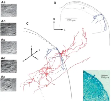
The vertebral column was quickly cut out and immersed in oxygenated artificial cerebrospinal fluid at room temperature. The lumbar spinal cord with unilateral L4 and L5 dorsal roots was dissected, and the pia mater was removed in the region of interest with forceps and scissors, to provide access for the recording pipettes. The spinal cord was glued with cyanoacrylate adhesive to a plate made of gold (the dorsolateral surface was up) and transferred to the recording chamber. All measurements were performed at 22˚C to 24˚C. Lamina I neurons were visualized through the intact white matter using the oblique infrared light-emitting diode illumination technique 37,47 in the region between the dorsolateral funiculus and dorsal root entry zone.32 Lamina I neurons could be clearly distinguished from deeper located lamina II neurons whose somata were densely packed within their layer.45 Lamina I small neurons differed from large cells (Figs. 1Aa-b) described in our previous studies; their apparent largest soma diameters did not exceed 15 µm when viewed with oblique infrared light-emitting diode illumination technique (Figs. 1Ac-e). Post hoc analysis of all successfully labelled neurons confirmed their location in lamina I and small size.
2.3. Recording
Whole-cell recordings were performed from lamina I neurons located in the L4 segment. Artificial cerebrospinal fluid contained (in millimolar) NaCl 115, KCl 3, CaCl2 2, MgCl2 1, NaH2PO4 1, NaHCO3 25, and glucose 11 (bubbled with 95% O2/5% CO2). The pipettes were pulled from thick-walled glass (BioMedical Instruments, Germany) and fire polished (resistance, 4-5 MΩ). The pipette solution contained (in millimolar) KCl 3, K-gluconate 150, MgCl2 1, BAPTA 1, HEPES 10 (pH 7.3 adjusted with KOH, final [K+] was 160 mM), and 1% biocytin. In 8 experiments studying effect of nifedipine on plateau potentials, we used the spinal cord preparations without dorsal roots and these neurons were not included in the major statistics. In 4 of these 8 cases, the pipette solution contained (in millimolar) KCl 3, K-gluconate 150, MgCl2 1, EGTA 1, and HEPES 10 (pH 7.3 adjusted with KOH, final [K1] was 150 mM). The amplifier was an EPC10-Double (HEKA, Lambrecht, Germany). The signal was low-pass filtered at 2.9 kHz and sampled at 10 kHz. Offset potentials were compensated before seal formation. Liquid junction potentials were calculated and corrected for in all experiments using the compensation circuitry of the amplifier. The blockers nifedipine, CNQX, and DAP-5 were from Sigma-Aldrich.
Input resistance (RIN) was measured in current-clamp mode from a hyperpolarization evoked by an injection of a 500-millisecond current pulse of -10 pA or -20 pA. Resting membrane potential (RMP) was measured with a balanced amplifier input.41 Intrinsic firing patterns were classified as tonic, adapting, delayed, and rhythmic, according to descriptions given for the superficial dorsal horn neurons.18,24,26,33,34 Tonic neurons were able to support regular discharge of action potentials during depolarization evoked by 500-millisecond current pulse injections. Adapting neurons fired several spikes that were confined to the beginning of depolarization. Delayed-firing neurons exhibited a considerable time delay before the first spikes appeared. Rhythmic neurons continuously discharged spikes at zero current injection 18, 24; RMP could therefore not be determined for neurons from this group. Plateau-generating neurons responded to a depolarizing current pulse injection with tonic or
adapting firing followed, after the pulse termination, by a prolonged depolarization with or without spike discharges.
Primary afferent inputs were evoked by stimulating 2 ipsilateral roots, L4 and L5, through suction electrodes as described by Pinto et al 31,32 using an isolated pulse stimulator (2100; A-M Systems). The root lengths were 6 to 8 mm. Each suction electrode had its own reference electrode, and there was no cross-stimulation between the roots. A 50-µs pulse of increasing amplitude was applied to recruit Aß/Aδ fibers and a 1-millisecond pulse to activate all Aß/Aδ and C fibers. Monosynaptic excitatory postsynaptic currents (EPSCs) were identified on the basis of low failure rates and small latency variations as described previously. 23,32 The afferent conduction velocity (CV) was calculated by dividing the conduction distance by the conduction time. The former included the length of the root from the opening of the suction electrode to the dorsal root entry zone and the estimated pathway within the spinal cord. The spinal pathway was measured from video images and calculated as the sum of the rostrocaudal and mediolateral distances between the cell body and corresponding dorsal root entry zone. The conduction time was calculated for a monosynaptic EPSC from its latency with a 1-millisecond allowance for synaptic transmission. In the case of a disynaptic inhibition (through an intercalated neuron), allowance was made for 2 serial synaptic transmissions, from the primary afferent to the intercalated neuron (1 millisecond) and from the intercalated neuron to the recorded neuron (1 millisecond), and for the spike initiation in the intercalated neuron (2 milliseconds).24 Afferent CVs for young animals were determined by recording at 22˚C to 24˚C compound action potential currents30 in L3-L5 dorsal roots (lengths, 5-8 mm) isolated from P15-P17 rats. The transition between the Aß and Aδ compound action potential current components corresponded to CV of 2.76±0.15 m/s (n=9). The slowest Aδ afferents had CVs of 0.74±0.14 m/s (n=7), whereas CV for the fastest C fibers was 0.41±0.03 m/s (n= 9). Based on these measurements, the following criteria were developed for identification of fibers mediating monosynaptic EPSCs in lamina I neurons. Aß-fiber– mediated EPSCs were evoked by 50 µs root stimulations and the afferent CV was higher than 2.8 m/s. Aδ-fiber EPSCs were evoked by 50-µs stimulations, and the afferent CV was between 2.8 m/s and 0.7 m/s. C- fiber EPSCs were evoked by 1-millisecond stimulations and the afferent CV was lower than 0.43 m/s. In some cases, EPSCs were mediated by fast afferents (CV>0.7 m/s); these required 1-millisecond stimulation and were classified as high-threshold Aδ (HT-Aδ) fibers. Fibers with CVs between 0.43 m/s and 0.7 m/s were considered as Aδ or C depending on the stimulation (50 µs or 1 millisecond, respectively) required to evoke an EPSC. In current-clamp mode, inputs (both monosynaptic and polysynaptic) were considered as suprathreshold if they evoked spike firing in 6 of 10 consecutive stimulations.
2.4. Histological processing and cell reconstruction
After fixation in 4% paraformaldehyde, the spinal cord was embedded in agar, and parasagittal serial sections of 100-µm thickness were prepared with a tissue slicer (VT 1000S; Leica, Wetzler, Germany). To reveal biocytin, the sections were permeabilized with 50% ethanol and treated according to the avidin-biotinylated horseradish peroxidase method (ExtrAvidin-Peroxidase, diluted 1:1000) followed by a diaminobenzidine chromogen reaction. Sections were counterstained with toluidine blue and mounted in DPX (Fluka; Sigma-Aldrich, St. Louis, MO). Dendrites and axons were identified as in our
Version: Postprint (identical content as published paper) This is a self-archived document from i3S – Instituto de Investigação e Inovação em Saúde in the University of Porto Open Repository For Open Access to more of our publications, please visit http://repositorio-aberto.up.pt/
A
0
1
/0
0
previous studies 45, 46; dendrites showed gradual tapering and occasional spines, whereas axons had even diameter and showed numerous branches and varicosities. The 2D and 3D reconstructions of neurons in Figures 1 and 4 were performed as described in Szucs et al.46 Contours of the preparation were created by drawing white and grey matter borders in the optical section containing the soma of the reconstructed neuron. Laminae I and II were plotted as 20-µm and 80-µm thick bands, respectively, on the dorsal edge of the grey matter. This lamination was based on analysis of the cell body distributions in the toluidine blue stained cross sections of the lateral dorsal horn (Fig. 1, inset), region where cells from our sample were located.
Unless otherwise stated, all numbers are given as mean ±SEM. The parameters were compared using a paired Student t test.
Figure 1. Imaging and reconstruction of lamina I small neurons. Living cell images comparing large (Aa and b) and
small (Ac-e) lamina I neurons. Small neurons chosen for recordings had the apparent largest cell body diameters not exceeding 15 µm. (Ab–d) show fusiform-like somata of large and small neurons, respectively. Horizontal axis in the photographs corresponds to the mediolateral spinal cord axis. The exact classification of a neuron was done after the post hoc analysis of biocytin labelled somatodendritic domain. (B and C) 3D reconstruction of a representative neuron of the fusiform type (cell 29, Table 1). (B) Transverse view of the dendritic tree of the neuron (blue) in the spinal cord cross section. Note that 2 dendrites enter lamina III. (C), Perspective view of the axon (red) and the somatodendritic domain (blue) of the same neuron. Dendrites showed gradual tapering and occasional spines, whereas the axon had even diameter, showed numerous branches, and terminated in laminae I-III. The axon of this local circuit neuron also entered the dorsolateral funiculus where it gave origin to rostrally and caudally directed branches resembling propriospinal collaterals. D, dorsal; L, lateral; R, rostral; C, caudal. Inset, Cross section of the lateral part of the dorsal horn (L4 segment; P18) stained with toluidine blue used to establish the boundaries of lamina II (LII) in our preparation.
3. RESULTS
Neurons with a small body size (apparent largest diameter ≤15 µm) were visually identified on the dorsal surface of the spinal grey matter (Figs. 1Ac-e) for the whole-cell recording and filing with biocytin. Recorded neurons (n= 47) were subjected to detailed analysis, and their physiological and morphological properties are summarized in Table 1. Cell bodies were recovered in 23 cases and all were proven to be located in lamina I. Of these 23 neurons, 12 showed successful labelling of their both dendritic and axonal trees, 5 of the dendritic trees, and 2 of the axons. Thus, somatodendritic organization could be analysed in 17 neurons, and most (n=10) had fusiform morphology with a mediolateral orientation of the dendritic tree (Figs. 1B and C), 4 neurons were flattened, and 3 were multipolar.20 Multipolar neurons differed from flattened neurons by the presence of at least one ventrally protruding dendrite. The cell body areas measured in parasagittal sections (212.2±13.5 µm2,n=17) were in the range given for the population of lamina I small neurons.1 The axons recovered (n=14) had diverse structures but all possessed numerous local branches, typical for interneurons, that terminated in laminae I-III.46 The axon extended mostly in the rostrocaudal direction, although over shorter distances than in the case of lamina I large local circuit neurons.46 As a typical example of the cells studied here, a small local circuit neuron with a fusiform somatodendritic morphology and rostrocaudally oriented local axon, reconstructed in 3D, is shown in Figure 1C. In this case and in some other neurons, the axon entered the dorsolateral funiculus where it gave origin to rostrally and caudally oriented branches resembling propriospinal collaterals.
Physiological properties and morphological features of lamina I small neurons. Firing Pattern
RMP RIN
Cell Primary afferent inputs Anatomical features Monosynaptic excitatory input Disynaptic inhibitory input Somatodendritic type
(orientation of dendrites) Axon distribution (laminae) L4 root L5 root L4 root L5 root
Tonic
-68.7 ± 1.2 mV 1.75 ± 0.20 GΩ
13 C HT-Aδ C C - -
14 C HT-Aδ, C C C - -
15 - - - - flattened rostrocaudal (I-III) with few ventral branches
16 - Aδ Aδ, C Aδ, C fusiform (rostrocaudal) rostrocaudal
17 - Aδ, C - - fusiform (mediolateral) -
27 C - - - fusiform rostrocaudal, rostral branch (I-III)
30 Aδ, C += Aβ, Aδ, C += - - only soma recovered -
31 Aβ, C += - = - Aδ multipolar rostrocaudal (I-III), rostral and caudal collaterals in DLF
35 Aδ, C Aδ, HT-Aδ, C - - - -
36 - C Aδ C - -
37 Aδ, C + C += Aδ, C - - -
39 C C Aβ Aδ multipolar -
42 Aδ, HT-Aδ, C = Aδ, C += Aδ Aδ, C only soma recovered -
44 Aδ, C Aβ, Aδ, HT-Aδ, C += Aβ, Aδ Aβ - -
45 C Aδ, C Aδ Aδ - -
47 Aδ, HT-Aδ, C Aδ, C C C - -
Adapting
-66.0 ± 5.5 mV 1.04 ± 0.27 GΩ
7 C C = - - fusiform (mediolateral) rostrocaudal (I-III), collaterals in DLF
11 - HT-Aδ, C - - - -
12 - - - -
18 C C - - flattened -
21 - Aδ, C Aδ Aδ flattened rostrocaudal (I-III)
29 Aβ, C Aδ, C - - fusiform (mediolateral) rostrocaudal (I-III), rostral and caudal collaterals in DLF
34 C C - - fusiform (mediolateral) rostral, caudal and ventral branches
Delayed
-69.0 ± 2.5 mV 1.38 ± 0.25 GΩ
3 - - - -
6 Aδ, C Aβ, Aδ, C - - only soma and axon recovered rostrocaudal (I-III)
38 Aβ, Aδ, C Aδ, C - - - -
40 Aδ Aδ - - - -
41 Aδ + Aδ, C += C - flattened rostrocaudal (I-III)
46 C Aδ, C C C - -
Rhythmic
1.94 ± 0.32 GΩ
5 C C - C - -
20 - - - - only soma recovered -
23 Aδ, C - Aδ, C Aδ, C - -
25 C Aδ Aδ, C Aδ, C - -
28 - C Aδ C only soma and axon recovered rostrocaudal (I-III)
43 - - C - fusiform (mediolateral) - Plateau -62.5 ± 4.9 mV 1.33 ± 0.08 GΩ 1 - C - - - - 2# - - - -
8 Aδ, C = Aβ, Aδ, C += - - - -
9# - - Aδ Aδ - -
19# - C Aδ Aδ multipolar rostrocaudal (I-III)
22 - - Aδ Aδ - -
24 Aδ Aδ - Aδ - -
26 C += Aδ, HT-Aδ, C += - - fusiform (mediolateral) -
32 Aδ, HT-Aδ += - - - fusiform (mediolateral) mediolateral
33# Aδ - Aδ, C - only soma recovered -
u. c. 4 - - - -
10 C HT-Aδ, C = - - fusiform (mediolateral) mediolateral and ventral
The neurons were grouped according to their intrinsic firing patterns. Each cell is specified by its serial code number. Monosynaptic excitatory inputs: all types of fibers mediating direct inputs to a given neuron are indicated. It should be noted that polysynaptic excitatory inputs, some of which were suprathreshold, are not included in this Table. Note that because of C-fiber-driven inhibition, in some cells (37 and 41), the multiple discharges were evoked by root (L4) stimulation at Aδ but not C-fiber intensities. Disynaptic inhibitory inputs; the afferents mediating identified disynaptic inhibitory inputs are indicated (inputs which were not considered as disynaptic are not included).
* Root stimulation at Aδ-fiber intensity evoked multiple spike discharges. † Root stimulation at C-fiber intensities evoked multiple spike discharges.
‡ Indicates cells that showed firing adaptation during the depolarizing current injection (Fig. 2Fb), in the group of plateau-generating neurons. DLF, dorsolateral funiculus; HT-Aδ, high-threshold Aδ; RMP, resting membrane potential; RIN, input resistance.
3.1. Intrinsic firing properties
Five different patterns of intrinsic firing were observed in small local circuit neurons (Fig. 2A). Tonic neurons (n=16, Fig. 2B) had RMP of 268.7±1.2 mV and RIN of 1.75±0.20 GΩ. Six of these neurons were successfully labelled with biocytin showing diverse somatodendritic morphologies: fusiform (n=3), multipolar (n=2), and flattened (n=1). Adapting neurons (n=7, Fig. 2C, RMP= -66.0±5.5 mV, RIN=1.04±0.27 GΩ) showed fusiform (n=3) and flattened (n=2) morphologies. Delayed firing neurons (n=6, Fig. 2D) had RMP of -69.0±2.5 mV and RIN of 1.38±0.25 GΩ; 1 labelled neuron was flattened. We also recorded from 6 rhythmic neurons (Fig. 2E). They were the only type of small neurons showing ongoing intrinsic discharge, which was also preserved in the presence of the blockers of the fast transmitter receptors as previously described for other types of lamina I neurons.18,24 Small cells in our sample showed rhythmic firing at frequencies of 5 to 10 Hz at zero current injection. Rhythmic firing could be stopped by injecting persistent hyperpolarizing current; RIN measured under these conditions was 1.94±0.32 GΩ. One neuron from the rhythmic group had a fusiform appearance. A subpopulation of neurons (n=10) generated a plateau potential, a prolonged membrane depolarization observed after the termination of the depolarizing current pulse injection (Figs. 2Fa
and b). Its duration varied from cell to cell ranging from 0.25 to 10 seconds. Plateau potentials were
observed in neurons discharging until the end of the depolarizing current injection (Fig. 2Fa) and in those showing firing adaptation (Fig. 2Fb). Plateau potentials could evoke ongoing firing (Fig. 2Fa) or keep membrane depolarized without discharges (Fig. 2Fb). Small depolarizing currents (+10 pA to +30 pA) were sufficient to evoke plateau potentials, whereas membrane responses to hyperpolarizing current injections (-10 pA and -20 pA) exhibited pronounced inward rectification (Figs. 2Fa and b). The RMP in this group of neurons was -62.5±4.9 mV and RIN was 1.33±0.08 GΩ. Single spikes evoked by a short current pulse were typically followed by an after-depolarization (Fig. 2Fc). The amplitude and duration of the plateau potentials increased with duration of the depolarizing pulse (Fig. 2Fd). Three plateau-generating neurons were successfully recovered showing fusiform (n=2) and multipolar (n=1) morphologies.
Plateau potentials described previously for deep dorsal horn neurons were suppressed by the L-type Ca2+ channel blocker nifedipine.28,35 We studied the effect of nifedipine on plateau-generating neurons (n=8) in the spinal cord preparations without dorsal roots; these neurons were not labelled with biocytin and were not included in the major statistics. In voltage-clamp mode, plateau- generating neurons showed inward rectification (Fig. 2Fe). The plateau potentials were completely blocked by 10-µM nifedipine in 7 cases (Fig. 2Ff). In the train of spikes generated during the current injection, the interspike potentials became more negative in the presence of nifedipine (Fig. 2Ff,
asterisk). In agreement with this, nifedipine blocked the after-depolarization of single spikes evoked
by a short pulse (n=3, Fig. 2Fg). It could be therefore concluded that the L-type Ca2+ channels were responsible for the generation of the plateau potentials in lamina I neurons.
Thus, lamina I small neurons demonstrated diverse morphologies and some unique intrinsic firing properties; however, no clear correlation between these parameters could be established. For example, fusiform cells showed tonic, adapting, rhythmic, or plateau-generating patterns. Multipolar
Version: Postprint (identical content as published paper) This is a self-archived document from i3S – Instituto de Investigação e Inovação em Saúde in the University of Porto Open Repository For Open Access to more of our publications, please visit http://repositorio-aberto.up.pt/
A
0
1
/0
0
neurons could be tonic firers or plateau potential generators. In a similar way, flattened neurons exhibited tonic, adapting, and delayed patterns.
3.2. Monosynaptic primary afferent inputs
Primary afferent inputs from the L4 and L5 dorsal roots were recorded in 47 neurons located in the spinal segment L4. Segmental (L4) and the adjacent caudal (L5) roots were chosen as those with the strongest inputs to the L4 lamina I neurons.32 Based on their CVs and activation thresholds, 4 types of afferents projecting monosynaptically to small local circuit neurons were identified (Fig. 3). Aß-fiber–mediated EPSCs were activated by a 50-µs pulse showing low threshold of 10 to 70 µA (mean, 32±8 µA; 6 roots) and shortest latency (Fig. 3A). The Aß inputs were observed in 7 neurons (Table 1, identified: multipolar, n= 1; fusiform, n=1) and originated from either L4 (n= 3) or L5 (n= 4) roots (Fig.
3E). Aδ-fiber EPSCs had threshold of 10 to 80 µA (mean, 34±3 µA; 32 roots) and longer latency (Fig. 3B) and were recorded in larger number of neurons (n= 23; Table 1, identified: fusiform, n= 5;
flattened, n= 2; and multipolar, n= 1). In most cases, a neuron received monosynaptic Aδ inputs from both roots (Fig. 3E). C-fiber–mediated EPSCs required a 1-millisecond pulse stimulation of the root (mean threshold, 29 ±2 µA; 44 roots), showed longest latencies (Fig. 3C) and represented the most frequent type of input to the small local circuit neurons (n= 33, Table 1, identified: fusiform, n= 7; flattened, n= 3; multipolar, n=3) from both roots (Fig. 3E). In addition, we recorded monosynaptic EPSCs mediated by the afferents with the Aδ range CVs but requiring a 1-millisecond pulse stimulation (mean threshold, 31±5 µA; 9 roots), HT-Aδ inputs (Fig. 3D). These arose from either L4 or L5 roots (Fig. 3E) and were observed in 10 neurons (Table 1, identified: fusiform, n=3).
In the overall population of the neurons tested (n= 47), 38 (80.9%) received monosynaptic inputs from primary afferents (Fig. 3F). Among them, 14 neurons showed monosynaptic inputs from only one specific type of fibers, either C (n=10, 21.3%) or Aδ (n= 4, 8.5%), and 24 (51.1%) were integrating complex inputs from several types of afferents (Fig. 3F).
Version: Postprint (identical content as published paper) This is a self-archived document from i3S – Instituto de Investigação e Inovação em Saúde in the University of Porto Open Repository For Open Access to more of our publications, please visit http://repositorio-aberto.up.pt/
A
0
1
/0
0
Figure 2. Intrinsic firing properties of small neurons. (A) Histogram showing the numbers of neurons with 5
different patterns of intrinsic firing. Two cells could not be classified (u.c., unclassified). (B) A tonic neuron. Here and in the following parts, the stimulation protocol is shown below the current-clamp recordings and the dashed line indicates 0 mV. Injected current pulses, -10, +10, +30, and +50 pA. (C) An adapting neuron. Injected current pulses, -10, +10, +20, and +60 pA. (D) A delayed-firing neuron. Injected currents, -10, +10, +50, +70, and +90 pA. (E) A rhythmic neuron; intrinsic firing at zero current injection. (Fa–g) Recordings from plateau-generating neurons. (Fa) A neuron showing spike discharges until the end of the depolarizing current injection and the plateau potential supporting ongoing firing. Note the pronounced inward rectification seen at hyperpolarizing current injections. Injected currents, -20, -10, and +10 pA. (Fb) A neuron showing firing adaptation during the depolarizing current injection and a plateau potential without spike discharges. Injected currents, -20, -10, and +10 pA. (Fc) Single spike evoked in a plateau-generating neuron by a short current pulse (10 milliseconds, +80 pA) is followed by a pronounced after-depolarization (ADP). (Fd) The amplitude and duration of the plateau potential were increased with duration of the depolarizing pulse (50, 150, 200, and 300 milliseconds; +40 pA). (Fe) Voltage-clamp recordings of subthreshold membrane currents in a plateau-generating neuron. Holding potential, -70 mV. Voltage pulses from -120 to -60 mV with a 10-mV increment. Dashed line, 0 pA. (Ff) The plateau potential was completely blocked by 10 µM nifedipine. Both traces were evoked by a +50-pA current pulse. Note more negative interspike potentials (*) seen in the presence of nifedipine. (Fg) Nifedipine blocks ADP after a single spike evoked by a short current pulse (10 milliseconds, +100 pA) in a plateau-generating neuron.
Figure 3. Monosynaptic primary afferent inputs. Recordings of monosynaptic excitatory postsynaptic currents
(EPSCs) (indicated by arrowheads) mediated by Aß (A), Aδ (B), C (C), and high-threshold Aδ (HT-Aδ) (D) afferents. Holding potential, -70 mV. Both 50-µs and 1-millisecond stimulating pulses had amplitude of 100 µA. Note that HT-Aδ-fiber EPSCs are not activated by a 50-µs pulse. (E) Histogram showing the number of neurons receiving each type of input from the L4 and L5 dorsal roots. (F) Histogram describing total monosynaptic primary afferent inputs to the neurons from both roots.
3.3. Aß-fiber–driven EPSPs
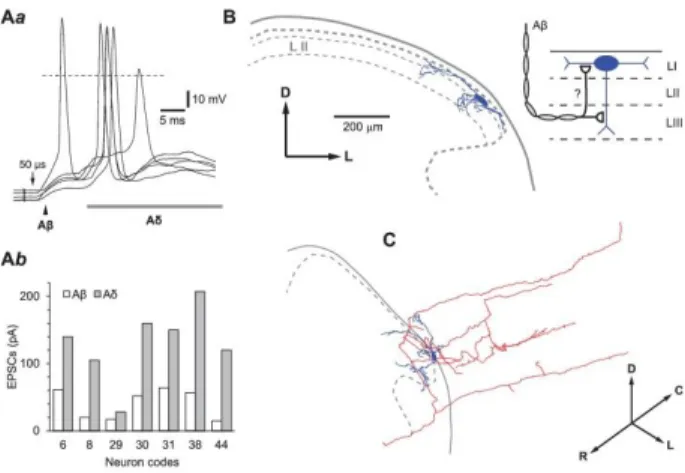
All neurons with monosynaptic Aß inputs (n= 7) also received direct inputs from other afferents, Aδ (n= 6), C (n= 7), and HT-Aδ (n= 1) (Fig. 3F). The Aß excitatory postsynaptic potentials (EPSPs) alone were subthreshold in all cases; however, they induced depolarization, which facilitated spike initiation by the following monosynaptic and polysynaptic Aδ-fiber–driven EPSPs (Fig. 4Aa). In agreement with this, the amplitudes of the Aß EPSCs (40.9±8.4 pA, n=7) were significantly smaller
than those of the Aδ EPSCs (130.0±21.0 pA, n= 7, P< 0.01, Fig. 4Ab). Two neurons with monosynaptic Aß-fiber inputs were labelled with biocytin to a degree allowing analysis of their somatodendritic organization (Figs. 1B and C, and Figs. 4B and C). Both these cells with somata located in lamina I had ventrally protruding dendrites reaching lamina III, the region of the Aß afferent fiber terminations.
3.4. Aδ-fiber inputs
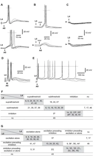
From a functional point of view, Aδ-fiber–mediated inputs to lamina I small neurons showed large diversity. Fifteen neurons received suprathreshold Aδ inputs (monosynaptic and/or polysynaptic) either from both roots (n= 8, Figs. 5A and F) or from one of the roots (n=7, Figs. 5B and F). In other 8 neurons, stimulation of both roots evoked overall inhibitory responses (Figs. 5C and F).
In 9 neurons, from the group with suprathreshold inputs, stimulation of Aδ afferents from the L4 (n=6) and/or L5 (n=7) roots could evoke multiple spikes, which appeared as short bursts of 2 to 3 spikes (Fig. 5D), after-discharges during hundreds of milliseconds (not shown), or as a combination of both short bursts and after-discharges (Fig. 5E). According to the intrinsic firing properties, 5 of these neurons were tonic and 3 belonged to a category of plateau-generating neurons. Therefore, both intrinsic firing properties and synaptic inputs contributed to the evoked multiple spike discharges. Anatomically, this group of cells included neurons with fusiform (n=2), multipolar (n=1), and flattened (n=1) somatodendritic morphologies.
It should be noted that in 5 neurons, the Aδ-fiber inputs (root L4, n=4; root L5, n=2) evoked a time-locked firing with a variation of the spike initiation time of less than 2 milliseconds (not shown). Such neurons with a reliable and time-locked firing can serve, for example, as intercalated neurons mediating an Aδ- fiber–driven disynaptic inhibition (Fig. 8).
We also studied whether a given lamina I neuron receives similar Aδ-fiber inputs from both roots. Two parameters were analysed; the overall efficacy of the monosynaptic and poly-synaptic inputs and the relative latency of appearance of excitatory and inhibitory components. The data are summarised in Figures 5F and G where each neuron is indicated by its serial code number.
According to their efficacy, inputs were classified as supra-threshold, subthreshold, and inhibitory (Fig. 5F). Regardless of the type of inputs, those arising from the L4 and L5 roots were similar in most neurons (22 of 34, 64.7%; Fig. 5F, grey diagonal). In 7 other neurons (of 34, 20.6%) with suprathreshold input from one root and subthreshold from the other one (Fig. 5F), the postsynaptic responses had similar time course (Fig. 5B).
Based on the relative latency of appearance of excitatory and inhibitory inputs, 3 types of responses were identified; excitation alone (Figs. 5A and 3B), excitation preceding inhibition (not shown), and inhibition either preceding excitation (Fig. 5B), or alone (Fig. 5C). Regardless of the type of response, most neurons (20 of 34, 58.8%) again showed similar inputs from both roots (Fig. 5G, grey diagonal). Noteworthy, most inhibitory inputs preceding excitation were found to be disynaptic (indicated by asterisks, Fig. 5G).
Thus, Aδ inputs to lamina I small neurons showed large diversity; however, in most cases, responses evoked in a given neuron by stimulating the L4 and L5 roots were similar.
Version: Postprint (identical content as published paper) This is a self-archived document from i3S – Instituto de Investigação e Inovação em Saúde in the University of Porto Open Repository For Open Access to more of our publications, please visit http://repositorio-aberto.up.pt/
A
0
1
/0
0
Figure 4. Aß-fiber–driven excitatory postsynaptic potentials (EPSPs). (Aa), Monosynaptic Aß-fiber–mediated
EPSPs (indicated by an arrowhead) followed by a polysynaptic Aδ EPSPs recorded in a lamina I small local circuit neuron after stimulation of the L4 dorsal root. Note that the Aß EPSP is subthreshold in 4 of 5 consecutive stimulations, but its depolarization facilitated spike initiation by the following Aδ-fiber–driven EPSPs. (Ab), The histogram comparing amplitudes of Aß- and Aδ-fiber–driven excitatory postsynaptic currents (EPSCs) in 7 neurons with monosynaptic Aß inputs. The Aδ-fiber EPSCs were both monosynaptic and polysynaptic. Anatomical 3D reconstruction of the neuron from (Aa) is shown in (B) and (C) (cell 31, Table 1). (B), Transverse view of the somatodendritic organization (blue). Note the ventrally protruding branch of dendrite reaching lamina III. (C), Perspective 3D view of the soma, dendrites (blue), and the axon (red) of the same neuron. The axon entered the dorsolateral funiculus giving origin to rostrally and caudally running propriospinal branches. Schematic drawing, A putative organization of the monosynaptic Aß-fiber inputs to a lamina I neuron (see Discussion). D, dorsal; L, lateral; R, rostral; C, caudal.
3.5. C and HT-Aδ fiber–mediated EPSPs
Monosynaptic responses to stimulation of C afferents were recorded in a total of 33 neurons (root L4, n=25; root L5, n=28). C fiber–driven monosynaptic and/or polysynaptic EPSPs evoked spike firing in 24 neurons (Fig. 6A; L4 root, n=18; L5 root, n=21). In 9 of them, the monosynaptic C-fiber EPSP component evoked a time-locked firing with a spike initiation time variation below 2 milliseconds (Fig. 6B; L4, n=5; L5, n=5); such neurons, provided that they are inhibitory, could mediate a C fiber– driven disynaptic inhibition (Fig. 8).
In 11 neurons, root stimulation at C-fiber intensity (L4, n=6; L5, n=9) evoked long-lasting EPSPs and ongoing after-discharges for hundreds of milliseconds (Fig. 6C); 5 of these cells were tonic and 3 generated intrinsic plateau potentials. Six biocytin labelled cells from this group showed fusiform (n=4), multipolar (n=1), and flattened (n=1) somatodendritic morphologies.
Monosynaptic HT-Aδ fiber EPSCs were observed in 10 cells (root L4, n=3; root L5, n=7) together with inputs mediated through other types of afferents. In 9 of them, HT-Aδ EPSP component elicited spike firing (Fig. 6D). Most cells with HT-Aδ inputs were tonic neurons (n=6), 2 generated plateau potentials and 1 showed adapting pattern (1 cell could not be classified).
3.6. Suppression of primary afferent inputs
We tested action of specific blockers of glutamate AMPA receptors (CNQX, 10 µM) and N-methyl-D-aspartate receptors (DAP-5, 50 µM) on primary-afferent–driven monosynaptic and polysynaptic responses evoked in lamina I small neurons by stimulating dorsal roots (n=19). EPSCs were completely suppressed by CNQX in 13 cases (not shown), whereas in 6 cases, a small EPSC component remained and was not affected by an addition of DAP-5 (Fig. 7, upper traces). Afferent-driven polysynaptic inhibitory postsynaptic currents (IPSCs) were incompletely blocked by CNQX in 5 cases, but were suppressed by adding DAP-5 (Fig. 7, lower traces). Thus, it could be concluded that, although transmission from primary afferents is mostly mediated through AMPA receptors, a minor contribution of N-methyl-D-aspartate and/or other receptors cannot be excluded.
3.7. The role of disynaptic inhibition
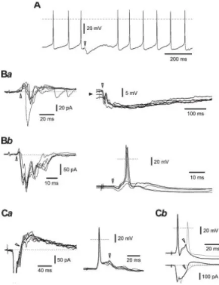
We have recently described a low-threshold afferent–driven disynaptic inhibition (through 1 intercalated neuron) of lamina I projection neurons and large local circuit neurons.24 Its characteristic features were a low failure rate and a small latency variation. Here, we observed several types of Aß-, Aδ-Aß-, and C-afferent–driven disynaptic inhibition of small local circuit neurons (Fig. 8). In rhythmic neurons (n=5), the Aδ- and C- afferent–mediated inhibition (root L4, n=4; root L5, n=4) could cause an interruption of firing for several hundreds of milliseconds (Fig. 8A). In another group of neurons (n 5 10), stimulation of dorsal roots (L4, n=9; L5, n=7) evoked responses where inhibitory postsynaptic potentials temporary preceded EPSPs and either prevented spike firing (Fig. 8Ba) or reduced firing probability (Fig. 8Bb). In most cases (neurons, n=12; root L4, n=9; root L5, n=10), however, the disynaptic inhibition followed monosynaptic excitatory inputs (Figs. 8Ca and b); such inhibitory postsynaptic potentials reduced a postspike membrane de- polarization and, in neurons with a strong afferent input, could prevent initiation of the second spike or after-discharges.
Version: Postprint (identical content as published paper) This is a self-archived document from i3S – Instituto de Investigação e Inovação em Saúde in the University of Porto Open Repository For Open Access to more of our publications, please visit http://repositorio-aberto.up.pt/
A
0
1
/0
0
Figure 5. Aδ-fiber inputs. (A) A neuron receiving suprathreshold Aδ inputs from both L4 and L5 roots.
Monosynaptic excitatory postsynaptic potentials (EPSPs) are indicated by arrowheads. Five consecutive traces. (B) A neuron with a suprathreshold input from the L4 root and a subthreshold input from the L5 root. Note that EPSPs are preceded by inhibitory postsynaptic potentials. Five consecutive traces. (C), A neuron with inhibitory inputs from both roots. (D) A neuron generating a short burst of 2 spikes on a stimulation of Aδ fibers in the L4 root. Five consecutive traces. (E) Long-lasting Aδ-fiber–driven EPSPs evoking short bursts of 2 spikes followed by an ongoing discharge during hundreds of milliseconds. Stimulated root, L4. (F) Comparison of the overall efficacy of Aδ inputs, both monosynaptic and polysynaptic, to lamina I neurons (n=34) from the L4 and L5 roots. Each neuron is indicated by its serial code number. Rhythmic neuron. Neurons on the diagonal (grey) have similar inputs from both roots. No, no input from the root. (G) Comparison of Aδ inputs from the L4 and L5 roots based on the latency of excitatory and inhibitory components. Each neuron is indicated by its serial code number. For the neurons receiving inhibitory inputs: * and *, disynaptic inhibition from the L4 and L5 root, respectively. Neurons on the diagonal (grey) have similar inputs from both roots. No, no input from the root.
Figure 6. C-fiber– and high-threshold Aδ (HT-Aδ) fiber–mediated excitatory postsynaptic potentials (EPSPs). (A) Suprathreshold monosynaptic EPSPs mediated by C afferents from the L4 root. Five consecutive traces. (B) A time-locked spike firing evoked by C-fiber–driven EPSPs (root, L4). Five consecutive traces. A variation of the spike initiation time was less than 2 milliseconds (inset). (C) Long-lasting EPSPs and ongoing spike discharges for hundreds of milliseconds evoked by stimulation of C fibers from the L5 root. (D) Monosynaptic HT-Aδ fiber EPSPs (2 components) evoked in lamina I neuron after stimulation of the L5 root (cell 13, Table 1). Five consecutive traces. Monosynaptic components are indicated by solid arrowheads. All responses are evoked by a 1-millisecond pulse stimulation of dorsal roots.
4. DISCUSSION
We did whole-cell recordings from lamina I small local circuit neurons in the isolated spinal cord preparation and analysed their anatomical features and physiological properties. The major findings of this study are that these neurons (1) have different somatodendritic morphologies, with a prevalence of fusiform one, (2) show diverse intrinsic firing properties including generation of rhythmic discharges and plateau potentials, (3) receive strong inputs from thin Aδ and C afferents, which can induce long-lasting membrane depolarizations and after-discharges, (4) can receive direct input from low-threshold Aß afferents, and (5) are under control of afferent-driven disynaptic inhibition. Thus, there are distinct classes of small local circuit neurons, which have their specific properties and play different roles in the processing of nociceptive information within lamina I.
4.1. Morphology
Lamina I neurons are integrated into the superficial dorsal horn network .14,16,19,39,40,48 The neurons studied here have mean cell body area of 212 µm2, which corresponds to a group of lamina I small cells.1 They are clearly distinct from large local circuit neurons we studied previously23,24 whose mean cell body areas measured under similar conditions were 351 µm246 and 434 µm222. Small local circuit neurons had, in most cases, fusiform but also flattened and multipolar, somatodendritic morphologies. The dominance of fusiform shape was not typical for the population of lamina I large
Version: Postprint (identical content as published paper) This is a self-archived document from i3S – Instituto de Investigação e Inovação em Saúde in the University of Porto Open Repository For Open Access to more of our publications, please visit http://repositorio-aberto.up.pt/
A
0
1
/0
0
interneurons, which, in most cases, belonged to multipolar or flattened groups.46 High percentage of fusiform neurons in our sample is in agreement with studies showing preferable location of this type of interneurons in the middle and lateral lamina I,20 the region best accessible for recordings with our experimental technique.32 Small neurons from our sample did not show correlation between their dendritic architecture and intrinsic firing properties. It cannot be excluded however that neurons with a mediolateral orientation of their somata and dendritic trees studied here may have properties different from those of rostrocaudally oriented cells.20
Dendritic trees of most lamina I interneurons are limited to laminae I-II, with an exception of some fusiform (type IB) and multipolar (type IIB) cells whose dendrites protrude ventrally and can reach lamina III.20 This anatomical arrangement may explain why most neurons in our sample received direct inputs from thin Aδ- and C-afferent fibers and some neurons from low-threshold Aß afferents. It is interesting to note that both successfully labelled neurons with Aß inputs had ventral dendrites reaching lamina III, the region of Aß-fiber terminations. It is also possible, however, that some lamina I neurons without deep ventral dendrites may receive direct Aß-fiber inputs, since large calibre afferents, after entering the spinal grey matter and traversing superficial laminae, turn dorsally and arborize within the superficial dorsal horn.44 This arrangement of dorsally recurving “flame”-shaped arbores extending into lamina I is established before birth10 and maintained in adulthood.4
We obtained intracellular labelling of the axons of small local circuit neurons, which branched extensively and terminated in laminae I-III suggesting that these cells can target other neurons in the superficial dorsal horn. It was also shown that lamina I local circuit neurons synapse on anterolateral-tract projection neurons.23 In addition, projection neurons receive Aß- and Aδ- fiber–driven disynaptic inputs,24 which can be conveyed, at least in part, through lamina I small inhibitory neurons with direct afferent inputs. Thus, local circuit neurons may play important role in the signal integration by relaying the Aß-, Aδ-, and C-afferent inputs to projection neurons.
Figure 7. Suppression of glutamatergic transmission. Upper traces, Excitatory postsynaptic currents (EPSCs)
evoked in a lamina I neuron by stimulating L4 dorsal root with a 1-millisecond current pulse in control, 10-µM CNQX, 10-µM CNQX plus 50-µM DAP-5, and after recovery. The CNQX block was incomplete and the degree of block was not increased after addition of DAP-5. Lower traces, Inputs recorded in another lamina I neuron after stimulation of the L5 dorsal root (a 1-millisecond stimulation). Polysynaptic inhibitory postsynaptic currents persisted in the presence of CNQX, but were blocked after addition of DAP-5. Holding potentials were -70 mV. Monosynaptic EPSCs are indicated by filled arrowheads.
Figure 8. Disynaptic afferent driven inhibition. (A) Effect of a disynaptic inhibition on discharge of rhythmic
neuron. The L5 dorsal root was stimulated with a 50-ms current pulse. A disynaptic inhibitory postsynaptic potential (IPSP) (here and in the following parts indicated by an open arrowhead) caused an interruption of intrinsic rhythmic discharge. (Ba) Aδ-fiber–mediated inhibition preceding excitatory inputs and preventing firing. Voltage-clamp: holding potential, 270 mV; stimulated dorsal root, L5 (a 50 ms pulse). Current-clamp: disynaptic IPSPs prevent membrane depolarization by the following excitatory postsynaptic potentials (EPSPs). Filled arrowhead indicates 270 mV. (Bb) High-threshold Aδ (HT-Aδ) fiber–mediated inhibition preceding excitatory C inputs and controlling spike firing. Voltage-clamp: holding potential, 270 mV; stimulated dorsal root, L5 (a 1 millisecond pulse). Current-clamp: disynaptic IPSPs reduce efficacy of the following EPSPs in evoking spike (3 spikes were evoked in 5 consecutive stimulations). (Ca) C-fiber–driven disynaptic inhibition after the monosynaptic excitatory inputs. Voltage-clamp: holding potential, 260 mV; stimulated dorsal root, L4 (a 1 millisecond pulse). Current-clamp: disynaptic IPSPs reduce membrane depolarization after the spike and thus prevent generation of the second spike (5 consecutive traces). (Cb) Current- clamp recording from another neuron where an Aδ-fiber–driven IPSP prevented initiation of the second spike (black trace); the latter is generated when the IPSP failed (grey trace). Below, voltage-clamp (holding potential 270 mV) recordings from the same neuron showing the monosynaptic excitatory postsynaptic current followed by the disynaptic inhibitory post- synaptic current (IPSC) (black trace) and the trace with the IPSC failure (grey trace). Nonconsecutive traces. A 50-ms pulse stimulation of the L5 root.
4.2. Intrinsic firing properties
Intrinsic firing properties determine how synaptic input to a neuron is converted to the action potential discharge pattern.13,21 Tonic, adapting, and delayed firing patterns were previously described in a number of studies for lamina I neurons.15,24,33,34 Intrinsic rhythmic firing, occurring independently of fast synaptic transmission, was previously observed under our experimental conditions in a half of lamina I large local circuit neurons.24 Rhythmic discharges can be transiently interrupted on the primary afferent stimulation activating disynaptic inhibitory pathways (Fig. 8A and Ref. 22). Rhythmic neurons can be both inhibitory and excitatory 18, 46 and may generate spontaneous network activity in the dorsal horn.38 In the neonatal spinal cord, rhythmic network activity in lamina I can be essential for development of spinal sensory circuitries.18 Besides, rhythmic
Version: Postprint (identical content as published paper) This is a self-archived document from i3S – Instituto de Investigação e Inovação em Saúde in the University of Porto Open Repository For Open Access to more of our publications, please visit http://repositorio-aberto.up.pt/
A
0
1
/0
0
transmission mechanisms, metabotropic Ύ-aminobutyric acidB receptors in a large population of dorsal horn neurons. Our results show that a subpopulation of lamina I small neurons generate plateau potentials. This pattern of activity was not seen in our samples of lamina I anterolateral tract projection neurons and large local circuit neurons24 and seems to be unique for the population of small cells. Plateau potentials were previously described for the deep dorsal horn neurons and are considered as an intrinsic component of the synaptically induced windup.6,28,29,35 As in the deep dorsal horn neurons, plateau potentials in lamina I neurons were blocked by nifedipine indicating a major role of the voltage-gated L-type Ca2+ channels (CaV1.2 and CaV1.3 α1 subunits7) in maintaining membrane depolarization after termination of the intracellular current injection. However, in lamina I small neurons, plateau potentials had low threshold and could be evoked by an injection of small (10-30 pA) depolarizing currents. On primary afferent stimulation, plateau-generating neurons could show long-lasting after-discharges. Therefore, in the superficial dorsal horn network, these local circuit neurons may function as sensitive amplifiers of the nociceptive afferent input and be involved in the spinal sensitization to pain, especially in neuropathies related to the L-type Ca2+ channel upregulation in dorsal horn neurons.11
4.3. Primary afferent inputs
We show that small local circuit neurons receive direct inputs from several types of primary afferents, including the low-threshold Aß afferents. This feature may be specific for small neurons as larger cells in our sample did not show direct Aß inputs.24,32 In naive animals, Aß-fiber–mediated EPSPs were insufficient to elicit spike in lamina I neurons, but could facilitate spike initiation by the following Aδ-fiber EPSPs. It is possible however that Aß input to lamina I could be increased under pathological conditions, for example, after network reorganization following neonatal peripheral tissue damage.19 Thus, prewiring of Aß afferents to lamina I small neurons can provide a substrate for functional plasticity increasing the strength of the low-threshold afferent drive to the nociceptive processing network and play a crucial role in development of allodynia.
The Aδ and C afferents intensively supply lamina I neurons.5, 15,22,32,50 We have found that in many neurons, the Aδ inputs from 2 adjacent dorsal roots (L4 and L5) were similar regardless of their overall excitatory or inhibitory nature. This may reflect the fact that these afferents could innervate the same peripheral point and transmit similar information to the central neuron.31 Both Aδ- and C-fiber inputs could evoke long-lasting depolarizations and after-discharges in lamina I neurons as was previously described for deep dorsal horn neurons.17,29,36 Since most of these cells were tonic or plateau-generating neurons, it is likely that an appropriate combination of synaptic inputs and intrinsic firing properties is required for generation of primary- afferent–driven after-discharges.
Some lamina I small neurons received direct inputs from HT-Aδ afferents. These may represent a special class of nociceptors, eg, high-threshold mechanoreceptors.9 We have recently shown that some HT-Aδ afferents are of somatic origin.22 Further elucidation of functions of HT-Aδ fibers, however, will require study of their peripheral terminal receptors.
Low-threshold afferent-driven disynaptic inhibition of lamina I projection neurons has recently been proposed as a postsynaptic mechanism of pain control24 complementary to the classical presynaptic mechanism described in the gate theory of pain.27 In this study, we have further found that small local
circuit neurons in lamina I show several types of disynaptic inhibition, which can be mediated through Aß, Aδ, and C afferents. Depending on its latency and the intrinsic firing properties of the receiving neuron, the disynaptic inhibition can produce different effects including: (1) a short-term interruption of rhythmic firing22 (2) prevention of spike initiation by weak excitatory inputs, (3) regulation of the firing threshold and probability, and (4) prevention of generation of the second spike or after-discharges in some neurons with strong primary afferent inputs. Peripherally driven inhibition was also reported for other types of dorsal horn neurons.13,21,53 Inputs from all these inhibitory pathways are critically important for normal signal processing because their loss, downregulation, or pharmacological block cause hyperalgesia, allodynia, and pain syndromes.2,12,42,52
In conclusion, small local circuit neurons in the spinal lamina I belong to different morphological classes and receive direct excitatory as well as disynaptic inhibitory inputs from Aß-, Aδ-, and C-fiber afferents. Their intrinsic firing properties allow integration and/or amplification of the primary afferent inputs in the superficial dorsal horn. These properties reflect a functional diversity of signal modalities processed by the spinal nociceptive network.
Conflict of interest statement
The authors have no conflicts of interest to declare.
This work is funded by FEDER funds through the Operational Competitiveness Programme-COMPETE and by national funds through FCT—Fundação para a Ciência e a Tecnologia under the project FCOMP-01-0124-FEDER-029623 (PTDC/NEU-SCC/ 0347/2012).
Acknowledgements
The authors thank Leonor Araújo for excellent technical assistance. The support by the János Bolyai Research Scholarship of the Hungarian Academy of Sciences and the Hungarian Brain Research Program (KTIA_NAP_13-2-2014-0005) to P. Szucs is acknowledged.
Article history:
Received 29 July 2015Received in revised form 9 October 2015 Accepted 14 October 2015
References
[1] Al Ghamdi KS, Polgar E, Todd AJ. Soma size distinguishes projection neurons from neurokinin 1 receptor-expressing interneurons in lamina I of the rat lumbar spinal dorsal horn. Neuroscience 2009;164:1794–804. [2] Beyer C, Roberts LA, Komisaruk BR. Hyperalgesia induced by altered glycinergic activity at the spinal cord. Life
Version: Postprint (identical content as published paper) This is a self-archived document from i3S – Instituto de Investigação e Inovação em Saúde in the University of Porto Open Repository For Open Access to more of our publications, please visit http://repositorio-aberto.up.pt/
A
0
1
/0
0
[3] Bice TN, Beal JA. Quantitative and neurogenic analysis of the total population and subpopulations of neurons defined by axon projection in the superficial dorsal horn of the rat lumbar spinal cord. J Comp Neurol
1997;388:550–64.
[4] Boada MD, Woodbury CJ. Myelinated skin sensory neurons project extensively throughout adult mouse substantia gelatinosa. J Neurosci 2008;28:2006–14.
[5] Dahlhaus A, Ruscheweyh R, Sandkuhler J. Synaptic input of rat spinal lamina I projection and unidentified neurones in vitro. J Physiol 2005;566 (pt 2):355–68.
[6] Derjean D, Bertrand S, Le Masson G, Landry M, Morisset V, Nagy F. Dynamic balance of metabotropic inputs causes dorsal horn neurons to switch functional states. Nat Neurosci 2003;6:274–81.
[7] Dobremez E, Bouali-Benazzouz R, Fossat P, Monteils L, Dulluc J, Nagy F, Landry M. Distribution and regulation of L-type calcium channels in deep dorsal horn neurons after sciatic nerve injury in rats. Eur J Neurosci 2005; 21:3321–33.
[8] Duggan AW, Morton CR, Zhao ZQ, Hendry IA. Noxious heating of the skin releases immunoreactive substance P in the substantia gelatinosa of the cat: a study with antibody microprobes. Brain Res 1987;403: 345–9.
[9] Fang X, McMullan S, Lawson SN, Djouhri L. Electrophysiological differences between nociceptive and non-nociceptive dorsal root ganglion neurones in the rat in vivo. J Physiol 2005;565(pt 3):927–43.
[10] Fitzgerald M, Butcher T, Shortland P. Developmental changes in the laminar termination of A fibre cutaneous sensory afferents in the rat spinal cord dorsal horn. J Comp Neurol 1994;348:225–33.
[11] Fossat P, Dobremez E, Bouali-Benazzouz R, Favereaux A, Bertrand SS, Kilk K, Leger C, Cazalets JR, Langel U, Landry M, Nagy F. Knockdown of L calcium channel subtypes: differential effects in neuropathic pain. J Neurosci 2010;30:1073–85.
[12] Foster E, Wildner H, Tudeau L, Haueter S, Ralvenius WT, Jegen M, Johannssen H, Hosli L, Haenraets K, Ghanem A, Conzelmann KK, Bosl M, Zeilhofer HU. Targeted ablation, silencing, and activation establish glycinergic dorsal horn neurons as key components of a spinal gate for pain and itch. Neuron 2015;85:1289–304. [13] Graham BA, Brichta AM, Callister RJ. In vivo responses of mouse superficial dorsal horn neurones to both current injection and peripheral cutaneous stimulation. J Physiol 2004;561(pt 3):749–63.
[14] Graham BA, Brichta AM, Callister RJ. Moving from an averaged to specific view of spinal cord pain processing circuits. J Neurophysiol 2007; 98:1057–63.
[15] Grudt TJ, Perl ER. Correlations between neuronal morphology and electrophysiological features in the rodent superficial dorsal horn. J Physiol 2002;540(pt 1):189–207.
[16] Kato G, Kawasaki Y, Koga K, Uta D, Kosugi M, Yasaka T, Yoshimura M, Ji RR, Strassman AM. Organization of intralaminar and translaminar neuronal connectivity in the superficial spinal dorsal horn. J Neurosci 2009;29:5088– 99.
[17] King AE, Thompson SW, Urban L, Woolf CJ. The responses recorded in vitro of deep dorsal horn neurons to direct and orthodromic stimulation in the young rat spinal cord. Neuroscience 1988;27:231–42.
[18] Li J, Baccei ML. Pacemaker neurons within newborn spinal pain circuits. J Neurosci 2011;31:9010–22. [19] Li J, Kritzer E, Craig PE, Baccei ML. Aberrant synaptic integration in adult lamina I projection neurons following neonatal tissue damage. J Neurosci 2015;35:2438–51.
[20] Lima D, Coimbra A. A Golgi study of the neuronal population of the marginal zone (lamina I) of the rat spinal cord. J Comp Neurol 1986;244:53–71.
[21] Lopez-Garcia JA, King AE. Membrane properties of physiologically classified rat dorsal horn neurons in vitro: correlation with cutaneous sensory afferent input. Eur J Neurosci 1994;6:998–1007.
[22] Luz LL, Fernandes EC, Sivado M, Kokai E, Szucs P, Safronov BV. Monosynaptic convergence of somatic and visceral C fiber afferents on projection and local-circuit neurons in lamina I: a substrate for referred pain. PAIN 2015;56:2042–51.
[23] Luz LL, Szucs P, Pinho R, Safronov BV. Monosynaptic excitatory inputs to spinal lamina I anterolateral-tract-projecting neurons from neighbouring lamina I neurons. J Physiol 2010;588(pt 22):4489–505.
[24] Luz LL, Szucs P, Safronov BV. Peripherally driven low-threshold inhibitory inputs to lamina I local-circuit and projection neurones: a new circuit for gating pain responses. J Physiol 2014;592(pt 7):1519–34.
[25] Mantyh PW, Allen CJ, Ghilardi JR, Rogers SD, Mantyh CR, Liu H, Basbaum AI, Vigna SR, Maggio JE. Rapid endocytosis of a G protein- coupled receptor: substance P evoked internalization of its receptor in the rat striatum in vivo. Proc Natl Acad Sci U S A 1995;92:2622–6.
[26] Melnick IV, Santos SFA, Szokol K, Szucs P, Safronov BV. Ionic basis of tonic firing in spinal substantia gelatinosa neurons of rat. J Neurophysiol 2004;91:646–55.
[27] Melzack R, Wall PD. Pain mechanisms: a new theory. Science 1965;150: 971–9.
[28] Morisset V, Nagy F. Ionic basis for plateau potentials in deep dorsal horn neurons of the rat spinal cord. J Neurosci 1999;19:7309–16.
[29] Morisset V, Nagy F. Plateau potential-dependent windup of the response to primary afferent stimuli in rat dorsal horn neurons. Eur J Neurosci 2000; 12:3087–95.
[30] Pinto V, Derkach VA, Safronov BV. Role of TTX-sensitive and TTX- resistant sodium channels in Adelta- and C-fiber conduction and synaptic transmission. J Neurophysiol 2008;99:617–28.
[31] Pinto V, Szucs P, Derkach VA, Safronov BV. Monosynaptic convergence of C- and Adelta-afferent fibres from different segmental dorsal roots on to single substantia gelatinosa neurones in the rat spinal cord. J Physiol 2008b;586(pt 17):4165–77.
[32] Pinto V, Szucs P, Lima D, Safronov BV. Multisegmental A{delta}- and C-fiber input to neurons in lamina I and the lateral spinal nucleus. J Neurosci 2010;30:2384–95.
[33] Prescott SA, De Koninck Y. Four cell types with distinctive membrane properties and morphologies in lamina I of the spinal dorsal horn of the adult rat. J Physiol 2002;539(pt 3):817–36.
[34] Ruscheweyh R, Ikeda H, Heinke B, Sandkuhler J. Distinctive membrane and discharge properties of rat spinal lamina I projection neurones in vitro. J Physiol 2004;555(pt 2):527–43.
[35] Russo RE, Hounsgaard J. Plateau-generating neurones in the dorsal horn in an in vitro preparation of the turtle spinal cord. J Physiol 1996;493 (pt 1): 39–54.
[36] Russo RE, Hounsgaard J. Burst-generating neurones in the dorsal horn in an in vitro preparation of the turtle spinal cord. J Physiol 1996;493(pt 1): 55–66.
Version: Postprint (identical content as published paper) This is a self-archived document from i3S – Instituto de Investigação e Inovação em Saúde in the University of Porto Open Repository For Open Access to more of our publications, please visit http://repositorio-aberto.up.pt/
A
0
1
/0
0
[37] Safronov BV, Pinto V, Derkach VA. High-resolution single-cell imaging for functional studies in the whole brain and spinal cord and thick tissue blocks using light-emitting diode illumination. J Neurosci Methods 2007; 164:292–8.
[38] Sandkuhler J, Eblen-Zajjur AA. Identification and characterization of rhythmic nociceptive and non-nociceptive spinal dorsal horn neurons in the rat. Neuroscience 1994;61:991–1006.
[39] Santos SF, Luz LL, Szucs P, Lima D, Derkach VA, Safronov BV. Transmission efficacy and plasticity in glutamatergic synapses formed by excitatory interneurons of the substantia gelatinosa in the rat spinal cord. PLoS One 2009;4:e8047.
[40] Santos SF, Rebelo S, Derkach VA, Safronov BV. Excitatory interneurons dominate sensory processing in the spinal substantia gelatinosa of rat. J Physiol 2007;581(pt 1):241–54.
[41] Santos SFA, Melnick IV, Safronov BV. Selective postsynaptic inhibition of tonic-firing neurons in substantia gelatinosa by mu-opioid agonist. Anesthesiology 2004;101:1177–83.
[42] Sivilotti L, Woolf CJ. The contribution of GABAA and glycine receptors to central sensitization: disinhibition and touch-evoked allodynia in the spinal cord. J Neurophysiol 1994;72:169–79.
[43] Spike RC, Puskar Z, Andrew D, Todd AJ. A quantitative and morphological study of projection neurons in lamina I of the rat lumbar spinal cord. Eur J Neurosci 2003;18:2433–48.
[44] Szentagothai J. Neuronal and synaptic arrangement in the substantia gelatinosa rolandi. J Comp Neurol 1964;122:219–39.
[45] Szucs P, Luz LL, Lima D, Safronov BV. Local axon collaterals of lamina I projection neurons in the spinal cord of young rats. J Comp Neurol 2010; 518:2645–65.
[46] Szucs P, Luz LL, Pinho R, Aguiar P, Antal Z, Tiong SY, Todd AJ, Safronov BV. Axon diversity of lamina I local-circuit neurons in the lumbar spinal cord. J Comp Neurol 2013;521:2719–41.
[47] Szucs P, Pinto V, Safronov BV. Advanced technique of infrared LED imaging of unstained cells and intracellular structures in isolated spinal cord, brainstem, ganglia and cerebellum. J Neurosci Methods 2009;177: 369–80.
[48] Todd AJ. Neuronal circuitry for pain processing in the dorsal horn. Nat Rev Neurosci 2010;11:823–36. [49] Todd AJ, Koerber R. Neuroanatomical substrates of spinal nociception. In: Wall and Melzack’s textbook of pain. McMahon SB, Koltzenburg M, editors. Edinburgh, United Kingdom: Churchill Livingstone, 2006. [50] Vikman KS, Rycroft BK, Christie MJ. Switch to Ca21-permeable AMPA and reduced NR2B NMDA receptor-mediated neurotransmission at dorsal horn nociceptive synapses during inflammatory pain in the rat. J Physiol 2008;586:515–27.
[51] Willis WD, Coggeshall RE. Sensory mechanisms of the spinal cord. New York, NY: John Wiley & sons, 1991. [52] Yaksh TL. Behavioral and autonomic correlates of the tactile evoked allodynia produced by spinal glycine inhibition: effects of modulatory receptor systems and excitatory amino acid antagonists. PAIN 1989;37: 111–23. [53] Yoshimura M, Nishi S. Primary afferent-evoked glycine- and GABA- mediated IPSPs in substantia gelatinosa neurones in the rat spinal cord in vitro. J Physiol 1995;482(pt 1):2 9–38.