Channel Properties of Na
x
Expressed in
Neurons
Masahito Matsumoto1, Takeshi Y. Hiyama1,2, Kazuya Kuboyama1, Ryoko Suzuki1, Akihiro Fujikawa1, Masaharu Noda1,2*
1Division of Molecular Neurobiology, National Institute for Basic Biology, Okazaki, Japan,2School of Life Science, SOKENDAI (The Graduate University for Advanced Studies), Okazaki, Japan
*madon@nibb.ac.jp
Abstract
Naxis a sodium-concentration ([Na+])-sensitive Na channel with a gating threshold of ~150
mM for extracellular [Na+] ([Na+]o)in vitro. We previously reported that Naxwas
preferential-ly expressed in the glial cells of sensory circumventricular organs including the subfornical organ, and was involved in [Na+] sensing for the control of salt-intake behavior. Although Naxwas also suggested to be expressed in the neurons of some brain regions including the
amygdala and cerebral cortex, the channel properties of Naxhave not yet been adequately
characterized in neurons. We herein verified that Naxwas expressed in neurons in the
later-al amygdlater-ala of mice using an antibody that was newly generated against mouse Nax. To
in-vestigate the channel properties of Naxexpressed in neurons, we established an inducible
cell line of Naxusing the mouse neuroblastoma cell line, Neuro-2a, which is endogenously
devoid of the expression of Nax. Functional analyses of this cell line revealed that the [Na+
]-sensitivity of Naxin neuronal cells was similar to that expressed in glial cells. The cation
se-lectivity sequence of the Naxchannel in cations was revealed to be Na+Li+>Rb+>Cs+ for the first time. Furthermore, we demonstrated that Naxbound to postsynaptic density
pro-tein 95 (PSD95) through its PSD95/Disc-large/ZO-1 (PDZ)-binding motif at the C-terminus in neurons. The interaction between Naxand PSD95 may be involved in promoting the
sur-face expression of Naxchannels because the depletion of endogenous PSD95 resulted in a
decrease in Naxat the plasma membrane. These results indicated, for the first time, that
Naxfunctions as a [Na+]-sensitive Na channel in neurons as well as in glial cells.
Introduction
Naxis a sodium (Na) channel that was originally cloned independently from rat astrocytes [1],
the human heart [2], a mouse atrial tumor cell line [3], and rat dorsal root ganglia [4]. Naxis a
member of the voltage-gated Na channel family, but markedly differs in key regions for voltage sensing and inactivation [5–8]. The generation ofNax-knockout (Nax-KO) mice by insertion
of thelacZreporter gene in-frame allowed us to visualize the distribution ofNax-gene
expres-sion [9]. The dense signals oflacZwere shown to be limited to glial cells in some brain regions,
OPEN ACCESS
Citation:Matsumoto M, Hiyama TY, Kuboyama K, Suzuki R, Fujikawa A, Noda M (2015) Channel Properties of NaxExpressed in Neurons. PLoS ONE
10(5): e0126109. doi:10.1371/journal.pone.0126109
Academic Editor:Diego Alvarez de la Rosa, Universidad de La Laguna, SPAIN
Received:February 11, 2015
Accepted:March 19, 2015
Published:May 11, 2015
Copyright:© 2015 Matsumoto et al. This is an open access article distributed under the terms of the
Creative Commons Attribution License, which permits unrestricted use, distribution, and reproduction in any medium, provided the original author and source are credited.
Data Availability Statement:All relevant data are within the paper and its Supporting Information files.
Funding:This work was supported by MEXT/JSPS KAKENHI (Grant Numbers; 25830021 to MM; 26293043 to TYH; and 24220010 to MN) and the Okazaki ORION project. URL:http://www.jsps.go.jp/ english/e-grants/grants01.html. The funders had no role in study design, data collection and analysis, decision to publish, or preparation of the manuscript.
including the subfornical organs (SFO) and organum vasculosum of the lamina terminalis (OVLT), and median eminence in the central nervous system (CNS) [9,10]. Futhermore, the relatively weak expression oflacZwas observed in the neurons of some brain regions, including
the cerebral cortex in layer IV of the lateral area and the amygdala [9]. In the peripheral ner-vous system (PNS),Naxis expressed in non-myelinating Schwann cells and neurons in the
dor-sal root ganglia (DRG) [9,11].
Functional analyses have revealed that Naxis a Na+concentration ([Na+])-sensitive, but not
a voltage-sensitive Na channel with a threshold of ~150 mM for extracellular [Na+] ([Na+]o)in
vitro[12].Nax-KO mice did not stop ingesting salt even when dehydrated, while wild-type
mice avoided salt. This defect was recovered by the site-directed transfer of theNaxgene into
the SFO, suggesting that glial cells in the SFO are the primary site for [Na+] sensing in order to control salt-intake behavior [13]. These findings indicated that Naxis a sodium sensor that
de-tects increases in [Na+] in the blood and cerebrospinal fluid (CSF). As subsequent study re-vealed that glial cells expressing Naxin the SFO used lactate as the gliotransmitter to transmit
information on [Na+] increases in body fluids from glial cells to GABAergic neurons in the SFO [14].
Naxhas a PSD95/Disc-large/ZO-1 (PDZ)-binding domain at the carboxyl (C)-terminus
[15]; the C-terminal sequence of Nax(–Q–T–Q–I for the rat and mouse, and–Q–S–Q–I for
humans) fits a‘non-canonical’PDZ-binding motif (–X–S/T–X–I/A). PDZ-binding domains are protein-protein interaction modules that bind specifically to their target PDZ proteins. We screened for potential interacting proteins with the PDZ-binding motif at the C-terminus of Nax. Several PDZ proteins were identified by the PDZ-array overlay assay using the glutathione
S-transferase (GST)-fused protein with the C-terminal region of Nax[15]. Of these proteins,
we found that SAP97, a member of the membrane-associated guanylate kinase (MAGUK) fam-ily, was co-expressed with Naxin glial cells in the SFO [15]. Further analyses using C6
glioblas-toma cells revealed that SAP97 contributed to the stabilization of Naxat the plasma membrane
[15].
In the present study, we demonstrated that Naxwas expressed in some neurons in the
amyg-dala. We established a cell line from mouse neuroblastoma Neuro-2a cells that exogenously ex-pressedNaxwhen induced with a drug. Using this cell line, we demonstrated that the [Na+]
sensitivity of Naxin Neuro-2a cells was similar to that in C6 glioma cells. We also found that
Naxbound to PSD95 through its PDZ-binding motif at the C-terminus. The knockdown of
en-dogenous PSD95 led to a reduction in the cell-surface expression of Nax, suggesting that
PSD95 in neurons contribute to the stabilization of Naxat the plasma membrane.
Materials and Methods
Ethics statement
All experimental protocols with animals were approved by The Institutional Animal Care and Use Committee of National Institutes of Natural Sciences, Japan; approval numbers are 12A051, 13A082, and 14A149. All surgeries were performed under sodium pentobarbital anes-thesia, and all efforts were made to minimize suffering.
Experimental animals
Primary culture
The lateral amygdala was dissected from newborn mice (5–10 days), and dissociated cells with papain at 37°C for 1 h were collected by centrifugation (800 g for 5 min). They were then plated on a glass-bottomed dish coated with 100μg/ml poly-D-lysine and cultured with neurobasal
medium containing B-27 and GlutaMAX I (Life Technologies) in a humidified incubator at 37°C with 5% CO2for 3 days.
Cell lines
Mouse neuroblastoma Neuro-2a cells (CCL-131, ATCC), C6 rat glioma cells (CCL-107, ATCC), and HEK293T cells (human embryonic kidney cells) (CRL-3216, ATCC) were ob-tained from ATCC. Cells were grown and mainob-tained in Dulbecco's Modified Eagle Medium (DMEM) supplemented with 10% fetal calf serum (FCS). Regarding the differentiation of Neuro-2a cells, the medium was replaced with DMEM containing 1 mM dibutyryl cyclic aden-osine monophosphate (dbcAMP) or 20μM retinoic acid and cultured for 48 h.
C6M16 cells, a C6 cell line in which the expression of mouse Naxis inducible under the
con-trol of the tetracycline-responsive element (TRE), was described previously [14]. To induce the expression of Nax, a Tet-off adenoviral vector (Clontech) was added to the medium.
The C6Mf4 and N2a-Mf1cell lines, in which the expression of mouse Naxfused to the
FLAG tag at its amino terminus is inducible, were established as follows. pTRE-FLAG-mNax
was generated by inserting cDNA encoding the FLAG-tagged mouse Nax[15] into a pTRE
plasmid (Clontech). Neuro-2a and rat C6 cells were co-transfected with pTRE-FLAG-mNax
to-gether with pcDNA3.1, carrying the neomycin-resistance gene, and selected with 0.5 mg/ml G418. Two clones (C6Mf4 from C6 and N2a-Mf1 from Neuro-2a), which were isolated by lim-iting dilutions, were used in this study. The induced expression of full-length FLAG-Nax
pro-teins was verified by Western blotting.
Specific antibodies against mouse Na
xThe GST fusion protein with the interdomain II-III (amino acid residues 724–933) of mouse Naxwas expressed using the expression plasmid pGEX-Nax-ID2/3 inEscherichia colistrain
BL21, and purified by glutathione affinity chromatography. Antisera were prepared using rab-bits immunized with the purified protein and Freund’s complete adjuvant (Scrum Inc.). Immu-noglobulin fractions were obtained by precipitation with ammonium sulfate at 33% (w/v) saturation. The specific anti-mNaxfraction was prepared by passing through Sepharose (GE
Healthcare) conjugated with GST.
Immunohistochemistry
Mice were anesthetized, and transcardially perfused with a solution containing 137 mM NaCl, 2.7 mM KCl, and 10 mM phosphate buffer, pH 7.3 (PBS), and followed by 10% neutral forma-lin (Wako Pure Chemical Industries). Dissected brains were post-fixed overnight and embed-ded in paraffin. After removing paraffin, tissue sections (7-μm thick) were microwaved in
10 mM citrate buffer, pH 6.0 for 15 min, and treated with 3% H2O2in 150 mM NaCl, 10 mM
Tris-HCl, pH 7.4 (TBS) for 15 min. They were then blocked with a blocking buffer (4% skim milk and 0.1% Tween-20 in TBS), and then incubated with the anti-mNaxantibody. The
Immunocytochemistry
Cells were fixed by layering 5% formaldehyde in PBS containing 20% sucrose at 37°C for 30 min, blocked with the blocking buffer, and then incubated with anti-mNaxand mouse anti-β
-tubulin III in the blocking buffer. Bound antibodies were visualized with appropriate fluores-cent secondary antibodies. Fluorescence was observed with a wide-field fluorescence micro-scope (BZ8000, Keyence) or laser scanning confocal micromicro-scope (A1R, Nikon). The densitometric analysis of fluorescence intensity was performed as previously described [16]. The antibodies used are listed inS1 Table.
Reverse transcription polymerase chain reaction (RT-PCR) analysis
Total RNA was isolated from Neuro-2a cells with TRIzol Reagent (Life Technologies). cDNA was synthesized from DNase I-treated total RNA with Superscript III reverse transcriptase (Life Technologies) and subjected to PCR for mouse Nax. Mouse glyceraldehyde-3-phosphate
dehydrogenase (GAPDH) was used as a control to adjust the amount of mRNA. RT-PCR was performed using primers in the TaqMan Gene Expression assay for Nax
(ID#Mm008801952_m1) and GAPDH (ID#Mm99999915_g1) (Applied biosystems).
Western blotting
Cells (~107cells) were homogenized in Tris-buffered saline containing 1% Triton X-100 for the Western blot analyses of Neuro-2a cells. After centrifugation at 15,000 g for 15 min, the su-pernatant was separated by sodium dodecyl sulfate polyacrylamide gel electrophoresis
(SDS-PAGE) followed by transfer to a polyvinylidene fluoride (PVDF) membrane (Immobi-lon-P, Millipore). The blotted membrane was probed with the anti-mNaxantibody as a
prima-ry antibody, followed by detection with a corresponding horseradish peroxidase (HRP)-conjugated secondary antibody. Western blot analyses of the pull-down sample using an anti-PSD95 antibody (7E3-1B8, Calbiochem) was performed as described previously [17]. The anti-bodies used are listed inS1 Table.
GST pull-down experiment and mass spectrometry
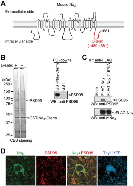
GST-Naxis a GST fusion protein at the C-terminus (amino acid residues 1489–1681) of mouse
Nax(GenBank accession no. NM_009135). pGEX-Naxwas prepared by subcloning NaxcDNA
from pTRE-mNax[14] into pGEX-6P (GE Healthcare) to express GST-Nax. The GST-Nax
pro-tein was expressed in theE.colistrain BL21, and purified by glutathione affinity
chromatogra-phy as described previously [15].
In pull-down experiments, glutathione Sepharose beads (20μl) were coated with GST fusion
proteins (2μg), and then incubated overnight at 4°C with synaptosomal lysate (200μg protein)
prepared from the adult rat cerebrum, as described previously [17]. After washing the beads, the bound proteins were solubilized, separated by SDS-PAGE, and stained with Coomassie Brilliant Blue. Specific bands were excised, subjected to in-gel tryptic digestion, and then ap-plied to matrix-assisted laser desorption ionization-time of flight mass spectrometry (MALDI--TOF MS) (Reflex III, Bruker Daltonics). Peptide mass fingerprinting was performed by a Mascot search (http://www.matrixscience.com/) against the NCBI nonredundant protein database.
Immunoprecipitation experiments
HEK293T cells were transfected with pFLAG-mNaxor pFLAG-mNax-T1679A [15] together
with DMEM containing 10% FBS under 5% CO2at 37°C for 2 days, and then lysed with lysis
buffer (1% Triton X-100 and 150 mM NaCl in 10 mM Tris-HCl, pH 7.4) containing protease inhibitors (Complete Protease Inhibitor Cocktail, Roche Applied Science). Cell extracts were incubated with an anti-FLAG M2 antibody, and the immunocomplexes were precipitated using protein G-Sepharose. After washing the beads, the bound proteins were separated by SDS-PAGE, and followed by Western blotting with anti-PSD95 and anti-mNaxantibodies as
described above. The antibodies used are listed inS1 Table.
RNA interference
Predesigned small interfering RNA (siRNA) against mouse PSD95 (SASI_Mm02_00304274) and control siRNA (MISSION siRNA Universal Negative Control, SIC-001) were purchased from Sigma-Aldrich. siRNAs were transfected into cells using Lipofectamine 2000 (Life tech-nologies), and these cells were then used for experiments after a 36-h culture.
Na
+imaging
Intracellular Na+imaging with sodium-binding benzofuran isophthalate acetoxymethyl ester (SBFI/AM; Molecular Probes) was performed as described previously [12]. The 145 mM Na+ -recording solution (isotonic solution) contained (in mM): 135 NaCl, 5 KCl, 2.5 CaCl2, 1
MgCl2, 20 HEPES, and 10 NaOH, titrated to pH 7.3 with HCl. NaCl was added to or removed
from the recording solution to achieve the appropriate [Na+].
Patch-clamp experiments
Patch-clamp experiments were performed as previously described with minor modifications [18]. The basal recording solution contained (in mM): 140 NaCl, 5 KCl, 2.5 CaCl2, 1 MgCl2, 5
HEPES, and 20 glucose. NaCl was added to or removed from the recording solution to achieve the appropriate [Na+]. In the experiments to test the ion selectivity of Naxchannel, NaCl in the
recording solution was replaced with an equivalent amount of the test salt. The pipette solution contained (in mM): 120 K-gluconate, 20 TEA-Cl, 2 MgCl2, 2 Na2ATP, 1 EGTA, and 10 HEPES
(pH 7.3). Cells were voltage clamped at—60 mV during the recordings. In order to detect Na+ -dependent currents, extracellular solutions were changed using the fast application method with a double-barreled application pipette [19]. The pipette was operated by a piezoelectric device (PZ-150M, Burleigh Instruments). [Na+]oat the half-maximal response (C1/2) of the
[Na+]o-dependence curve was determined by curve fitting using the equation: I = IMax/{1 + exp
[(C1/2—C)/a]}, where I is the current density and C is [Na+]o. The value, IMax= 1.0 was used
for the calculation. The half maximal‘C1/2’and value‘a’were determined by curve fitting.
Statistical Analysis
Data were tested for significance with Kyplot software (Kyens). p<0.01 was considered
signif-icant. Data are shown as the mean ± SE.
Results
Expression of Na
xin neurons
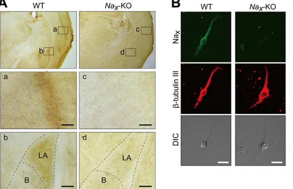
In order to verify the expression of Naxin the mouse cortex and amygdala [9] by
immunohis-tochemistry, we newly generated antibodies using the interdomain II-III of mouse Nax
(anti-mNax, see theMaterials and Methodsfor details). Using this anti-mNax, we successfully
de-tected the expression of Naxin the cortex and amygdala (Fig 1A, WT), which was previously
restricted to the lateral part (Fig 1A and 1B). The signals in these loci were absent inNax-KO
mice, indicating the specificity of the signals (Fig 1A and 1D;Nax-KO). Immunocytochemistry
using a primary culture of the mouse lateral amygdala further showed that Naxwas expressed
in neurons because they co-localized with the neuronal marker,β-tubulin III (Fig 1B).
Establishment of Na
x-expressing neuronal cells
To characterize Naxchannel properties in neuronal cells, we attempted to establish stable cells
expressing Naxusing neuronal cell lines. We examined whether the mouse neuroblastoma
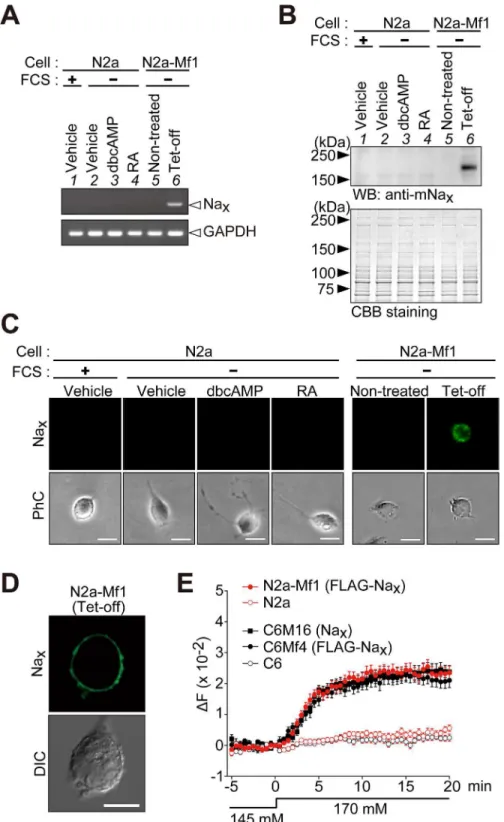
Neuro-2a cell line was devoid of the endogenous expression of Naxby RT-PCR (Fig 2A, lane
1). The expression of Naxwas not detected in Neuro-2a cells even when they were cultured in
serum-depleted (differentiation-inducing) medium or in medium containing dbcAMP or reti-noic acid (Fig 2A; lanes 2–4).
We first established C6Mf4 cells, a C6 cell line expressing FLAG-tagged mouse Nax, and
compared the [Na+]-sensitive responses of the FLAG-tagged Naxin C6Mf4 to those of
non-tagged Naxin C6M16 cells [14] using Na+-imaging experiments. The expression of Nax
chan-nels was inducible in these cells under the control of TRE (see theMaterials and Methods, [14]). When [Na+]owas increased from 145 mM to 170 mM, C6Mf4 cells expressing
FLAG-tagged Naxexhibited increases in intracellular Na+concentrations ([Na+]i), as did C6M16 (Fig
2E, C6M16 and C6Mf4). The time courses of these cells were similar, indicating that the FLAG tag did not affect the gating of Naxchannels.
Because Neuro-2a was found to be available for the host cell as described above, we then at-tempted to establish a cell line using Neuro-2a cells, which are inducible for the expression of
Fig 1. Lateral amygdala neurons express Naxchannels.(A) Immunohistochemical staining of the coronal sections of adult wild-type (WT) andNax
-knockout (Nax-KO) mice with an anti-mNaxantibody. The lower panels are magnified views of the square areas inside the upper panel.
Immunohistochemical brown staining was observed in the cortex (a) and lateral amygdala (b) in WT mice, but not inNax-KO mice (c and d). LA, Lateral amygdala; B, Basal amygdala. Scale bars, 50μm. (B) Double immunofluorescence staining of primary cultured cells obtained from the lateral amygdala of WT andNax-KO mice with anti-mNax(green) and anti-β-tubulin III (red, a neuronal marker) antibodies. DIC, differential interference contrast image. Scale
bars, 20μm.
Fig 2. Establishment of mouse neuroblastoma Neuro-2a cells that inducibly express Nax.(A) A
RT-PCR analysis of the expression of Nax(upper) and glyceraldehyde 3-phosphate dehydrogenase
(GAPDH, lower). Lane 1, parental Neuro-2a (N2a) cells cultured in DMEM containing 10% FCS; lanes 2–4, Neuro-2a cells cultured in serum-free medium with vehicle (lane 2), 1 mM dbcAMP (lane 3), or 20μM retinoic acid (RA) (lane 4); lanes 5 and 6, a stable transfectant of Neuro-2a cells with pTRE-FLAG-mNax(N2a-Mf1)
maintained under serum-free conditions (lane 5) and N2a-Mf1 cells infected with the Tet-off adenovirus for the expression of FLAG-mNax(lane 6). The parental N2a cells were Nax-negative. GAPDH was amplified
FLAG-tagged Nax. When Neuro-2a cells were treated with the Tet-off vector, the expression of
Naxwas detected by RT-PCR (Fig 2A, lanes 5 and 6). These RT-PCR results were confirmed by
a Western blot analysis (Fig 2B) and immunocytochemistry using a wide-field fluorescence mi-croscope (Fig 2C). When we used a confocal microscope to observe the immunostained cells, Naxsignals were mainly observed at the plasma membrane, indicating the cell-surface
expres-sion of Nax(Fig 2D): Neuro-2a cells endogenously expressed PSD95, which promotes the
cell-surface expression of Nax(see below). We named the cell line thus obtained N2a-Mf1.
We examined the function of Naxin N2a-Mf1 using FLAG-tagged Nax. When [Na+]owas
increased from 145 mM to 170 mM, N2a cells expressing FLAG-tagged Naxshowed increases
in [Na+]i(Fig 2E, N2a-Mf1), indicating that Naxopened in a [Na+]-dependent manner also in
neurons. The [Na+]-sensitive responses of these cells were very similar, suggesting that the channel properties of Naxwere not affected by the host cell (Fig 2E, compare N2a-Mf1 with
C6Mf4).
Their parental Neuro-2a and C6 cells, which were Nax-negative, did not show this increase
in [Na+]i. These results indicated that Naxchannels expressed in neurons, as well as those in
glial cell, were functional and responded to increase in [Na+]o.
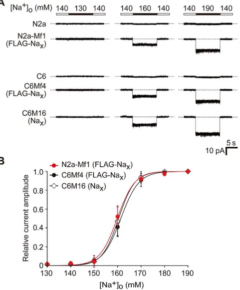
Na
+sensitivity of Na
xin neuronal cells is similar to that in glial cells
We measured the current response of Naxexpressed in N2a-Mf1 to [Na+]ochanges using a
patch-clamp method with a voltage-clamp configuration. When the“high Na+solution”
([Na+]o= 160 and 190 mM) was applied to the cells, inward currents were observed in Nax
-ex-pressing cells (Fig 3A, N2a-Mf1, middle and right columns). It was not inactivated under the high Na+solution conditions, but disappeared rapidly when [Na+]owas returned to the basal
level ([Na+]o= 140 mM). No inward currents were observed when [Na+]owas lowered from
the control amount of 140 mM to 130 mM (Fig 3A, N2a-Mf1, left column). These responses were very similar to those observed in C6Mf4 and C6M16 (Fig 3A, C6Mf4 and C6M16), but were not observed in Neuro-2a or C6 cells (Fig 3AN2a and C6). We further examined the rela-tionship between the relative current amplitude and [Na+]o(Fig 3B). The response curve of the
[Na+]odependency of Naxobserved in Nax-expressing Neuro-2a cells was similar to that in
Nax-expressing C6 cells (the half maximums of the curves for N2a-Mf1, C6Mf4, and C6M16
were 159.8, 161.4, and 160.4, respectively).
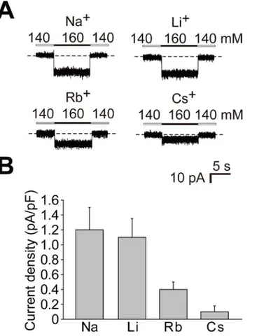
Ion selectivity of the cation-sensitive response of Na
xchannels
We examined current responses to different monovalent ions using N2a-Mf1 cells expressing Naxchannels. When extracellular Na+at 160 mM was completely replaced with lithium ions
(Li+), rubidium ions (Rb+), or cesium ions (Cs+), the current density (current amplitude nor-malized with cell capacitance) decreased in this order (Fig 4). The current amplitude for each
The lower panel shows Coomassie Brilliant Blue (CBB) staining to verify the amount of protein applied. (C
andD) Anti-Naximmunocytochemistry of cells cultured as inAusing a wide-field fluorescence microscope
(C) and confocal laser scanning microscope (D). PhC, phase contrast; DIC, differential interference contrast image. Scale bars, 20μm for (C) and 10μm for (D). (E) Na+imaging to confirm the functional expression of FLAG-tagged Naxin N2a-Mf1 cells. C6M16 cells expressing non-tagged mouse Naxand C6Mf4 cells
expressing FLAG-tagged Naxwere analyzed to determine whether the FLAG tag affected the gating of Nax
channels. C6M16, C6Mf4, and N2a-Mf1 cells showed similar [Na+]
o-sensitive responses. Their parental N2a
and C6 cells, which were Nax-negative, did not show [Na+]o-sensitive responses. The ordinate shows the
change observed in the fluorescence ratio (ΔF, 340/380 nm). The fluorescence ratio at 0 min was set as the zero point on the ordinate. The extracellular perfusion solution was changed from the 145 mM Na+solution to the 170 mM Na+solution at 0 min. Data represent the mean±SE (n = 25 for each). Uncropped images of gels and blots are shown inS3 Fig.
ion species remained unchanged when the concentration (160 mM for each cation) was main-tained, but immediately disappeared when the extracellular concentration was returned to the basal level (140 mM for each cation). Therefore, these current properties were similar to those for Na+.
Na
xinteracts with PSD95 through its PDZ-binding motif
Naxon the plasma membrane of glial cells is known to be stabilized by binding to SAP97
through its C-terminus [15]; therefore, we assumed that other PDZ proteins exist in neurons that interact with the C-terminus of Naxand promote the cell-surface expression of Naxin
neu-rons. We performed pull-down experiments using the NaxC-terminal region fused with GST
(GST-Nax-Cterm, seeFig 5A) from the synaptosomal fraction of the cerebral cortex of adult
Fig 3. Comparison of Na+sensitivity of Naxexpressed in neuronal Neuro-2a and glial C6 cells.(A) A
comparison of the sodium sensitivity of Naxchannels between Neuro-2a and C6 transfectants by whole-cell
patch-clamp recording. Representative whole-cell current responses by the application of hypotonic (130 mM) or hypertonic (160 and 190 mM) solution of [Na+]
owere shown. (B) Relationships between the relative current
amplitude and [Na+]
o. Each current amplitude was normalized to the amplitude of the current elicited by a
solution change to 190 mM [Na+]o. Data represent the mean±SE.
rats. Several specific bands bound for GST-Nax-Cterm were detected in the pulled-down
frac-tion (Fig 5B, left panel). The main band at 95 kDa was identified as PSD95 by mass spectrome-try. We confirmed interactions between the C-terminal region of Naxand PSD95 by Western
blotting of the pull-downed sample with GST-Nax-Cterm (Fig 5B, right panel). We also
identi-fied an interaction between the full-length Naxand full-length PSD95 by immunoprecipitation
using cell extracts from HEK293T cells in which the expression vectors of FLAG-tagged Nax
and PSD95 were co-transfected (Fig 5CandS1 Fig). PSD95 was not immunoprecipitated with mouse Naxwith a mutation at the PDZ-binding motif (FLAG-Nax-T1679A) (Fig 5C). This
re-sult indicated that PSD95 bound to Naxthrough the C-terminal PDZ-binding motif of Nax, as
was the case for SAP97 [15]. Double immunostaining of sections of the mouse brain showed that Naxand PSD95 were co-expressed at the cellular level in Thy1-positive neurons in the
amygdala (Fig 5D).
PSD95 promotes the stability of Na
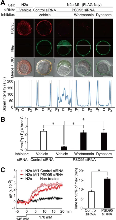
xat the plasma membrane
In parental Neuro-2a cells, endogenous PSD95 was detected in the intracellular region (Fig 6A, N2a; see alsoS2 Fig, Non-treated). We examined the subcellular distribution of endogenous PSD95 and heterologously expressed Nax. In N2a-Mf1 cells that expressed Nax, PSD95 was
clearly observed at the plasma membrane in addition to the cytoplasm, whereas Naxwas
local-ized to the plasma membrane (Fig 6A and 6B, N2a-Mf1, Control siRNA).
Fig 4. Ion selectivity of the cation-sensitive response of the Naxchannel.(A) Representative whole-cell
current responses in N2a-Mf1 expressing FLAG-Naxelicited by the addition of 20 mM of the test cations as
chloride salt. (B) Summary of whole-cell current responses inA. Data represent the mean±SE. n = 5.
Fig 5. Naxbinds to PSD95 via its C-terminal PDZ-domain binding motif.(A) Schematic diagram of
mouse Nax. The C-terminal region used for the pull-down experiment is indicated by a red line. Numbers refer
to amino acid residues. (B) GST pull-down assays. Left, CBB-stained gel of pull-down samples. The asterisk indicates the main band, which was specifically detected in the pull-down samples using GST-Nax-Cterm.
The main band at 95 kDa was identified as PSD95 by mass spectrometry. Right, Western blot analyses of pull-down samples from the synaptosome fraction of the rat cortex using GST-Nax-Cterm. Western blotting
was performed with an anti-PSD95 antibody. (C) Binding of full-length Naxto PSD95 in HEK293T cells. The
expression construct for PSD95 was transfected into HEK293T cells, together with the control vector (Mock), FLAG-tagged wild-type Nax, or its FLAG-tagged PDZ-binding-motif mutant. The upper box shows the
sequence of the PDZ-binding motif of mouse Naxand its mutant. The lower panels show
immunoprecipitation. The amounts of protein immunoprecipitated with anti-FLAG M2 were analyzed by Western blotting using anti-PSD95 and anti-mNaxantibodies. The amounts of protein expressed in the cell
extract were shown inS1 Fig. (D) Immunohistochemical staining of the lateral amygdala in the Thy1-YFP mouse with anti-mNax(green) and anti-PSD95 (red). The fluorescence signals of YFP are indicated in blue.
Fig 6. PSD95 promotes the stability of Naxchannels at the plasma membrane in neuronal cells.(A)
We previously demonstrated that SAP97 contributed to the stabilization of Naxat the
plas-ma membrane in glial cells. To determine whether the surface expression of Naxchannels was
affected by endogenous PSD95, we transfected siRNA to knockdown PSD95 in N2a-Mf1 cells. Along with a decrease in the expression of PSD95 (S2 Fig), the surface expression of wild-type Naxwas found to be markedly decreased (Fig 6A and 6B, N2a-Mf1, PSD95 siRNA, Vehicle).
An incubation with wortmannin, an inhibitor of endocytosis [20], or dynasore, an inhibitor for dynamin-dependent endocytosis [21], markedly ameliorated the surface expression of Nax(Fig
6A and 6B, Wortmannin and Dynasore). These results indicated that binding to PSD95 pro-moted the stabilization of Naxat the plasma membrane.
We next investigated whether the reduction in cell surface Naxby the treatment with PSD95
siRNA resulted in the suppression of Na+influx by Nax. When [Na+]owas increased from 145
mM to 170 mM, N2a-Mf1 cells showed increases in [Na+]i, and this level eventually reached an
equilibrium point between Na+influx by Naxand Na+export by Na+/K+-ATPase (Fig 6C, left
panel). However, when the expression of PSD95 was knocked down with siRNA, the time taken to reach the plateau of [Na+]iwas markedly prolonged (Fig 6C, right panel).
Discussion
In the present study, we confirmed that Naxwas expressed in the neurons of the lateral
amyg-dala. Functional analyses of Naxexogenously expressed in neuronal cells revealed that the
[Na+]-sensitivity of Naxwas similar to that expressed in glial cells. Furthermore, we
demon-strated that Naxbound to PSD95 through its PDZ-binding motif at the C-terminus in neurons.
The interaction between Naxand PSD95 was crucial for the surface expression of Nax.
As showen inFig 2, the expression of Naxwas not detected in Neuro-2a cells not only by
RT-PCR (Fig 2A), but also by immunocytochemistry using our anti-mouse Naxantibody (Fig
2B and 2C), irrespective of their differentiation state. [Na+]-sensitive responses were not ob-served in Neuro-2a cells by our Na+-imaging and electrophysiological experiments (Figs2E
and3A). Naxproteins and [Na+]-sensitive responses only appeared when exogenous Naxwas
expressed (Figs2and3). These results clearly indicated that Neuro-2a cells did not express Naxendogenously.
However, a very recent study reported that the immunocytochemical signals of Naxwere
de-tected in Neuro-2a cells [22]. This group postulated that Naxwas expressed in neurons in the
rat median preoptic nucleus (MnPO) using their antibodies [23]. We examined the expression of Naxby immunohistochemistry using our antibodies to rat Naxand mouse Nax[12 and the
present study, respectively]: The specificities of our antibodies were confirmed using tissues and tissue lysates fromNax-KO mice. We did not detect any Naxsignals in the MnPO in the
rat or mouse brain (S4 Fig), as we have previously discussed [24]. This result is consistent with our previous findings in whichlacZsignals were negative in the MnPO inNax-KO mice [9].
Furthermore, they claimed that the Na+leak currents observed in rat MnPO neurons have [Na+]-independent conductance [25]. However, we previously demonstrated that Naxhad a
or PSD95 siRNA. Upper panels: Immunostaining with anti-PSD95 and anti-mNaxantibodies. In order to
inhibit endocytosis, cells expressing Naxwere treated with 100 nM wortmannin or 200μM dynasore for 6 h.
These cells were then fixed, permeabilized, and stained with anti-mNax. Scale bars, 10μm. Lower graphs:
Fluorescence intensity profiles along the white lines in the upper panels. The profile was divided into three parts (P1: C: P2 = 15: 70: 15 in length). a.u., arbitrary unit. (B) The relative fluorescence intensity of the membrane region to the central region inA. Data represent the mean±SE (n = 8 for each);*p<0.01,
ANOVA followed by Scheffe’s test. (C) Reduced Na+influx in N2a-Mf1 cells in the absence of PSD95. Left panel: Na+imaging of N2a-Mf1 cells transfected with PSD95 or control siRNA, or non-treated N2a cells. Data represent the mean±SE (n = 21 for each). Right panel: Summary of the time taken to reach 95% of the
plateau level. Data represent the mean±SE (n = 21 for each);*p<0.01, two-tailed t test.
[Na+]o-dependent gating property [6,12]. Collectively, these results indicated that the signals
and currents that they described were not derived from Nax.
We herein showed that the cation selectivity sequence of Naxwas Na+Li+>Rb+>Cs+
(Fig 4). This sequence was similar to those of voltage-gated sodium channels (Nav) in
myelinat-ed nerves in a previous study [26]. Naxpassed certain amounts of Rb+and Cs+(Fig 4B), while
the permeability of Navfor Rb+and Cs+was nearly negligible [26]. An ion selectivity filter has
been postulated to exist on the extracellular side of the pore of the sodium channelα-subunit: an outer ring with the amino acid sequence EEMD and inner ring with DEKA [27,28]. These two rings were conserved in all Nav(Nav1.1–1.9). In contrast, those in Naxwere EEID and
DENS, respectively, suggesting that the relatively larger permeability to Rb+and Cs+in Nax
may be caused by these differences. The best way to estimate the ion selectivity of channel per-meability is to determine the perper-meability ratios for each ion. Measurements of precise reversal potentials for each condition are required to calculate permeability ratios [28]; however, we could not measure precise reversal potentials because the currents were very small.
Electrophysiological analyses of purified Naxin a planar phospholipid bilayer are needed to
further characterize Nax.
Taken together with our previous findings [15], PSD95 and SAP97 both contributed to the surface expression of Naxin neurons and glial cells, respectively. Both PSD95 and SAP97 are
members of the membrane-associated guanylate kinase (MAGUK) family, which form a scaf-fold for the clustering of receptors, ion channels, and associated signaling proteins [29]. As shown inFig 5D, Naxin the lateral amygdala co-localized with PSD95 clusters, which appeared
to exist along dendrites, suggesting that PSD95 played a role in the stabilization of Naxat
syn-apses. On the other hand, Naxin the SFO was localized to perineuronal lamellate processes
that extended from glial cells (ependymal cells and astrocytes) [10], suggesting that SAP97 con-tributed to this localization in glial cells. SAP97 was reported to be expressed at the postsy-napses of GABAergic interneurons in the lateral amygdala [30]. Therefore, SAP97 may also be expressed in Nax-positive neurons and play a role in the stabilization of Naxat the plasma
membrane not only in glial cells, but also in neurons.
Several ion channels have been shown to interact with PSD95 via their C-terminal PDZ-binding motifs: Voltage-gated K channels (Kv1.4, Kv1.5, and Kv4.2), the inward rectifier K channels (Kir channels; Kir2.1, Kir2.3, and Kir5.1), the Na+-sensitive K channel (Slo2), the acid-sensing ion channel (ASIC3), and ligand gated glutamate receptor NMDA receptor (NR2) [31–39]. Together with these channel proteins, Naxchannels may exist in the postsynaptic
den-sity of excitatory synapses in the lateral amygdala and be functionally coupled to these channels through its ion transport. However, it is unlikely that [Na+]oaround synapses increased in the
amygdala under normal conditions. A certain level of endothelins (ETs) has been shown to ac-tivate Naxunder physiological [Na+]oconditions [18,40]. The opening of Naxchannels by ET
signaling may depolarize the postsynaptic membrane in neurons through the influx of Na+. The physiological roles of Naxin brain neurons including the amygdala will be the subject of
future investigations.
Supporting Information
S1 Fig. Supplemental data related toFig 5C.(A) Western blotting of the total cell extracts used in the immunoprecipitation with anti-mNax(left) and anti-PSD95 (right) antibodies. (B)
The original blot images of the Western blotting of the immunoprecipitates with anti-mNax
(left) and anti-PSD95 (right) antibodies presented inFig 5C. Red squares indicate the areas used in the main figures.
S2 Fig. Depletion of PSD95 in N2a-Mf1 cells by siRNA.Reduction of PSD95 by PSD95 small interfering RNA (siRNA) in N2a-Mf1 cells was verified by Western blotting with anti-PSD95 antibody (left). The right panel shows CBB staining to verify the amount of protein applied. The expression of PSD95 was reduced in N2a-Mf1 cells transfected with PSD95 siRNA but not with control siRNA.
(TIF)
S3 Fig. Original images presented inFig 2A and 2B, andFig 5B.(A) Original gel images pre-sented inFig 2A. (B) Original blot image presented inFig 2B. (C) Original blot image pre-sented inFig 5B. Red squares indicate the areas used in theFig 5B.
(TIF)
S4 Fig. Immunohistochemical staining of rat and mouse brains with anti-Naxantibodies.
Immunohistochemical staining of the coronal sections of rat and mouse brains, containing the median preoptic nucleus (MnPO) (A) and median eminence (B) with anti-rat Nax[12] and
anti-mNaxantibodies. Immunohistochemical staining was performed as described in S5 File.
Neither rat nor mouse MnPO was negative for Nax(A). On the other hand, the median
emi-nence was clearly stained with both antibodies (B). AC, anterior commissure. Scale bars, 200μm.
(TIF)
S1 File. Supporting method forS4 Fig.
(DOCX)
S1 Table. Antibodies used for this study.
(XLSX)
Acknowledgments
We thank N. Nakanishi, Y. Isoshima, S. Miura, and T. Hashimoto for their technical assistance, and A. Kodama for her secretarial assistance. Confocal images were acquired at the Spectrogra-phy and Bioimaging Facility, NIBB Core Research Facilities.
Author Contributions
Conceived and designed the experiments: TYH AF MN. Performed the experiments: MM TYH KK RS AF. Analyzed the data: MM TYH. Wrote the paper: MM TYH MN.
References
1. Gautron S, Dos Santos G, Pinto-Henrique D, Koulakoff A, Gros F, Berwald-Netter Y. The glial voltage-gated sodium channel: cell- and tissue-specific mRNA expression. Proc Natl Acad Sci USA. 1992; 89: 7272–7276. PMID:1379737
2. George AL Jr Knittle TJ, Tamkun MM. Molecular cloning of an atypical voltage-gated sodium channel expressed in human heart and uterus: evidence for a distinct gene family. Proc Natl Acad Sci USA. 1992; 89: 4893–4897. PMID:1317577
3. Felipe A, Knittle TJ, Doyle KL, Tamkun MM. Primary structure and differential expression during devel-opment and pregnancy of a novel voltage-gated sodium channel in the mouse. J Biol Chem. 1994; 269: 30125–30131. PMID:7982916
4. Akopian AN, Souslova V, Sivilotti L, Wood JN. Structure and distribution of a broadly expressed atypical sodium channel. FEBS Lett. 1997; 400: 183–187. PMID:9001394
5. Goldin AL, Barchi RL, Caldwell JH, Hofmann F, Howe JR, Hunter JC, et al. Nomenclature of voltage-gated sodium channels. Neuron. 2000; 28: 365–368. PMID:11144347
7. Noda M. The subfornical organ, a specialized sodium channel, and the sensing of sodium levels in the brain. Neuroscientist. 2006; 12: 80–91. PMID:16394195
8. Noda M, Hiyama TY. The NaxChannel: What It Is and What It Does. Neuroscientist. 2014 Jun 24. doi: 10.1177/1073858414541009
9. Watanabe E, Fujikawa A, Matsunaga H, Yasoshima Y, Sako N, Yamamoto T, et al. Nav2/NaG channel is involved in control of salt-intake behavior in the CNS. J Neurosci. 2000; 20: 7743–7751. PMID: 11027237
10. Watanabe E, Hiyama TY, Shimizu H, Kodama R, Hayashi N, Miyata S, et al. Sodium-level-sensitive so-dium channel Naxis expressed in glial laminate processes in the sensory circumventricular organs. Am J Physiol Regul Integr Comp Physiol. 2006; 290: 568–576. PMID:16223844
11. Watanabe E, Hiyama TY, Kodama R, Noda M. Naxsodium channel is expressed in non-myelinating Schwann cells and alveolar type II cells in mice. Neurosci Lett. 2002; 330: 109–113. PMID:12213645
12. Hiyama TY, Watanabe E, Ono K, Inenaga K, Tamkun MM, Yoshida S, et al. Naxchannel involved in CNS sodium-level sensing. Nat Neurosci. 2002; 5: 511–512. PMID:11992118
13. Hiyama TY, Watanabe E, Okada H, Noda M. The subfornical organ is the primary locus of soudium-level sensing by Naxsodium channels for the control of salt-intake behavior. J Neurosci. 2004; 24: 9276–9281. PMID:15496663
14. Shimizu H, Watanabe E, Hiyama TY, Nagakura A, Fujikawa A, Okado H, et al. Glial Naxchannels con-trol lactate signaling to neurons for brain [Na+] sensing. Neuron. 2007; 54: 59
–72. PMID:17408578
15. Matsumoto M, Fujikawa A, Suzuki R, Shimizu H, Kuboyama K, Hiyama TY, et al. SAP97 promotes the stability of Naxchannels at the plasma membrane. FEBS Lett. 2012; 586: 3805–3812. doi:10.1016/j. febslet.2012.09.018PMID:23022437
16. Hiyama TY, Matsuda S, Fujikawa A, Matsumoto M, Watanabe E, Kajiwara H, et al. Autoimmunity to the sodium-level sensor in the brain causes essential hypernatremia. Neuron. 2010; 66: 508–522. doi:10. 1016/j.neuron.2010.04.017PMID:20510856
17. Fujikawa A, Chow JP, Shimizu H, Fukada M, Suzuki R, Noda M. Tyrosine phosphorylation of ErbB4 is enhanced by PSD95 and repressed by protein tyrosine phosphatase receptor type Z. J Biochem. 2007; 142: 343–350. PMID:17646177
18. Hiyama TY, Yoshida M, Matsumoto M, Suzuki R, Matsuda T, Watanabe E, et al. Endothelin-3 expres-sion in the subfornical organ enhances the sensitivity of Nax, the brain sodium-level sensor, to suppress salt intake. Cell Metab. 2013; 17: 507–519. PMID:23541371
19. Johnson JW, Ascher P. Glycine potentiates the NMDA response in cultured mouse brain neurons. Na-ture. 1987; 325: 529–531. PMID:2433595
20. Gong Q, Weide M, Huntsman C, Xu Z, Jan LY, Ma D. Identification and characterization of a new class of trafficking motifs for controlling clathrin-independent internalization and recycling. J Biol Chem. 2007; 282: 13087–13097. PMID:17331948
21. Kirchhausen T, Macia E, Pelish HE. Use of dynasore, the small molecule inhibitor of dynamin, in the regulation of endocytosis. Methods Enzymol. 2008; 438: 77–93. doi:10.1016/S0076-6879(07)38006-3 PMID:18413242
22. Berret E, Smith PY, Henry M, Soulet D, Hébert SS, Toth K, et al. Extracellular Na+levels regulate for-mation and activity of the Nax/alpha1-Na+/K+-ATPase complex in neuronal cells. Front Cell Neurosci. 2014; 8: 413. doi:10.3389/fncel.2014.00413eCollection 2014. PMID:25538563
23. Nehmé B, Henry M, Mouginot D, Drolet G. The Expression Pattern of the Na+Sensor, Na
xin the Hydro-mineral Homeostatic Network: A Comparative Study between the Rat and Mouse. Front Neuroanat. 2012; 6: 26 doi:10.3389/fnana.2012.00026eCollection 2012. PMID:22833716
24. Noda M, Hiyama TY. Sodium sensing in tha brain. Pflugers Arch. 2014 Dec 10. doi: 10.1007/s00424-014-1662-4
25. Tremblay C, Berret E, Henry M, Nehmé B, Nadeau L, Mouginot D. Neuronal sodium leak channel is re-sponsible for the detection of sodium in the rat median preoptic nucleus. J Neurophysiol. 2011; 105: 650–660. doi:10.1152/jn.00417.2010PMID:21084682
26. Hille B. The permeability of the sodium channel to metal cations in myelinated nerve. J Gen Physiol. 1972; 59: 637–658. PMID:5025743
27. Noda M. Structure and function of sodium channels. Ann N Y Acad Sci. 1993; 707: 20–37. PMID: 9137539
28. Hille B. Ion channels of excitable membranes: Sinauer Associates; 2001. pp. 21.
30. Polepalli JS, Sullivan RK, Yanagawa Y, Sah P. A specific class of interneuron mediates inhibitory plas-ticity in the lateral amygdala. J Neurosci. 2010; 30: 14619–14629. doi:10.1523/JNEUROSCI.3252-10. 2010PMID:21048119
31. Imamura F, Maeda S, Doi T, Fujiyoshi Y. Ligand binding of the second PDZ domain regulates clustering of PSD-95 with the Kv1.4 potassium channel. J Biol Chem. 2002; 277: 3640–3646. PMID:11723117
32. Eldstrom J, Doerksen KW, Steele DF, Fedida D. N-terminal PDZ-binding domain in Kv1 potassium channels. FEBS Lett. 2002; 531: 529–537. PMID:12435606
33. Wong W, Newell EW, Jugloff DG, Jones OT, Schlichter LC. Cell surface targeting and clustering inter-actions between heterologously expressed PSD-95 and the Shal voltage-gated potassium channel, Kv4.2. J Biol Chem. 2002; 277: 20423–20430. PMID:11923279
34. Nehring RB, Wischmeyer E, Döring F, Veh RW, Sheng M, Karschin A. Neuronal inwardly rectifying K+ channels differentially couple to PDZ proteins of the PSD-95/SAP90 family. J Neurosci. 2000; 20: 156–
162. PMID:10627592
35. Inanobe A, Fujita A, Ito M, Tomoike H, Inageda K, Kurachi Y. Inward rectifier K+channel Kir2.3 is local-ized at the postsynaptic membrane of excitatory synapses. Am J Physiol Cell Physiol. 2002; 282: C1396–C1403. PMID:11997254
36. Tanemoto M, Fujita A, Higashi K, Kurachi Y. PSD-95 mediates formation of a functional homomeric Kir5.1 channel in the brain. Neuron. 2002; 34: 387–397. PMID:11988170
37. Uchino S, Wada H, Honda S, Hirasawa T, Yanai S, Nakamura Y, et al. Slo2 sodium-activated K+ chan-nels bind to the PDZ domain of PSD-95. Biochem Biophys Res Commun. 2003; 310: 1140–1147. PMID:14559234
38. Eshcol JO, Harding AM, Hattori T, Costa V, Welsh MJ, Benson CJ. Acid-sensing ion channel 3 (ASIC3) cell surface expression is modulated by PSD-95 within lipid rafts. Am J Physiol Cell Physiol 2008; 295: C732–739. doi:10.1152/ajpcell.00514.2007PMID:18579798
39. Kornau HC, Schenker LT, Kennedy MB, Seeburg, PH. Domain interaction between NMDA receptor subunits and the postsynaptic density protein PSD-95. Science. 1995; 269: 1737–1740. PMID: 7569905