Universidade de Aveiro 2017
Departamento de Química
Antoine
Simões Almeida
Avaliação do potencial inflamatório de aerossóis
orgânicos inaláveis presentes em uma atmosfera
urbana
Assesment of the inflammatory potential of inhalable
organic aerosols present in an urban atmosphere
III Universidade de Aveiro 2017 Departamento de Química
Antoine
Simões Almeida
Avaliação do potencial inflamatório de aerossóis
orgânicos inaláveis presentes em uma atmosfera
urbana
Assesment of the inflammatory potential of inhalable
organic aerosols present in an urban atmosphere
Dissertação apresentada à Universidade de Aveiro para cumprimento dos requisitos necessários à obtenção do grau de Mestre em Bioquímica, ramo de Bioquímica Clínica, realizado sob a orientação científica da Doutora Regina Maria Brandão de Oliveira Duarte, equiparada a Investigadora Principal do Centro de Estudos do Ambiente e do Mar (CESAM) da Universidade de Aveiro, e do Doutor Bruno Miguel Rodrigues das Neves, Professor Auxiliar convidado da Faculdade de Farmácia da Universidade de Coimbra
V
o júri
presidente Prof. Doutora Maria do Rosário Gonçalves dos Reis Marques Domingues
Professora Associada com Agregação do Departamento de Química da Universidade de Aveiro
Doutora Regina Maria Brandão de Oliveira Duarte
Equiparada a Investigador Principal do Centro de Estudos do Ambiente e do Mar da Universidade de Aveiro
Prof. Doutora Rita Maria Pinho Ferreira
VII
agradecimentos Aos meus orientadores, Doutora Regina Duarte e Professor Doutor Bruno Neves, pelo apoio, dedicação, contribuição e pela disponibilidade demonstrada durante a realização deste trabalho.
Ao Professor Doutor Artur Silva, pela disponibilidade para a aquisição dos espetros de RMN uni- e bidimensionais e pela sua contribuição e ajuda na interpretação destes mesmos espetros.
À Professora Doutora Rita Ferreira, pela disponibilidade e colaboração na realização dos ensaios de atividade antioxidante in chemico.
Ao Professor Doutor Armando Duarte, pela disponibilidade de meios, recursos laboratoriais e financeiros que muito contribuíram para os resultados alcançados neste trabalho, bem como para a respetiva divulgação.
Aos colegas de laboratório, em especial ao Doutor João Matos, pela sua disponibilidade e ajuda prestadas na realização deste trabalho, nomeadamente na aquisição dos espetros de fluorescência e na análise de carbono orgânico dissolvido nos extratos aquosos das amostras de partículas atmosféricas Ao Centro de Estudos do Ambiente e do Mar (CESAM) da Universidade de Aveiro pelo suporte financeiro, nomeadamente na aquisição de material e reagentes utilizados na realização deste trabalho.
IX
palavras-chave Aerossóis atmosféricos; Matéria particulada fina; Carbono orgânico solúvel em água; Partículas inaláveis; Imunidade inata; Inflamação; Stress oxidativo
resumo A matéria particulada (sigla inglesa, PM) é definida como uma suspensão de partículas sólidas ou líquidas num gás (i.e., atmosfera), sendo estas tipicamente classificadas de acordo com o seu tamanho: PM10 (diâmetro aerodinâmico < 10μm), PM2.5 (diâmetro aerodinâmico < 2.5μm) e PM1.0 (diâmetro aerodinâmico <1.0μm). Estas partículas podem ser emitidas diretamente para a atmosfera através de fontes antropogénicas ou naturais, ou formadas in situ na atmosfera através de processos físico-químicos. A PM tem impacto relevante não só em processos atmosféricos e climáticos, mas também na saúde humana, onde se destaca o aumento de risco de doenças pulmonares e cardiovasculares, o aumento de suscetibilidade a infeções, e efeitos carcinogénicos e mutagénicos. Particularmente, ao nível pulmonar, os principais efeitos da PM descritos na literatura incluem a indução de stress oxidativo e da inflamação, podendo levar a danos pulmonares. O conhecimento dos efeitos nocivos das PM para a saúde pública resulta fundamentalmente de estudos realizados com as partículas, sendo a contribuição da fração de matéria orgânica solúvel em água (sigla inglesa, WSOM) pouco abordada. Neste sentido, o presente trabalho procura caracterizar a fração WSOM de PM2.5, recolhida durante o período de outono, e avaliar os seus efeitos em macrófagos. A caracterização estrutural, com recurso à técnica de espectroscopia de ressonância magnética nuclear de protão (RMN 1H), mostra que as amostras WSOM recolhidas no período noturno apresentam um maior conteúdo de estruturas aromáticas e oxigenadas do que as amostras recolhidas no período diurno, com estas últimas a apresentarem maior conteúdo em estruturas alifáticas. Adicionalmente, a caracterização das amostras WSOM com recurso à espectroscopia de RMN bidimensional (2D) permitiu identificar diferentes componentes estruturais e determinar possíveis fontes destas estruturas. Os ensaios biológicos de exposição aguda (24h) de macrófagos às amostras WSOM demonstraram que tanto as amostras diurnas como noturnas induzem um aumento da transcrição da enzima destoxificante Hmox, como das moléculas pro-inflamatórias Il1b, Il6 e Nos2. Observou-se também que os extratos apresentam atividade antioxidante in chemico e in vitro. Em ensaios de exposição prolongada (3 semanas) a concentrações fisiologicamente relevantes observou-se um pequeno aumento na transcrição dos genes estudados. No entanto, verificou-se que estes macrófagos apresentavam uma capacidade limitada de resposta a um agente inflamatório como o Lipopolissacarídeo bacteriano (LPS).
De uma forma geral, os resultados obtidos indicam que a exposição prolongada a WSOM presente nas partículas atmosféricas pode provocar um ligeiro estado inflamatório a nível pulmonar, limitando, no entanto, a capacidade de resposta dos macrófagos a agentes invasores, o que pode originar um aumento da suscetibilidade a infeções.
XI
keywords Atmospheric aerosols; Fine particulate matter; Water-soluble organic carbon; Inhalable particles; Innate immunity; Inflammation; Oxidative stress.
abstract Airborne particulate matter (PM), are defined as solid or liquid particles suspended in a gas (i.e., atmosphere), and are typically distributed in three size-ranges: PM10 (coarse PM, aerodynamic diameter < 10μm), PM2.5 (fine PM, aerodynamic diameter < 2.5μm) and PM1.0 (ultrafine PM, aerodynamic diameter < 1.0μm). These particles can be emitted directly into the atmosphere from both anthropogenic and natural sources, or formed in situ in the atmosphere through physico-chemical processes. The PM plays a significant role in diverse atmospheric and climatic processes, as well as on human health by increasing the risk of cardiovascular and pulmonary diseases, the susceptibility to infections, and having carcinogenic and mutagenic effects. Particularly, in the lungs, the main effects of PM described in the literature include the induction of oxidative stress and inflammation, occasionally leading to lung injury. The knowledge of the harmful effects of PM on public health is fundamentally due to studies carried out with particles, being the contribution of the fraction of water-soluble organic matter (WSOM) largely unknown. Therefore, the present work aims to characterize the WSOM fraction of PM2.5, collected during autumn season, and assess its effects on macrophages. The structural characterization of the WSOM from atmospheric particles, by means of proton nuclear magnetic resonance (1H NMR) spectroscopy, showed that samples collected during the night have higher content of aromatic and oxygenated structures than those collected during the day, with the later exhibiting higher content of aliphatic structures. Furthermore, the structural characterization of the WSOM samples by means of two-dimensional (2D) NMR spectroscopy also allowed the identification of different structural components and the tentative assignment of their sources. The biological assays on the effect of an acute exposure (24h) to the WSOM samples showed that both day and night samples induced an increase in the transcription of the detoxifying enzyme Hmox, and the pro-inflammatory molecules Il1b, Il6 and Nos2. It was also observed that these extracts present antioxidant activity in chemico and in vitro. In the biological assays of prolonged exposure (3 weeks) at physiologically relevant concentrations, it was observed a small increase in the transcription of the studied genes. However, the obtained data show that these macrophages present a limited capacity to respond to an inflammatory agent such as bacterial lipopolysaccharide (LPS).
In general, the obtained results show that a prolonged exposure to WSOM present in atmospheric particles can induce or worsen an inflammatory response at the pulmonary level, leading however, to a limited capacity of response of the macrophages to invading agents which could increase the susceptibility to infections.
VIII
X
Table of contents……….………….………....……… VIII List of figures.………..……….…..………..……...…… XII List of tables……….……….………..………….. XX List of abbreviations …………...…………..….………....………...… XXIV
Chapter 1. Introduction…………...………..……….... 1
1.1 Atmospheric particles: sizes, composition, and their effect on human health……... 3
1.1.1 An overview of the classification and chemical composition of atmospheric particle………..……….…….... 3
1.1.2 Chemical composition of inhalable fine particulate matter……....……....….... 7
1.1.3 Effects of atmospheric particulate matter on human health……..………. 8
1.2 The innate immune response to inhaled atmospheric particles …………...…….. 10
1.2.1 Inflammation. A look on the body defense mechanism...………...….. 10
1.2.2 Immune players and inflammatory mediators in respiratory system…... 12
1.2.3 Airways inflammation and oxidative stress as a result of inhalation of particulate matter………..……….……….... 15
1.3 Objectives of the M.Sc. thesis……..…...……...…... 18
Chapter 2. Materials and methods………..………...………...…….…… 21
2.1 Atmospheric aerosol sampling campaigns………..…..……….. 23
2.2 Extraction of aerosol WSOC fractions………..……….……. 23
2.3 Excitation-emission matrix (EEM) fluorescence spectroscopy of the WSOC samples………..………... 24
2.4 UV-vis spectroscopy of the WSOC samples………...…… 24
2.5 Dissolved Organic Carbon (DOC) analysis of the WSOC samples…..…….…….. 24
2.6 WSOC samples preparation for NMR and biological studies…………..…..…….. 24
2.7 1D and 2D NMR spectroscopy………..………..……….... 25
XI
2.9 Cell viability assays………..……….….………. 26
2.10 Nitric oxide production………..……….………...…. 26
2.11 Evaluation of NO Scavenging activity………..…………..……… 27
2.12 In vitro antioxidant activity………..………... 27
2.13 Analysis if gene expression by q-PCR………..……….. 27
2.14 Analysis of NF-κB activation by immunofluorescence……..……..……….. 28
2.15 In chemico antioxidant activity………..………. 28
2.16 Statistical analysis……….……….. 28
Chapter 3. Structural characterization and biological effects of aerosol organic matter………..………..……….………….………... 31
3.1 Diurnal trend of fine urban aerosol and its WSOM fraction……...……… 33
3.2 Spectroscopic characterization of atmospheric aerosol WSOM samples…..….…… 34
3.3 Biological activity of WSOM………..………..………...……. 46
Chapter 4. Conclusions……….……..……….… 57
References………..……….……...……….... 61
Annexes………..……….... 71
A1. Meteorological conditions during the sampling campaigns……..…………...……. 73
A2. UV-vis spectroscopy of the WSOM samples………..…………..……… 74
A3. EEM fluorescence spectroscopy of fine urban aerosol WSOM samples…..………. 75
XII
XIV
Figure 1. Schematic representation of primary and secondary sources of atmospheric particles………..…………..………..………….. 4 Figure 2. A typical aerosol mass size distribution profile showing a typical segmentation of
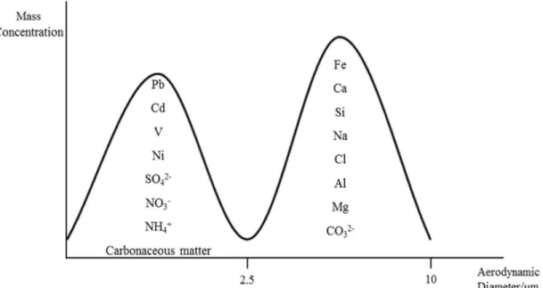
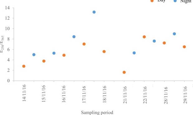
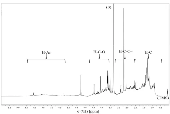
chemical species into fine (aerodynamic diameter < 2.5µm) and coarse (2.5 < aerodynamic diameter < 10 µm) modes (adapted from (Samara and Voutsa, 2005; Seinfeld and Pandis, 2006))……….……….……… 5 Figure 3. SEM images of different atmospheric particles with aerodynamic diameter less than 2.5 μm (photos by courtesy of ORGANOSOL research project, PTDC/CTE-ATM/118551/2010, https://organosolproject.wordpress.com/)... 6 Figure 4. Schematic representation of the chemical composition of PM2.5 in Europe (adapted from (Putaud et al., 2010))………....… 7 Figure 5. Schematic representation of main events of the inflammatory process. Figure made using the Servier Medical Art (“Powerpoint image bank | Servier,” )….. 11 Figure 6. Atmospheric concentrations of PM2.5 collected during the sampling campaign. 33 Figure 7. WSOM content in PM2.5 total mass collected in each sampling period……... 34 Figure 8. EEM fluorescence spectra of the aerosol WSOM samples collected in different time periods: day (up, 16/11/16) and night (down, 16/11/16 to 17/11/16)..…… 35 Figure 9. E250/E365 ratios of the WSOM extracts from PM2.5 collected during the sampling campaign.………..………..………... 36 Figure 10. 1H NMR spectrum of aerosol WSOM collected during day periods. Four spectral
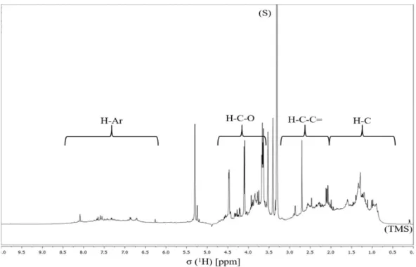
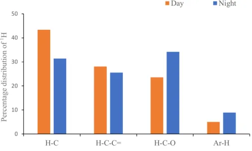
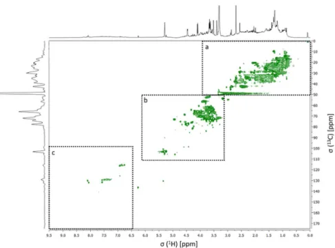
regions are identified at the top of the spectrum: H-C, H-C-C=, H-C-O, and Ar-H. Resonance signals: Solvent (S) – MeOH-d4, and Tetramethylsilane (TMS) - 0.03% (v/v).…………..……….………. 37 Figure 11. 1H NMR spectrum of the aerosol WSOM collected during night periods. Four spectral regions are identified at the top of the spectrum: H-C, H-C-C=, H-C-O, and Ar-H. Resonance signals: Solvent (S) – MeOH-d4, and Tetramethylsilane (TMS) - 0.03% (v/v)……….………. 38 Figure 12. Percentage distribution of the 1H functional groups in aerosol WSOM sample6s collected during the day and night time periods…………..………... 39 Figure 13. 1H-13C HSQC spectrum of the WSOM sample collected during day time period. Section A: aliphatic region; Section B: oxygenated aliphatic region; Section C: aromatic region………..…………..……….………… 40
XV
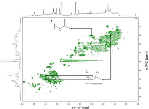
Figure 14. Expanded aliphatic region of the 1H-13C HSQC spectrum of the WSOM sample collected during day time periods, with some tentative structure assignments………..……….………...………. 41 Figure 15. Expanded oxygenated aliphatic region of the 1H-13C HSQC spectrum of the WSOM sample collected during day time periods, with some tentative structure assignments………..……….……… 41 Figure 16. Expanded aromatic region of the 1H-13C HSQC spectrum of the WSOM sample collected during day time periods, with some tentative structure assignments………..……….……….... 42 Figure 17. 1H-13C HSQC spectrum of the WSOM sample collected during night time periods. Section A: aliphatic region; Section B: oxygenated aliphatic region; Section C: aromatic region……….…………..…….………...….... 42 Figure 18. Expanded aliphatic region of the 1H-13C HSQC spectrum of the WSOM sample
collected during night time periods, with some tentative structure assignments ………..…………..……….. 43 Figure 19. Expanded oxygenated aliphatic region of the 1H-13C HSQC spectrum of the
WSOM sample collected during night time periods, with some tentative structure assignments ………..………..………..… 43 Figure 20. Expanded aromatic region of the 1H-13C HSQC spectrum of the WSOM sample collected during night time periods, with some tentative structure assignments ………..………...………... 44 Figure 21. WSOM extracts do not significantly affect Raw 264.7 macrophages viability.
Cells were exposed to different concentrations of WSOM during 24h and then viability assessed by the rezasurin assay. Data is presented as percentage of control (non-treated cells/medium) and represent the mean ± SD from at least 3 independent experiments. * significantly different (one-way ANOVA p<0.05) when compared to control………….……….………….... 46 Figure 22. WSOM extracts induced NO production on raw 264.7 macrophages. Cells were
exposed to different concentrations of WSOM and then their NO production was assessed by Griess assay. Data is presented as concentration of NO and represent mean ± SD from at least 3 independent experiments Multiple group comparisons
XVI
(C vs conditions) was performed by one-way ANOVA analysis,with a Dunnet´s post test. Significance levels are as follows: *P<0.05, **P<0.01, ***P<0.001,and ****P<0.0001…………..………..………..………….… 47 Figure 23. WSOM extracts induced an increase in the gene transcription of the studied genes on Raw 264.7 macrophages. Cells were exposed to different concentrations of WSOM extracts during 4h and then the mRNA was extracted and the gene transcription was assessed by q-PCR. Data is presented as gene expression and represent the mean ± SD from at least 2 independent experiments. Multiple group comparisons (C vs conditions) was performed by one-way ANOVA analysis,with a Dunnet´s post test. Significance levels are as follows: *P<0.05, **P<0.01, ***P<0.001,and ****P<0.0001……..………...….. 48 Figure 24. WSOM extracts do not induce ROS production on Raw 264.7 macrophages and abolish the LPS-induced oxidative stress. Cells were exposed to a set of different conditions and the ROS production was assessed by an in vitro antioxidant activity assay. Hoechst – blue (cell nucleus); H2DCFDA – green (ROS); magnification of 63x……….. 50 Figure 25. WSOM extracts have antioxidant activity. The antioxidant activity was assessed in chemico with the DPPH antioxidant activity assay. Multiple group comparisons (C vs conditions) was performed by one-way ANOVA analysis,with a Dunnet´s post test. Significance levels are as follows: *P<0.05, **P<0.01, ***P<0.001,and ****P<0.0001……….……...…... 50 Figure 26. WSOM extracts caused a decrease in the LPS-induced NO production of raw 264.7 macrophages at higher concentrations. Cells were treated with different concentrations of WSOM extracts and then stimulated with LPS, the NO production was then assed via a Griess assay. Data is presented as concentration of NO and represent the mean ± SD from at least 3 independent experiments. Multiple group comparisons (C vs conditions) was performed by one-way ANOVA analysis,with a Dunnet´s post test. Significance levels are as follows: *P<0.05, **P<0.01, ***P<0.001,and ****P<0.0001...………..…… 51 Figure 27. WSOM do not present NO scavenging activity. Scavenging activity of WSOC was assed via the SNAP assay. Data is presented as percentage of control (non-treated cells/medium) and represent the mean ± SD from at least 3 independent
XVII
experiments. Multiple group comparisons (C vs conditions) was performed by one-way ANOVA analysis,with a Dunnet´s post test. Significance levels are as follows: *P<0.05……..……….…………..………..………… 51 Figure 28. WSOM extracts induced alterations in the LPS-induced gene transcription of the studied genes on Raw 264.7 macrophages. Cells were exposed to different concentrations of WSOM extracts and then stimulated with LPS, then the mRNA was extracted and the gene transcription was assessed by q-PCR. Data is presented as gene expression and represent the mean ± SD from at least 3 independent experiments. Multiple group comparisons (LPS vs conditions) was performed by one-way ANOVA analysis,with a Dunnet´s post test. Significance levels are as follows: *P<0.05, **P<0.01, ***P<0.001,and ****P<0.0001………..………..……... 52 Figure 29. WSOM exposure do not prevent the LPS-induced translocation of NF-kB to the nucleus on Raw 264.7 macrophages. Cells were exposed to the WSOM extracts during 24h and then stimulated with LPS for an additional 30 min. NF-kB translocation was assessed by an immunocytochemical assay. Alexa Fluor 555 - red; Alexa Fluor 488 - green; DAPI - blue magnification of 63x…..……..…. 54 Figure 30. Prolonged exposure to physiologically relevant quantities of WSOM extracts induced alterations in the gene transcription and the LPS-induced gene transcription of the studied genes on Raw 264.7 macrophages. Cells were exposed for 3 weeks to physiologically relevant quantities of WSOM extracts, then the mRNA was extracted and the gene transcription was assessed by q-PCR. Data is presented as gene expression and represent the mean ± SD from at least 3 independent experiments. Multiple group comparisons (control vs conditions) was performed by one-way ANOVA analysis,with a Dunnet´s post test. Significance levels are as follows: *P<0.05, **P<0.01, ***P<.001,and ****P<0.0001. +significantly different (t test p<0.05) when compared to LPS; ++significantly different (t test p<0.01) when compared to LPS………..……….. 55 Figure A1. UV-visible spectra of the aerosol WSOM samples collected in different time periods: day (up) and night (down)………..……...……….. 74
XVIII
Figure A2. EEM fluorescence spectra of the aerosol WSOM samples collected in different time periods: day (left, 14/11/16) and night (right, 14/11/16 to 15/11/16)……. 75 Figure A3. EEM fluorescence spectra of the aerosol WSOM samples collected in different time periods: day (left, 15/11/16) and night (right, 15/11/16 to 16/11/16)….... 75 Figure A4. EEM fluorescence spectra of the aerosol WSOM samples collected in different time periods: day (left, 16/11/16) and night (right, 16/11/16 to 17/11/16)……. 76 Figure A5. EEM fluorescence spectra of the aerosol WSOM samples collected in different time periods: day (left, 17/11/16) and night (right, 17/11/16 to 18/11/16)…... 76 Figure A6. EEM fluorescence spectra of the aerosol WSOM sample collected during the day period (18/11/16)……….…... 76 Figure A7. EEM fluorescence spectra of the aerosol WSOM samples collected in different time periods: day (left, 21/11/16) and night (right, 21/11/16 to 22/11/16)..….. 77 Figure A8. EEM fluorescence spectra of the aerosol WSOM samples collected in different time periods: day (left, 22/11/16) and night (right, 22/11/16 to 23/11/16)……. 77 Figure A9. EEM fluorescence spectra of the aerosol WSOM samples collected in different time periods: day (left, 28/11/16) and night (right, 28/11/16 to 29/11/16)..….. 77 Figure A10. EEM fluorescence spectra of the aerosol WSOM sample collected during the day period (29/11/16)………...…………..…... 78 Figure A11. 1H-13C HMBC spectrum of the aerosol WSOM sample collected during day time periods………..………..……….. 78 Figure A12. 1H-1H COSY spectrum of the aerosol WSOM sample collected during day time
periods………..………...…………. 79 Figure A13. 1H-13C HMBC spectrum of the aerosol WSOM sample collected during night
time periods………..……….…... 79 Figure A14. 1H-1H COSY spectrum of the aerosol WSOM sample collected during night
XX
XXII
Table 1. Summary of the main biological responses to PM exposure………..…... 16 Table A1. Meteorological data collected during the sampling period……….…… 73
XXIV
XXVI
COX
Cyclooxygenase
DEP
Diesel exhaust particles
DNA
Deoxyribonucleic acid
EC
Elemental carbon
EDX
Energy dispersive X-ray
IL
Interleukin
MAPK
Mitogen-activated protein kinases
NF-kB
Nuclear factor-kB
NOS
Nitric oxide synthase
OC
Organic carbon
PAF
Platelet-activating factor
PAHs
Polycyclic aromatic hydrocarbons
PM
Particulate matter
PM
1.0Particulate matter with aerodynamic diameter less than 1.0 µm
PM
2.5Particulate matter with aerodynamic diameter less than 2.5µm
PM
10Particulate matter with aerodynamic diameter less than 10 µm
RNS
Reactive nitrogen species
ROS
Reactive oxygen species
SEM
Scanning electron microscopy
SOA
Secondary organic aerosols
SOD
Super oxide dismutase
TGF-β
Transforming growth factor-β
TNF
Tumor necrosis factor
VOCs
Volatile organic compounds
WSOC
Water soluble organic carbon
WSOM
Water soluble organic matter
1
Chapter 1
Introduction
3
1.1. Atmospheric particles: sizes, composition, and their effect on human
health
1.1.1 An overview of the classification and chemical composition of atmospheric particles
Atmospheric aerosols, also referred in the literature as airborne particulate matter (PM), are defined as solid or liquid particles suspended in a gas (i.e., atmosphere), whose diameters range from over 100μm to about 1nm (Seinfeld and Pandis, 2006). In relation to their size, the PM are usually distributed among three size ranges: PM10, PM2.5 and PM1.0. The PM10 is also called as “coarse PM”, and it includes air particles whose aerodynamic diameter measure less than 10μm. The PM2.5 is usually named “fine PM”, and it comprises air particles whose aerodynamic diameters is less than 2.5μm, and finally the PM1.0 is considered as “ultrafine PM” with an aerodynamic diameter less than 1.0μm. Due to their small sizes, the atmospheric particles with diameters less than 2.5μm can pass through the human body’s physical barriers, being therefore inhalable and thus reach the lungs (Miller et al., 1979). This makes the study of the interactions between these particles and the organism particularly relevant.
As schematically illustrated in Figure 1, atmospheric particles can be emitted directly into the atmosphere as a result of anthropogenic activities, such as traffic emissions (i.e. exhaust emissions, brakes and tire deterioration), wood burning in domestic fireplaces, industrial processes, and resuspension of soil dust caused by road traffic. Atmospheric aerosols can also be emitted from natural sources, such as volcano eruptions, sea spray from wave bursting, soil particles resuspension due to wind erosion, as well as from the biosphere (e.g. pollen release, and volatile organic compounds (VOCs) emissions from trees and plants) (Gelencsér et al., 2007; Schauer et al., 1996). These particles released directly into the atmosphere are typically classified as primary aerosols. In turn, secondary aerosols comprise the particles formed in the atmosphere through gas-to-particle conversion processes, including the oxidation of precursor gases, such as sulphur dioxide, nitrogen oxides, and biogenic and anthropogenic VOCs. Once in the atmosphere, the primary atmospheric particles can also undergo chemical and physical transformations, by condensation of vapor species, evaporation, coagulation with other particles, and chemical reactions (Atkinson, 2000).
4
In terms of chemical composition, PM is generally composed of variable amounts of carbonaceous material, consisting of elemental carbon (EC) and organic carbon (OC), as well as trace metals (e.g. Cu, Zn, Sr, Ni, Cd, Pb), crustal elements (e.g. Al, Si, Mg, Mn, Fe, Ca) and inorganic ions such as sulphate (SO42-), nitrate (NO3-), ammonium (NH4+), carbonate (CO32-), sodium (Na+), and chloride (Cl-) (Calvo et al., 2013) (Figure 2). The EC has a primary origin and it is mainly derived from fossil-related sources, whereas the main sources of the OC component differ between seasons (i.e., summer and winter). In warmer conditions, the main contributor to the OC are secondary organic aerosols (SOA) originated from precursors emitted from non-fossil sources, with some minor contribution from fossil fuel combustion sources. In cold seasons, the main source of OC are primary emissions from biomass burning, alongside some contribution from fossil fuel combustion sources. Both OC and EC are preferentially present in fine-size particles (Gelencsér et al., 2007). As for the sources of trace metals, which are also preferentially found in fine-size PM, they arise from anthropogenic activities including traffic, residual oil combustion, and industrial processes. The crustal material (e.g. CO32-, Si, Al, Fe, Ca, and Mn) and sea spray (e.g. Mg, Na, and Cl), are usually found in the coarse PM fraction. Wind erosion, primary emissions, mechanical disruption, sea spray, and volcanic eruptions, they all contribute to the atmospheric concentrations of these species (Gelencsér et al., 2007; Seinfeld and Pandis, 2006; Senesi et Figure 1. Schematic representation of primary and secondary sources of atmospheric
5
al., 2009). The inorganic ions SO42-, NO3-, and NH4+ are present mainly in the fine-size mode, and typically have a secondary origin (Senesi et al., 2009).
Figure 2. A typical aerosol mass size distribution profile showing a typical segmentation of chemical species into fine (aerodynamic diameter < 2.5µm) and coarse (2.5 < aerodynamic diameter < 10 µm) modes (adapted from (Samara and Voutsa, 2005; Seinfeld and Pandis, 2006)).
Once in the atmosphere, the PM will undergo some specific processes that lead to their removal from the atmosphere. For the PM close to the ground (altitudes under 100m), the main deposition mechanism is dry deposition (e.g. settling and impaction on surfaces), whereas the PM present at higher altitudes are predominantly removed by wet deposition processes (i.e. deposition caused by precipitation processes, such as rain or snow) (Seinfeld and Pandis, 2006). The residence time of PM in the atmosphere depends on its size: the deposition of coarse PM occurs within some hours after their emission due to their large size, leading to their deposition close to the emission sources. On the other hand, fine PM have residences times in the order of days (from a few days to a week) due to their small sizes, which leads to their deposition on locations far away from their emission sources (Seinfeld and Pandis, 2006). The residence time of PM has important implications on their own physico-chemical characteristics, since during their atmospheric transportation, the PM can undergo chemical transformations, such as photo-oxidation (secondary aerosols formation) that alters the chemical properties of the particles (Seinfeld and Pandis, 2006).
6
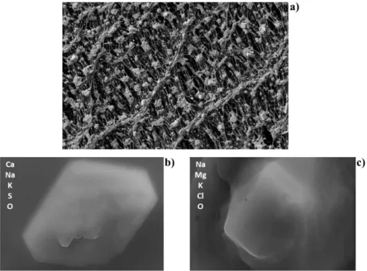
The PM can also have various shapes, as shown in Figure 3. This figure shows photos obtained from scanning electron microscopy (SEM) analysis of different atmospheric particles: Figure 3a exhibits a SEM image of a section of a Teflon filter with atmospheric PM deposited onto its surface; Figures 3b and 3c show the SEM images of two different atmospheric particles and their main elemental composition obtained by means of energy dispersive X-ray (EDX) spectroscopy - in particular, Figure 3c clearly shows the cubic shape of a sodium chloride crystal.
Figure 3. SEM images of different atmospheric particles with aerodynamic diameter less than 2.5μm (photos by courtesy of ORGANOSOL research project, PTDC/CTE-ATM/118551/2010, https://organosolproject.wordpress.com/).
7
1.1.2 Chemical composition of inhalable fine particulate matter
As previously described, PM2.5 are composed roughly of a carbonaceous fraction, trace metals, crustal elements, and inorganic ions. The contribution of each of these components to the total mass of the particle depends on a set of factors, such as the distance from the emission source as well as the emission source itself (Calvo et al., 2013; Putaud et al., 2010). Figure 4 shows a schematic representation of the average composition of PM2.5 samples in Europe.
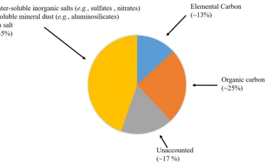
In PM2.5, the EC accounts on average to 13% of the mass of the air particles, ranging from 6% to 25%. The lowest EC contents are usually found in remote areas, such as mountain tops and rural areas, whereas the highest EC values are typically reported for urban and industrial areas, with percentage values as high as 25% of the aerosol mass being measured in road tunnels (Putaud et al., 2010). As for the OC component, the average mass percentage of this organic fraction is about 25%, ranging between 20% and 30%, but it can reach percentage values up to 36% in locations with lower EC emissions. The OC component is further comprised of a water-insoluble organic carbon and water-soluble organic carbon (WSOC) fractions, with the latter accounting for 10 to 80% of the OC fraction (Duarte et al., Figure 4. Schematic representation of the chemical composition of PM2.5 in Europe (adapted
8
2016; Duarte and Duarte, 2011). As recently reviewed by Duarte and co-workers (Duarte et al., 2016), the WSOC fraction is particularly important due to its important role in several atmospheric processes. The WSOC might potentially affect the properties that determine the PM ability to act as cloud condensation nuclei (Dinar et al., 2006; Padró et al., 2010). It has been also suggested that the WSOC might contribute to the absorption of solar radiation, and thus atmospheric heating and global climate change (Dinar et al., 2008; Mladenov et al., 2010). Furthermore, the wet deposition fluxes of atmospheric WSOC indicate that it may be an important temporal source of OC to surface waters and, thus, playing an important role in the global carbon cycle (Jurado et al., 2008). From the public health perspective, there are evidences on the association between aerosol OC and adverse health effects (Cassee et al., 2013), with the secondary OC component (more water-soluble) being suggested as a valuable air quality metric. Lower percentages of WSOC are usually found in urban areas, whereas higher WSOC contents are found in remote locations, such as mountain tops and rural areas (Anderson et al., 2008). This geographic distribution can be explained by the distance from the emission sources, with the PM2.5 samples collected in more remote locations having a higher WSOC content due to the longer distances travelled by these particles as compared to those collected at urban centers, thus allowing them to suffer chemical transformations (secondary aerosols formation), leading to an increase in the WSOC content (Duarte et al., 2016; Duarte and Duarte, 2011). The water-soluble inorganic salts (such as 𝑁O and 𝑆𝑂 ) account for about 40% (26% to 46%) of the PM2.5 mass, while the insoluble mineral dust accounts for about 4% of the PM2.5 mass, and the sea salt only accounts for about 1% of the mass particle (1% to 2%) (Putaud et al., 2010). The water-soluble inorganic salts are mainly formed through secondary processes in the atmosphere and, therefore, it is expected to observe a higher content of these compounds in rural environments due to a higher travel distance from the emission sources being subjected to more secondary processes in the atmosphere (Putaud et al., 2010). The atmospheric content of these compounds follow also a seasonal trend, with the 𝑆𝑂 being found in higher concentrations during warm seasons, whereas 𝑁O are found in higher concentrations during cold seasons (Seinfeld and Pandis, 2006).
1.1.3 Effects of atmospheric particulate matter on human health
The effects of atmospheric particles on human health have been studied throughout several epidemiological and experimental studies. These studies evidenced the noxious
9
effects of the exposure to high levels of PM, namely the increased risks of respiratory and cardiovascular diseases and lung cancer (Hoek et al., 2013; Kim et al., 2015; Pope et al., 2002). For instance, Pope and co-workers (Pope et al., 2002) studied the effects of long term exposure to fine particulate air pollution on lung cancer and cardiopulmonary related mortality. The authors concluded that an increase of 10μg m-3 in atmospheric concentration of PM2.5 was associated with an increase of 6% and 8% in cardiopulmonary and lung cancer mortality risk, respectively. On the other hand, in another study, the same group of authors also reported that a decrease in 10μg m-3 of the concentration of PM2.5 was associated with an increase in live expectancy of about 0.61±0.20 years (Pope et al., 2009).
Reviewing the toxicity and health impact of different PM constituents, Schwarze et al (Schwarze et al., 2006) concluded that metals appear to have an important role in the development of cardiovascular and pulmonary diseases. Additionally, and in spite the short experimental evidence of the effects of the crustal elements, epidemiological studies have shown that these particles may have adverse effects on respiratory morbidity. There has been also reported a high human health risk associated with the mutagenic and carcinogenic effects of the exposure to polycyclic aromatic hydrocarbons (PAHs) present in anthropogenic-related air particles (Mazzeo, 2011), as well as the oxidative stress-induced damage on deoxyribonucleic acid (DNA) caused by the exposure to diesel exhaust particles (DEP) (Risom et al., 2005). Finally, exposure to PM also affects the immune system, increasing the susceptibility to infections and pathogens, or aggravating preexisting lung diseases such as asthma (Bauer et al., 2012; Jang, 2012).
Overall, current data demonstrate the negative impact to PM exposure in terms of human health, and emphasize the importance of implementing mitigation strategies to diminish and limit the emission of fine particles into the atmosphere. In this regard, the European Union created the 2008/50/EC directive, which set the limit levels for the emission of PM in its member states. For the PM2.5 emissions, the European annual threshold by the year of 2015 was set at 20μg m-3, based upon measurements in urban background locations.
10
1.2. The innate immune response to inhaled atmospheric particles
The toxicity of inhaled atmospheric particles and their deleterious effects in human health is in great part due to the induction or exacerbation of a preexisting inflammatory condition. It is therefore important to briefly overview the inflammatory process and their innate immune effectors in order to understand how air particles affect body homeostasis.
1.2.1 Inflammation: a look on the body defense mechanisms
Inflammation is the organism primary mechanism of defense against infections (fungal, bacterial, parasitic or viral), damaged/necrotic cells, and foreign bodies (dirt, splinters or sutures). Even though it is a protective response with the aim of reestablish homeostasis, if exacerbated or sustained in time, inflammation has serious harmful effects to the host. This process can be sorted as acute or chronic depending on the cells involved in its development and resolution. The former is characterized by a short duration, ranging from minutes to a few days, by neutrophilic leukocyte accumulation, fluid and plasma proteins exudation and by a mild level of tissue injury with limited fibrosis. On the other hand, chronic inflammation is characterized by a longer duration, from days to years, by vascular proliferation, influx of lymphocytes and macrophages and severe tissue damage with progressive fibrosis. Additionally, in acute inflammation the systemic and local signs are prominent, whereas in chronic inflammation they are more subtle (Kumar et al., 2010; Serhan, 2010). The inflammatory process is orchestrated by innate immune cells such as neutrophils, macrophages, and dendritic cells and comprises a series of physiological alterations that manifest into the five cardinal signs of inflammation: pain, redness, swelling, heat, and loss of function. The first four signs were known for a long time being described by Celsus, a roman writer of the 1st century AD, whereas the latter (loss of function), was described by Rudolf Virchow in the 19th century (Kumar et al., 2010).
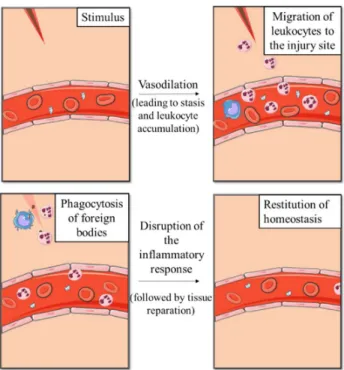
As illustrated in Figure 5, the kinetics of the inflammatory process can be resumed in four main events: phase 1: the vasodilation resulting from the release of inflammatory mediators such as histamine, causes an increase in vascular caliber and leads to an increase in blood flow which is responsible for redness and heat in the inflamed area; phase 2: the vascular dilation is quickly followed by an increase in the permeability of the microvasculature, leading to the extravasation of plasma fluid and proteins into the extravascular tissues; phase 3: this increase in permeability causes the erythrocytes to
11
become more concentrated in the blood, leading to an increase in blood viscosity - a condition known as stasis. As stasis develops, there is an accumulation of blood leukocytes (mainly neutrophils) in the vascular endothelium. Simultaneously, endothelial cells are activated by mediators produced at the infection site or sites of damaged tissue, which causes an increase expression of adhesion molecules. This allows leukocytes to adhere to the endothelium and then migrate into the interstitial tissue in order to reach the injury site; and phase 4: once on the injury site, phagocytic cells such as neutrophils, macrophages and dendritic cells, can ingest and eliminate microorganisms, foreign bodies and necrotic cells, contributing to the resolution of inflammation (Kumar et al., 2010; Serhan, 2010).
Figure 5. Schematic representation of main events of the inflammatory process. Figure made using the Servier Medical Art (“Powerpoint image bank | Servier,” )
As early mentioned, the inflammatory process needs to be tightly regulated in order to prevent damage to healthy tissues. Inflammation is orchestrated by inflammatory mediators, that are only produced in short bursts while the pro-inflammatory stimulus persists, they have short half-lives, and are degraded after their release. Additionally, neutrophils have short half-lives outside the blood vessels and undergo apoptosis within a few hours in the tissues and as the inflammation develops there are a variety of stop signals which are triggered in order to terminate the process (Nathan, 2002; Serhan and Savill,
12
2005). These stop signals lead to the activation of mechanisms enabling a change in the arachidonic acid metabolites that are being produced, from pro-inflammatory metabolites (leukotrienes) to anti-inflammatory metabolites (lipoxins). In addition, these mechanism also lead to the release of anti-inflammatory cytokines, such as transforming growth factor-β (TGF-β) and interleukin-10 (IL-10), production of anti-inflammatory lipid mediators derived from polyunsaturated fatty acids (resolvins and protectins), and neural impulses that inhibit the production of tumor necrosis factor (TNF) by macrophages (Serhan et al., 2008; Tracey, 2002).
1.2.2 Immune players and inflammatory mediators in respiratory system
In the lungs, innate immune response is orchestrated by lung leukocytes, and epithelial cells of the alveolar and conducting airways surfaces (Martin, 2005). In general, the predominant leukocytes acting in the inflammatory response are the neutrophils in the first 6 to 24 hours being then replaced by monocytes in the subsequent 24 to 48 hours. These monocytes differentiate then into macrophages at the tissue level (Kumar et al., 2010).
As previously described, the inflammatory response also relies on soluble mediators, such as proteins and lipids, to help control the different stages of the inflammation. These mediators can be sorted according to their origin in two main groups: the cell-derived and the plasma protein-derived mediators. Among the cell derived mediators are included the vasoactive amines, the arachidonic acid metabolites, the reactive oxygen species (ROS), the nitric oxide, the platelet-activating factor (PAF), the cytokines and the chemokines. The plasma protein-derived group is composed by the mediators from the complement system, the coagulation system, and the kinin system (Kumar et al., 2010).
Among cell-derived mediators, histamine assumes a particular relevance in the inflammatory process in the respiratory system. It’s produced mainly by mast cells that are present in connective tissue beneath the airway basement membrane (the bronchial lumen), near submucosal blood vessels and in the alveolar septa. Histamine is released in response to various stimuli (e.g. physical trauma) and causes bronchoconstriction, mucosal edema, and increased mucus secretion (Kumar et al., 2010; Martin, 2005). Another important group of inflammatory cell-derived mediators are the arachidonic acid metabolites, which includes the prostaglandins, leukotrienes, and lipoxins. Prostaglandins are produced mainly by mast cells, macrophages, and endothelial cells. These compounds result from the action of two
13
cyclooxygenases (COX), COX-1, which is constitutively expressed, and COX-2, which is inducible. The most important prostaglandins in the inflammatory response are the prostaglandins E2, D2, F2α, I2. and thromboxane, deriving each one from the action of a different specific enzyme. Some of the effects of these compounds include: platelet aggregation and vasoconstriction (thromboxane), vasodilation (prostaglandins D2 and I2), inhibition of platelet aggregation and potentiation of the permeability-increasing and chemotactic effects of other mediators (prostaglandin I2), contraction of bronchial smooth muscle and small arterioles (prostaglandin F2α), and chemoattraction of neutrophils (prostaglandin D2) (Kumar et al., 2010; Ricciotti and FitzGerald, 2011; Serhan et al., 2008). The leukotrienes, on the other hand, are produced by the action of the lipoxygenase enzyme. The leukotrienes are mainly produced by leukocytes, acting as chemoattractants for these cells and having vascular effects. In particular, the leukotriene B4 can act as a strong chemotactic agent and activator of neutrophils, leading to the aggregation and adhesion of the neutrophils to the venular endothelium, release of lysosomal enzymes, and generation of ROS. On the other hand, leukotrienes C4, D4, and E4 can cause intense vasoconstriction, bronchospasm and augmented vascular permeability, being more potent than the histamine in the latter effects (Kumar et al., 2010; Serhan et al., 2008). Finally, the lipoxins are also formed through the action of the lipoxygenase. However, unlike the leukotrienes, lipoxins are inhibitors of the inflammation. Leukocytes, mainly neutrophils, produce intermediate compounds in the lipoxin synthesis, which are then converted to lipoxins by platelets. Lipoxins primarily act in the inhibition of leukocyte recruitment and neutrophil chemotaxis, and adhesion to the endothelium (Kumar et al., 2010; Serhan et al., 2008).
Another cell-derived mediator of the inflammatory cascade is PAF, which is produced by a range of different cells, such as platelets, basophils, mast cells, neutrophils, endothelial cells and alveolar macrophages. This mediator can cause platelet aggregation, vasoconstriction and bronchoconstriction and, in extremely low concentration, it can also cause vasodilation and augmented permeability in venules, with a higher strength than that of histamine (up to 10000 times higher). In addition to these effects, PAF also causes an increase in the leukocyte adhesion to the endothelium, oxidative burst, chemotaxis, and it can boost the synthesis of other inflammatory mediators, particularly eicosanoids (e.g. prostaglandins and leukotrienes) (Kumar et al., 2010; Stafforini et al., 2003).
14
Among the cell-derived mediators there are also soluble proteins, named cytokines, which are mainly produced by activated lymphocytes and macrophages and whose purpose is to modulate the functions of other cells. A few examples of this group of soluble proteins include, IL-1, IL-6, TNF, and chemokines. TNF and IL-1 are two of the major effectors of the inflammatory process, exerting both local and systemic effects. The local effects of these mediators have actions on three main cell types: vascular endothelium, leukocytes, and fibroblasts. At the vascular endothelium, TNF and IL-1 stimulate the expression of adhesion molecules, coagulation and production of IL-1 and chemokines, while suppressing the anticoagulant activity. These soluble proteins also activate leukocytes and stimulate the production of other inflammatory cytokines. Finally, TNF and IL-1 stimulate the proliferation of fibroblasts and collagen synthesis by these cells. As for the systemic effects of these mediators, they include fever, leukocytosis, loss of appetite, positive modulation of the production of acute-phase proteins, and stimulation of sleep (Kumar et al., 2010).
Chemokines, a family of small proteins, constitute another important player in the inflammatory process, acting mainly as chemoattractant for leukocytes. These molecules are distributed according to four groups: i) α chemokines (e.g. IL-8), which are released by macrophages and act on the activation and chemotaxis of neutrophils, with some activity on monocytes and eosinophils. The release of these chemokines is stimulated by microbial products and other cytokines (IL-1 and TNF); ii) β chemokines (e.g. monocyte chemoattractant protein, eotaxin), which are chemoattractant to monocytes, eosinophils, basophils and lymphocytes; iii) γ chemokines (e.g. lymphotactin), which are specific chemoattractant for lymphocytes; and iv) CX3C chemokines (e.g. fractalkine), which acts as a potent chemoattractant for monocytes and T cells in its soluble form, however in its cell-surface bound form, it promotes adhesion of monocytes and T cells (Charo and Ransohoff, 2006; Sallusto and Mackay, 2004).
Other important players in the inflammatory process are the reactive oxygen and nitrogen species (ROS and RNS, respectively). ROS are oxygen derived free radicals, mainly superoxide anion, hydrogen peroxide and hydroxyl radical, produced by leukocytes through the activation of the NADPH oxidase system. When released, these mediators increase the expression of chemokines, cytokines and leukocyte adhesion molecules, amplifying the inflammatory response. ROS are also the main effectors in the destruction of phagocytosed microbes. The release of high levels of these mediators can cause massive cell damage to the
15
host; to prevent these damages, the organism uses a series of antioxidant mechanisms (Kumar et al., 2010; Salvemini et al., 2006). Nitric oxide, produced by the activity of inducible nitric oxide synthase (NOS2), is also an important mediator of inflammation, and when present in high levels, it also acts as a microbicide agent (Cirino et al., 2006; Laroux et al., 2001).
Finally, among the plasma protein-derived mediators, the complement system mediators C3a, C5a, and C4a act on the inflammatory response by stimulating histamine release from mast cells. Moreover, C5a is also a strong chemotactic agent for neutrophils, eosinophils, monocytes and basophils, and activates the lipoxygenase pathway in neutrophils and monocytes. The C3b connects to microbial cell walls, promoting the phagocytosis by macrophages and neutrophils. In turn, the kinin mediator bradykinin, is responsible for increasing the vascular permeability, and causing systemic effects such as pain (Kumar et al., 2010).
Since the respiratory system is the primary and major exposure site to inhaled air particles, the toxicological study of their interaction is of great relevance, particularly in respect to possible pro-inflammatory effects. This topic is therefore explored in the following section.
1.2.3 Airways inflammation and oxidative stress due to inhalation of air particles Understanding the interaction between the body and atmospheric air particles, as well as the mechanisms through which the air particles cause inflammation and oxidative stress is of major importance. One of the main theories that has been put forward is that particles lead to pro-inflammatory responses and other cellular effects by means of formation of ROS and induction of oxidative stress. However, there are also other mechanisms leading to the activation of non-oxidant pro-inflammatory mediators, such as the activation of channels, like the transient receptor potential cation channel subfamily V member 1 or TRPV1, directly by combustion particles (Øvrevik et al., 2015). Table 1 summarizes the main biological effects of the exposure to PM.
16
Table 1. Summary of the main biological responses to PM exposure Experimental
method Sample Main observations Reference
In vitro study on rat alveolar cell lines
PM2.5 and PM10
Oxidative stress induction due to the particle contents on Ni, Cr, Cu and WSOC.
(Daher et al., 2012) In vitro study on
macrophages PM
Dose dependent modulation of NO production due to alterations on NO synthase protein expression.
(Chauhan et al., 2004)
In vivo study on
rats PM2.5
Inflammation mediated by pathways regulated by ROS and NO.
Oxidative stress mainly due to inhibition of SOD activity.
Activation of metabolic enzymes leading to exacerbation of respiratory diseases.
(Li et al., 2015) In vivo study on rats Ambient and wood smoke air particles
Oxidative stress induction.
Stimulation of pro-inflammatory genes in the lungs. (Danielsen et al., 2010) In vitro study on alveolar epithelial cells and macrophages PM10
Cytotoxicity, pro-inflammatory response and oxidative stress induction.
When exposed in equal concentrations of rural and urban extracts; urban extracts produced a greater response.
(Michael et al., 2013) In vitro study on human bronchial epithelial cells Diesel exhaust particles
Pro-inflammatory response through activation of NF-kB and MAPK pathways.
(Totlandsdal et al., 2010) In vivo study on humans Diesel exhaust particles
Increased production of IL-8, endothelial adhesion molecules and neutrophil recruitment
(Stenfors et al., 2004)
17
Nowadays, there are several studies relating air particles inhalation and the induction of oxidative stress. Daher and co-workers, using rat alveolar cell lines, studied the redox activity of fine and coarse PM (Daher et al., 2012). The authors verified the existence of a strong correlation between cell ROS production and the PM content in Ni, Cr, Cu and WSOC, suggesting that these PM constituents are the main factors responsible for oxidative stress induction. Another recent in vitro study using macrophages, demonstrated that the capacity of PM to induce ROS is inversely correlated to PM size, and that photochemical aging as well as the content in Cu, Cr, Pb, Co, Ni and WSOC causes an increase in ROS production (Saffari et al., 2014). Chauhan and co-workers carried out an in vitro study to assess the effect of air particles in the nitric oxide production by macrophages (Chauhan et al., 2004). The authors reported that the exposure to inhalable air particles induce a dose dependent modulation of nitric oxide released, and concluded that this modulation was caused by an alteration in nitric oxide synthase protein expression (Chauhan et al., 2004). This set of studies suggest the potential of PM for inducing oxidative stress; however, beside the direct noxious effects of ROS and RNS, oxidative stress can also lead to the activation of a series of intracellular signaling pathways, such as MAPK transcription factors activator protein 1 (AP-1) and NF-kB. These signaling pathways control the expression of numerous pro-inflammatory effectors such as interleukins, COX-2, and inducible isoform of the nitric oxide synthase (NOS2), which are major players in pathological changes of the lungs (Li et al., 2008; Mazzoli-Rocha et al., 2010).
Beside the induction of oxidative stress, PM can also promote the production of pro-inflammatory mediators, leading to inflammation and damage of the airways. In an in vivo study on the effects of PM2.5 on rats, Li and co-workers reported that PM2.5 induced a lung inflammatory injury, accompanied by an increase in pro-inflammatory cytokine levels and inflammatory cell numbers (Li et al., 2015). The authors also verified that the oxidative stress caused by PM2.5 was due to both the inhibition of the super oxide dismutase (SOD) activity and the stimulation of the iNOS activity, leading to an increase in the nitric oxide levels. The authors concluded that PM2.5 induced inflammation in the rat lungs is mediated, in part, by the pathways mediated by ROS and nitric oxide and the activation of metabolic enzymes which exacerbate respiratory diseases (Li et al., 2015). Similarly, Danielsen and co-workers, using an in vivo study in rats, also verified that ambient and wood smoke air particles, induced an oxidative stress response and a stimulation of the expression of pro-inflammatory
18
genes in the lungs (Danielsen et al., 2010). Michael and co-workers, using an in vitro approach, verified that the cytotoxicity, oxidative stress, and pro-inflammatory responses in lung epithelial cells and macrophages significantly differed after exposure to equal concentrations of urban and rural PM, with the highest values being associated with urban air particles (Michael et al., 2013). This result confirms the hypothesis that PM composition and source constitute an important factor in PM-induced toxicity (Michael et al., 2013).
Additional studies have been reported in the literature focusing on both the effects of PM on the production of pro-inflammatory mediators, and in several pathways that play an important role in the immune system. In an in vitro study of the effects of DEP in human bronchial epithelial cells, Totlandsdal and co-workers reported that the pro-inflammatory response induced by DEP appeared to occur via both the activation of nuclear factor kB (NF-kB) and the p38 mitogen-activated protein kinase (MAPK) (Totlandsdal et al., 2010). Another in vitro study on the induction of cytokine expression in human bronchial epithelial cells by PM, showed an increase in the transcription of mRNA and the release of 1, IL-8, granulocyte-macrophage colony-stimulating factor, and leukemia inhibitory factor, leading to the enhancement of the inflammatory response in the lungs (Fujii et al., 2001). These results are corroborated by in vivo data showing that asthmatic and healthy humans exposed to DEP produce a vast profile of cytokines (Stenfors et al., 2004). According to this data, DEP had a direct effect on the IL-8 production, with an upregulation of endothelial adhesion molecules and neutrophil recruitment in healthy human airways, whereas the mildly asthmatic humans responded with an induction of epithelial IL-10 (Stenfors et al., 2004). The gathered data suggest that more work is required to clarify the different conclusions reported in the literature.
1.3. Objectives of the M.Sc. Thesis
The review presented in this Introduction section, has shown that atmospheric PM can have different chemical composition, depending on their local or regional sources, sampling location (i.e., remote, rural, or urban location and, therefore, linked to variations in residence time in the atmosphere), and even the seasonal period. It has been shown that the fine size range of PM, i.e., PM2.5, is composed mainly of EC, OC (consisting of a water-soluble and a water-inwater-soluble fraction), water-water-soluble inorganic salts, mineral dust, and sea salt. Moreover, the PM2.5 can exert an array of detrimental effects on the human health,
19
including the increasing risk of cardiovascular and pulmonary diseases, carcinogenic effects, oxidative damage to DNA, and effects on the immune system causing augmented susceptibility to infections and pathogens. In the airways, PM2.5 is known to induce oxidative stress and inflammation (e.g. through stimulation of the production and release of pro-inflammatory mediators), leading in some cases to lung injury.
An important finding of this review work is that atmospheric PM2.5 can have different effects on the lungs tissues depending on its chemical composition. The available literature on this issue suggests that water-soluble trace metals (e.g., Ni, Cu, Cr, Co and Pb) and WSOC have an important influence on the oxidative potential of airborne PM. Nevertheless, it should also be highlighted that the link between WSOC composition and PM-induced oxidative activity is still unknown, mostly because of the complex nature of the atmospheric particulate WSOC component. Accurate characterization of the WSOC fraction is therefore mandatory for better understanding the effect of this organic fraction on PM-associated pro-inflammatory potential, particularly in urban areas.
Thus, the main objective of this M.Sc. thesis is to assess the pro-inflammatory effects and oxidative potential of the WSOC fraction from atmospheric aerosol samples. To these aims, PM2.5 samples were collected at the campus of the University of Aveiro, which is classified as a near-city background site, located between 3 and 10 km from large pollution sources (e.g., an industrial complex, which includes the production of nitric acid, aniline, nitrobenzene and chlorate compounds, is located 10 km to the North of the city of Aveiro). The sampling campaign was conducted during the day and night periods in late autumn, in order to assess and compare the role played by different ambient conditions on the effect of WSOC on the PM-associated oxidative potential. The WSOC fractions were extracted from the collected PM2.5 samples following analytical procedures described in previous studies (e.g., (Duarte et al., 2016)). The structural features of the collected aerosol WSOC fractions were assessed by means of excitation-emission matrix (EEM) fluorescence, ultraviolet-visible (UV-Vis), and solution-state one-dimensional 1H nuclear magnetic resonance (NMR) and two-dimensional NMR (1H-1H Correlation Spectroscopy (COSY), and 1H-13C Heteronuclear Single Quantum Coherence (HSQC) and Heteronuclear Multiple Bond Correlation (HMBC)) spectroscopies. Finally, the biological effects of the WSOC fractions collected under the different ambient conditions were assessed on Raw 264.7 macrophage
20
cell line. The results were then related to the atmospheric concentrations and structural characteristics of the different WSOC fractions.
21
Chapter 2
Materials and Methods
23
2.1 Atmospheric aerosol sampling campaigns
The PM2.5 samples were collected on pre-fired (at 500 °C for 12h) quartz fibre filters (20.3 × 25.4 cm Whatman QM-A) with an airflow rate of 1.13 m3 min-1. The high-volume sampler (model TE-6070V, Tisch Environmental, Inc.) was provided with a pre-separator for excluding particles larger than 2.5 μm. This size-cut was accomplished using a size selective PM10 inlet (model TE-6001, Tisch Environmental, Inc.) and a PM2.5 single-stage impactor (model TE-231, Tisch Environmental, Inc.) and a back-up quartz filter. The total mass of the collected PM2.5 samples was determined by weighing the filter under controlled moisture conditions before and after sampling. The filters were then wrapped with aluminium paper and stored frozen until further analysis.
The sampling campaign was conducted between the 14th and 29th of November 2016 (late autumn), during the weekdays (i.e., from Monday to Friday) and in two distinct time periods: (i) day-time: 9:40 a.m. to 5:30 p.m. and (ii) overnight: 5:40 p.m. to 9:30 a.m. Throughout the sampling period no attempts were made to control any adsorption/desorption phenomena on the filters. Consequently, some volatilisation/condensation processes may occur on the filter and particles surface for semi-volatile organics. Also, oxidation by strong oxidants (e.g. ozone) of filter deposited organics may occur during aerosol collection. Therefore, in a similar fashion to what has been considered in previous works ((Duarte et al., 2007; Lopes et al., 2015), the measured concentrations and chemical features for oxygenated organic species, namely WSOC, should be considered an upper limit of the true atmospheric levels.
2.2. Extraction of aerosol WSOC fractions
An area of 315 cm2 of each filter was extracted with 150 mL of ultra-pure water (18.2 M Ω cm, filter area to water volume ratio of 2.1 cm2 mL-1) by mechanical stirring during 5 min plus ultrasonic bath during 15 min. The final slurry obtained was filtered through a hydrophilic polyvinylidene fluoride (PVDF) membrane filter (Durapore®, Millipore, Cork, Ireland) of 0.22 mm pore size. The filtered aqueous extracts were then collected into vials and stored frozen until further analysis.
24
2.3. Excitation–emission matrix (EEM) fluorescence spectroscopy of the
WSOC samples
The fluorescence spectra of each WSOC sample were recorded on a spectrophotometer JASCO (Tokyo, Japan) model FP-6500 using a 1 cm path-length quartz cuvette. Excitation and emission wavelength ranges were set from 220 to 450 nm and 250 to 600 nm, respectively, and their scanning intervals were set at 10 nm and 5 nm, respectively. The excitation and emission slit widths were fixed at 10 nm and the scan speed was set at 100 nm min-1. For each day of EEM analysis, a spectrum of ultra-pure water was acquired under the same experimental conditions and used as blank. The peaks due to water Raman scattering were eliminated from all WSOC samples spectra by subtracting the ultra-pure water blank spectra.
2.4. UV-vis spectroscopy of the WSOC samples
The UV–vis spectra (in the range of 200–600 nm) were registered on an UV–vis spectrophotometer Shimadzu (Dusseldorf, Kent, Germany) Model UV 2101PC in 2 cm path length quartz cells. Ultra-pure water was used as a blank.
2.5. Dissolved Organic Carbon (DOC) analysis of the WSOC samples
The DOC content of the aqueous extracts of the PM2.5 samples was measured by means of a Skalar (Breda, Netherlands) San++ Automated Wet Chemistry Analyzer, based on a UV-persulfate oxidation method (Lopes et al., 2006). The WSOC concentrations are expressed in µg C m-3.
2.6. WSOC samples preparation for NMR and biological studies
In order to have sufficient amount of WSOC samples for both NMR structural characterization and biological assay studies, the WSOC samples from each daily time period were batched together, on a total of two pooled WSOC samples representative of Day and Night time conditions. Subsequently, each pooled WSOC sample was concentrated in a rotary evaporator, at 35ºC, until a final volume of approximately 200 mL. This volume was equally separated (100 mL) into 2 round-bottom flasks (one for NMR analysis and the other