Spectroscopic characterization of
a
high-potential monohaem cytochrome
from
Wolinella
succinogenes,
a
nitrate-respiring organism
Redox and spin equilibria studies
Isabel MOURA’, Ming Y . LIU’, Cristina COSTA I , Ming C. LIU’, Govind PAT’, Antonio V. XAVIER’, Jean LeGALL’,
William J. PAYNE and Josi: J. G. MOURA
’
Centro de Quimica Estrulurdl, Universidade Nove de Lisboa
Departments of Biochemistry and Microbiology, University of Georgia, Athens (Received May 9, 1988) EJB 88 0528
When purified, a high-potential c-type monohaem cytochrome from the nitrate-respiring organism, Wollinellu
succinogenes (VPI 10659), displayed a minimum molecular mass of 8.2 kDa and 0.9 mol iron and 0.95 mol haem groups/mol protein. Visible light spectroscopy suggested the presence of an equilibrium between two ligand arrangements around the haem, i. e. an absorption band at 695 nm characteristic of haem-methionine coordination (low-spin form) coexisting with a high-spin form revealed by a band at 619 nm and a shoulder at 498 nm. The mid-point redox potential measured by visible redox titration of the low-spin form was approximately
+
100 mV. Binding cyanide ( K , = 5 x lo5 M-’) resulted in the displacement of the methionyl axial residue, and full conversion to a low-spin, cyanide-bound form.Structural features were studied by 300-MHz ‘H-NMR spectroscopy. In the oxidized state, the pH dependence of the haein methyl resonances (pH range 5 - 10) and the magnetic susceptibility measurements (using an
NMR method) were consistent with the visible light spectroscopic data for the presence of a high-spin/low-spin equilibrium with a transition pK, of 7.3. The spin equilibrium was fast on the NMR time scale. The haem methyl resonances presented large downfield chemical shifts. An unusually broad methyl resonance at around 35 ppm (pH = 7.5, 25°C) was extremely temperature-dependent [6(323K) - 6(273K) = 7.2 ppm] and was assigned to the S-CH3 group of the axial methionine. In the ferrous state only a low-spin form is present. The haem meso
protons, the methyl group and the methylene protons from the axial methionine were identified in the reduced form. The resonances from the aromatic residues (three tyrosines and one phenylalanine) were also assigned.
Detailed monitoring of the NMR-redox pattern of the monohaem cytochrome from the fully reduced up to the fully oxidized state revealed that the rate of the intermolecular electronic exchange process was approximately 6 x lo6 M-’ sK1 at 303 K and pH = 6.31.
A dihaem cytochrome also present in the crude cell extract and purified to a homogeneous state, exhibited a molecular mass of 21 kDa and contained 2.43 mol iron and 1.89 mol haem c moieties/mol cytochrome. The absorption spectrum in the visible region exhibited no band at 695 nm, suggesting that methione is not a ligand for either of the two haems. Recovery of only small amounts of this protein prevented more detailed structural analyzes
In denitrifying bacteria, nitrate and its reduction products, nitrite, nitric oxide and nitrous oxide, serve as terminal elec- tron acceptors in anaerobic respiration [l]. The electron trans- port system responsible for the denitrifying process is coin- plex. The different electron acceptors involved are not yet precisely placed in the electron transfer scheme. Independent or branched electron transfer chains lead the electrons towards the different nitrogen oxides during denitrification [l] and a number of b and c-type cytochromes have been detected [2]. Furthermore, certain bacteria carry out less ex- tensive denitrification than others [3]. Some reduce nitrate incompletely to nitrous oxide. Others reduce nitrite, nitric oxide and nitrous oxide, but not nitrate to dinitrogen [3].
Different still is Wolinellu succinogenes (VPI 10659), which exhibits capability for dissimilatory reduction of nitrate Correspondence 10 J. J . G. Moura, Cenlro de Quimica Estrutural, Complexo Interdisciplinar, Avenida Rovisco Pais, P-1096 Lisboa, Portugal
through nitrite to ammonia only. It produces no gaseous products but reduces both nitric and nitrous oxides [3]. The electron transport components of this organism thus appear uniquely interesting.
In addition to the previously purified hexahaem c-type cytochrome (an ammonia-yielding nitrite reductase) [4] we isolated and purified two other c-type cytochromes from nitrate-respiring W . succinogenes cells. One is a high-potential monohaem cytochrome present exclusively in the soluble frac- tion and the other a dihaem cytochrome located in both the soluble and the membrane fractions. The present article describes the purification of these cytochromes and a detailed spectroscopic study of the monohaem cytochrome. Our in- terest in this cytochrome arises from having noticed its unique physicochemical properties revealed by visible-light absorp- tion and NMR analyzes. The spectroscopic features are com- pared with those of cytochromes from other dissimilatory nitrate reducers.
674
MATERIALS AND METHODS
W. succinogcvzes (VPI 10659) was grown anaerobically under oxygen-free argon at 37°C in VSY-4 medium (pH 7.6)
[5] supplemented with 100 mM nitrate. Cells were harvested by centrifugation after 24 h, ruptured and fractionated as indicated in the text. Products employed in the purification procedures included Ultragel AcA-44 (obtained from LKB Instruments) and DEAE-Bio-Gel A and Bio-Gel HTP pur- chased from Bio-Rad Laboratories.
Ultraviolet/visible-light absorption spectra of the cyto- chromes were obtained with a Varian DMS 90 recording spectrophotometer. The number of c-type haem groups per molecule of protein was estimated by the pyridine haemo- chromogen techniques based on a red E~~~ nm = 29.1 mM-' cm-' of haem I' [6]. Analytical polyacrylamide disc gel
electrophoresis was performed according to the method of Brewer and Ashworth [7]. Minimal molecular masses were determined by SDS/polyacrylamide gel electrophoresis ac- cording to the method of Weber and Osborn [S]. Iron content was determined by plasma emission spectroscopy using the Jarrel-Ash model 750 Atomcomp. The modified Biuret method developed by Zamenhof and Chargoff [9] was used for the protein determination using horse cytochrome c as the standard. Amino acid analyzes were performed with a Beckman model 120 amino acid analyzer. Protein samples were hydrolyzed in 6 M HCI at 110°C for 24 h, according to the method of Moore and Stein [lo]. The values for threonine, serine and tyrosine were corrected for decomposition during hydrolysis.
Electron paramagnetic resonance (EPR) was carried out on a Bruker 200-tt spectrometer, equipped with an ESR-9 flow cryostat (Oxford Instruments Co., Oxford) and a Nicolet 1180 computer where mathematical manipulations were performed.
For the nuclear magnetic resonance (NMR) studies, the protein was dialyzed against deionized water at pH 7.0 at 4°C and lyophilized twice from 99.8% ' H 2 0 . The sample was dissolved in 99.8% 2 H z 0 and the pH was adjusted with either NaO'H or 'HC1. Quoted pH values are meter readings uncor- rected for the isotope effect. High-resolution N M R spectra were recorded in the Fourier-transform mode on a Bruker CXP 300 spectrometer (300 MHz) equipped with an Aspect 2000 computer. The temperature was controlled to + O S T with a standard Bruker B-VT-1000 variable temperature con- trol unit. All chemical shift values are quoted in parts per million (ppm from internal sodium 3-trimethylsilyl-(2,2,3,3- 'H4)propionate, positive values referring to low-field shifts. Reduction of the protein was achieved by addition of small amounts of sodium dithionite under an argon atmosphere, to delay auto-oxidation. The samples were reoxidized very slowly by introducing small amounts of air into the NMR tube with a Hamilton syringe through serum bottle caps.
Magnetic susceptibility measurements were performed by an NMR method [ l l ] using concentric cylindrical cells (Wilmad Glass Company Inc., WGS-SBL), the internal capil- lary containing only the solvent and the marker. The methyl proton resonance of sodium trimethylsilyl (2,2,3,3,-2H4)- propionate was used as susceptibility marker.
The paramagnetic contribution to the molar magnetic sus- ceptibility (&) of the oxidized form was determined as a function of pH. The differences
4f'
between the resonance positions of the marker in the protein and in the capillarysolution were measured and converted into magnetic suscepti- bilities using the relation:
3n A f '
xo
X M = - -
+
4 f;.
where xM is the molar magnetic susceptibility,fis 3 x 10' Hz and c is the concentration of the protein. The solvent and solution densities were assumed to be similar.
xo
is the mag- netic susceptibility of the solvent and was considered to be negligible. A correction value ofxD
= 1.7 x cm3/mol was used for the diamagnetic contribution of the protein [12].Purifi'cution o f t h e cytochromes
Frozen cells (400 g) were mixed with 10 mM Tris/HCl buffer (pH 7.6 at 4°C) to give 1:1.5 (mass/vol.) suspension and broken by passing three times through a MantonGaulin homogenizer at 62 MPa. A few crystals of DNase were added
to lessen the viscosity of the extract. After 20 min, the prep- aration was centrifuged at 13 200 x g in a Sorvall RC-SB re- frigerated centrifuge for 30 min. The pellet was discarded and the supernatant fluid treated with neutralized streptomycin sulfate (0.5 mg/mg protein). After stirring for 15 min, the preparation was centrifuged at I44000 x g for 2 h. The super- natant containing only soluble proteins served as the source of the high-potential monohaem c-type cytochrome, whereas the low-potential c-type cytochrome was extracted and purified from the pellet (membrane particles).
High-potential monohaem cytochrome
The soluble fraction was dialyzed against 10 mM Tris/ HCI buffer (pH = 7.6) for 18 h with three changes of dialysis
buffer. The monohaem cytochrome was next subjected to the chromatographic purification steps as indicated in Scheme 1,
in which all the elution buffer concentrations and columns sizes are indicated.
Low-potentiul dihaem cytochrome
The membrane fraction was washed three times with 200 ml 0.1 M Tris/HCl buffer and recovered each time by centrifuging at 144000 x g for 1 h. The supernatant fluids containing contaminating soluble proteins were discarded. The pellet from the last wash was suspended in 0.1 M Tris/ HCl buffer with a final volume of 200 ml. Sodium cholate was
Cell-free extract of W. succinogenes (after ultra-ccn trifugation and dialysis) DEAE-Bio-Gel A (6 x 30 cm)
Hydroxyapatite
1
(4 x 15 cm)(non-adsorbed fraction) washing with 10 mM Tris/HCI equilibrated with 0.1 M Tris/HCI non-adsorbed fraction from washing with 0.1 M Tris/HCI
1
Dialysis
1
DEAE-Bio-Gel A (3 x 15 cm) Hydroxyapatite
1
(4 x 15 cm)non-adsorbed fraction from washing with 10 mM Tris/HCl gradient (1 -50 mM phosphate) monohaem cytochrome was eluted at 20 mM phosphate
elution with 0.05 M Tris/HCl pure cytochrome
4
Sephadex G-50 (5 x 90 cm)
4
Cholate-solubilized fractions Hydroxyapatite (4.5 x 20 em)
equilibrated with 0.1 M Tris/HCI, gradient Tris/HCI(O.l - 0.01 M) gradient phosphate (0.01 -0.2 M) low
potential cytochrome eluted at 30 m M phosphate
1
Dialysis
1
DEAE-Bio-Gel A (2.5 x 25 em)
Gradient Tris/HCl (0.01 - 0.2 M) low-potential cyto- chrome eluted at 0.16 M
1
Concentration on a n Amicon Diaflo with YM5 membrane Ultrogel AcA-44 (2 x 90 em)
1
Scheme 2
1
elution with 0.05 M Tris/HCI: purc cytochroine
added ( 3 mg/ml) and, after the cholate had dissolved, the mixture was incubated at 4°C for 24 h. The preparation was centrifuged and the pellet again suspended and similarly ex- tracted with a higher concentration of sodium cholate (4 mg/ ml). The cholate extracts containing the low-potential c-type cytochrome and the nitrite reductase were combined and sub- jected to the chromatographic purification steps indicated in Scheme 2.
Redox titration of the high-potential cytochrome
The redox titration of the protein was performed under anaerobic conditions in an optical redox cell, modified slightly from the design of Dutton et al. [13]. The potentials were measured with a Fisher Acumet pH meter, model 620. The redox cell containing a volume of 5 ml was kept well stirred and under a continuous stream of argon gas. The final protein concentration was 20 pM. The redox mediator used was 2,6- dichloroindophenol in a final concentration of 2 pM. The potentials were varied by injecting appropriate volumes of deaerated 1 mM sodium dithionite and potassium ferricya- nide. The protein was dissolved in 100 mM potassium phos- phate buffer, pH 7.6, and titrated in both the oxidative and reductive directions to ensure reversibility. Optical spectra were recorded throughout the titration.
Determination q f the binding constant of cyanide to ferric high-potential cytochrome
A 430 pM solution W . succinogenes high-potential cyto-
chrome in 0.01 M TrislHCI, pH = 7, was titrdted with potas- sium cyanide (7 mM). The titration was carried out by in- jecting different amounts of potassium cyanide solution. The absorbance of the solution at 619 nm was measured after a 15-min incubation. Longer periods of incubation yielded no further changes. The pH value during the titration was maintained at 7.0.
Absorption changes measured at 619 nm were used to determine the ligand stoichiometry and equilibrium constant. The ligand stoichiometry ( n ) and the association constant (K,) were determined considering that for the equilibrium reaction :
Cyt-c
+
n(CN-) $ Cyt-c (CN-),. The binding parameters can be calculated as follows:where A. is the absorbance of the solution in the absence of cyanide,
Aloe
is the value of the absorbance of the solution when the cytochrome is completely ligated to CN- and A isthe absorbance of the solution for a given concentration of C N - .
The concentration of cyanide ion was calculated from the total concentration of cyanide, taking into account the pH and the acid dissociation constant of hydrogen cyanide:
RESULTS
Lo w-p o t en t iddihaem cy tochromes
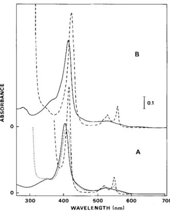
Inspection of the absorption spectra of the purified low- potential dihaem cytochrome indicated that, as the desig- nation implies (Fig. 1 A), sodium dithionine with a low redox potential (broken line) fully reduced this cytochrome, but the higher-potential sodium ascorbate (dotted line) did not. Partial, just perceptible, reduction by sodium ascorbate shown by the small rise in the 550-nm region) indicated that one of the haem groups has a redox potential only slightly higher than of sodium ascorbate. A purity index [ A s s o - A ~ ~ ~ nm
(reduced)/Azso nm (oxidized)] of 1.69 was calculated for this
cytochrome. Assay of a concentrated solution of the low- potential cytochroine by visible-light spectroscopy revealed no band at 695 nm, suggesting that no methionine ligand arrangement occurs for this cytochrome (Fig. 2). For com- parison the visible spectra of Desulfovibria vulgaris tetrahaem
lu 0 z K 0 v) m a
2
0 0 I I I P I 1 1 I I 1 I 1 1 I I 1 I 1 1 B 300 400 5 0 0 6 0 0 7 0 0 WAVELENGTH (nm)Fig. 1. Absorption spectru of'cytochvomesfrom W. succinogenes. (A) The low-potential c-type: (-) oxidized; (. .. .. .) ascorbate-re- duced; (- - - -) dithionite-reduced. The protein concentration was
2.5 pM in 0.1 M potassium phosphate buffer, pH 7.6. Solid sodium ascorbate o r sodium dithionitc was added for reduction of the cytochrome. (B) The high-potential c-type: (----) oxidized; (- - - -)
ascorbate-reduced. Thc protein concentration was 8.5 pM in 0.1 M potassium phosphate buffer, pH 7.6. Solid sodium ascorbate was added for reduction of cytochrome
676 .8 .6 2
2
$ .4 a 0 v) .2 500 600 7 0 0 800 WAVELENGTH (nm)Fig. 2. Absorption spectru a! the higher wavelength .fhr cytochromes f r o m W. succinogenes and other sources. (. . . .) Horse heart cyto- chrome c; (- - -) Desulfovibrio vulgaris cytochrome c 3 ; (-)
W . succinogenes high-potential c-type cytochrome; (- . - . -) W. suc-
cinogenes low-potential c-type cytochrome. Horse heart cytochrome c has an axial ligand arrangement of the histidine-methionine type and shows a peak in the 695-nm region. D. vulgaris cytochrome c3 has a histidine-histidine axial ligand arrangement and does not show the 695-nm peak. The spectrum of the W. succinogenes high-potential
c-type cytochrome resembles that of horse heart cytochrome c,
whereas that of W . succinogenes low-potential c-type cytochrome has features similar to those of the D. vulgaris cytochrome c 3
cytochrome c3 (c-tpye haem with bis-hystidinyl axial coordi- nation) and horse-heart cytochrome c (c-type haem with methionyl and hystidinyl axial ligands) are super-imposed in the figure.
The molecular mass determined by SDS/polyacrylamide gel electrophoresis was 11 kDa. Based on this value, we found 2.43 mol Fe and 1.89 mol haem c/mol cytochrome and con- cluded it to be a dihaem protein.
High-potential monohaem cytochromes
SDS/polyacrylamide gel electrophoresis revealed that the monohaem cytochrome has a niimiinal molecular inass of 8.2 kDa. Based on this value we found 0.87 mol Fe and 0.95 mol haem c/mol cytochrome.
Spectra in Fig. 1 B reflect the typical c-type cytochrome nature of this protein. A purity index [ A 5 5 2 - A 5 7 0
",,,
(reduced)/Azso nm (oxidized)] of 1.06 was determined. Table 1compares the absorption maxima and molar absorption coef- ficients of both cytochromes. In an attempt to elucidate the ligand arrangement around the haem iron atom of the purified cytochrome, spectral details of a concentrated preparation of the oxidized cytochrome were examined (Fig. 2). An absorp- tion maximum at 695 nm indicated the presence of a methionyl residue as an axial haem ligand. In addition to the typical absorption bands of a ferric c-type cytochrome, another band was found at 619 nm and a shoulder at 498 nm revealing the presence of a high-spin-state ferric haem iron. After addition of uotassium cvanide to the ferricvtochrome.
Table 1. Absorption muxima andmolur absorption coefficients of c-type
cytochromes f r o m W. succinogenes
The values were determining for purified cytochromes in 0.1 M so- dium phosphate buffer, p H = 7.6. The high-potential c-type cytochrome was reduced by addition of a few crystals of sodium ascorbate; the low-potential c-type cytochrome by addition of a few crystals of sodium dithionite
c-Type Oxidized Reduced
cytochrome species --______ 4 n a x i: Lax E nm High-potential 522.0 407.2 280.0 Low-poten tial 530.0 401.5 280.0 monohaem dihaem m M - ' em-' 7.5 80.1 19.2 20.7 228.1 22.1 nm mM-' em-' 552.0 20.3 523.2 12.6 416.5 104.0 550.6 57.0 552.0 30.5 418.0 349.2
Table 2. Amino acid composition of'c-type cyfochromesfrom W. suc- cinogenes
Amino acid Residues/molecule of
high-poten tial low-poten tial monohaem dihaem cytochrome cytochrome LY S His Arg ASP Thr Ser Glu Pro GlY Ala CYS Val Met Ile Leu TYr Phe TrP 11 1 1 7 3 5 7 1 9 13 2 4 2 2 6 3 1 0 3 5 4 13 6 5 12 10 5 12 4 5 7 4 5 1 4 0 Total 78 105
other bands as well 407 nm to 412 nm and 522 nm to 532 nm. These effects are comparable to those observed for mam- malian cytochrome c' [14] and for cytochrome css4 from
Alcaligenes Jaecalis [15] and indicate the displacement of the sixth ligand methionine by cyanide.
The amino acid composition of the high-potential mono- haem cytochrome differed from that of the low-potential dihaem cytochrome (Table 2). The number of cysteine resi- dues found for each cytochrome appeared sufficient, however, for the attachment of one and two haems in the mono and these bands no longer appeared. Cyanide induced shifts of dihaem proteins, respectively.
Mid-point redox potential
The mid-point oxidation/reduction potential was estimat- ed from measurements of the absorbance changes at 416.5 nm and 552 nm that were followed during the redox titration with dithionite and potassium ferricyanide reoxidation in the presence of dichlorophenolindophenol.
The absorbance changes were plotted as a function of the poised redox potential of the solution as shown in Fig. 3 (only shown for R = 552 nm). The experimental data were fitted for one electron in the Nernst equation. The mid-point redox potential determined was
+
105Cyanide binding
A 430-pM solution of W . succinogenes high-potential cytochrome was titrated with potassium cyanide at pH 7.0. The changes in the intensity of the band at 619nm were followed (Fig. 4). The calculated binding constant was 5.2 x lo5 M-’ (the fitted slope is 1.21).
The 695-nm band also changed in intensity during the titration indicating that cyanide bound both the high-spin and low-spin forms.
15 mV.
R E D O X POTENTIAL {mV)
Fig. 3. Redox titration of W. succinogenes high-potential cytochrome. Absorbance changes, A 5 5 2 (cr-band reduced) minus A560 (isosbestic
point, oxidized/reduced) as a function of the poised redox potential.
The theoretical curve shown is calculated from a Nernst equation withn = 1 9.29 8.35 7.53 7.06 6.07
Temperature and p H influences
on H - N M R studies of the high-potential cytochrome
Ferricytochrome
The ‘H-NMR spectra of the ferric form at different tem- peratures are shown in Fig. 5. As expected for a paramagnetic protein, a large number of resonances were seen downfield from 10 ppm. In analogy with other cytochromes c, the four haem methyl groups were expected to present large downfield hyperfine shifts due mainly to contact mechanism. The three- proton intensity resonances Mi ( i = 1.4) were assigned to haem methyl groups. The hyperfine shifts of the haem methyl groups were quite large when compared to those of low-spin cytochrome [16] (and references therein).
Varying temperature markedly effected the ferricyto- chrome spectrum for some selected resonances (Fig. 5). Resonance M5 was highly temperature-dependent [6(323 K - h(273 K) = 7.2 ppm at pH = 7.51 and showed an anti-Curie behaviour. Haem methyl 3 displayed an anti-Curie depen- dence, haem methyls 2 and 4 were almost temperature-inde- pendent, and haem methyl 1 was slightly Curie temperature- dependent.
Examination of the low-field region of the ‘H-NMR spectra of the ferricytochrome at different p H values revealed that all four haem methyl resonances moved downfield with
.z 0 -. 2 c 4 8 -.4 4- : -.6 4
-
&
-.8 5 -1 0 -1.2 -1.4 I 8 -5.6 -5.4 -5.2 - 5 -4.8 -4.6 -4.4 LOG [CN-]Fig. 4. Log/log plot of the response to binding of cyanide to W. suc-
cinogenes high-potential c-type cytochrome
I 50 40 30 20 10 0 -10 6.0 7.0 8.0 9.0 p. p. rn. P H c - 2 5
-
20 D E1’”
A 6.0 7.0 8.0 9.0Fig. 5 . 300-MHz H - N M R spectra (fc-type cytochrames of’ W. succinogenes. Left: the high-potential ferricytochrome assayed at 303 K and a t the indicated pH values. Right: pH dependence of selected resonances, as seen on left
-
-
-
- A p H = 7.53 25 20 15 10 I M 3 1 8 K J\
-IA
303Kk
I
i
2 9 3 K -' 283K 274K E F G 5 0 40 3 0 2 0 10 0 p.p. m. L 3.2 3.4 3.6 3.2 3.4 3.6 1 0 3 / T K 1 )Fig. 6. Influence oj'assuy temperaturr on the 300-MHz H - N M R spectra. Left: on the high-potential ferricytochrome from W. succinogenes (pH = 7.53). Right: chcmical shifts of selectcd resonances, as seen on left
Table 3. Magnetic ,susceptibility of the high-potential ferricytochrome from W. succinogenes
Cytochrome suiution was assayed at 308 K by the N M R method described in Materials and Mcthods
PH 1 0 - 3
xg,
P4.90 7.59 4.31
7.50 4.64 3.38
9.98 2.52 2.49
decreasing pH (Fig. 6). A ply value of '7.3 was associated with these variations. Interestingly a line-broadening of the haem methyl peaks occurred in the intermediate range of pH, but only one methyl resonance position was observed throughout the pH range. This indicates fast-to-intermediate chemical exchange between the two spin states on the NMR time scale. Magnetic susceptibility measurements at several pH values were compatible with an increase of the amount of a high- spin form of the cytochrome at lower pH (Table 3). True S = 512 and S = 312 spin systems have well defined magnetic values, i.e. 5.92 and 1 .'73, respectively. The experimental values obtained for the monohaem cytochrome clearly indi- cate that the contribution of the high-spin species depended largely on the pH value.
The changes in the visible absorption bands at 619 nm (high-spin) and 695 nm (methionine-low-spin) forms were also monitored at different pH values (Fig. 7). Two inflection points appeared, one at pH '7.3 (decrease of Ab19
",,,
at higher pH values) and another at pH 10.9. Measurement of the intensity of the 695-nm band is not easily interpretable due to the contribution of the tail of the 620-nm band, but intensity clearly increased at pH values > 7 and disappeared at pH values >10 (data not shown). These results are consistent with the p H dependence of the establishment of the high-spin/ low-spin equilibrium observed by NMR.The methyl resonance from the bound methionine axial ligand was not observed in the upfield spectral region. Thus, the high-spin state could be produced by loss of the sixth axial ligand (methionyl sulfur) to the ferric ion. The chemical exchange between the bound and unbound axial ligand could induce a large chemical shift in the methionine methyl reso- nance [15]. Resonance M 5 is a plausibel candidate for this
0.4 Y 0 z m K 0 u) a 0.3 * * *. - 0 ** om
. .
2 4 6 8 10 12 P HFig. 7. p H dependence oftlie intiwsity of the 619-nm absorbance band of the hi~h-potential,ferricytochrome jiom W. succinogenes
S-CH3 methionine, due to its chemical shift and strong tem- perature dependence.
Moore [I 61 reported a linear correlation between the chemical shift of methionine ligand methyl resonance and the sum of the chemical shifts of the accompanying four haem methyl resonances. Using this correlation, the value found for the chemical shift of methionine ligand methyl resonance was close to the one found for resonance M5. This support the previous assignment.
Ferrocytochrome
The haem meso-proton resonances (9.88, 9.59, 9.30 and 9.25 ppm) and the resonances originated from the bound axial methionine (S-CH3 at -3.72 ppm and methylene protons at -3.85, -1.66 and -0.70 ppm) were readily discernible (Fig. 8). The resonances at - 1.66 and -0.70 ppm belong to
the same methylene protons. The other methylene proton was not identified.
Cyanide complex ofjhrricytochrome
In the presence of cyanide at 303 K, the 'H-NMR spec- trum of ferricytochrome exhibited hyperfine-shifted methyl resonances at 26.4,24.6 and 18.9 pprn (data not shown). These observations were consistent with the visible spectroscopic data that also revealed binding of CN- in the oxidized form converting the haem iron to a low-spin form.
C H ~ - MET
1
I
A I 1 I I 0 -1 -2 -3 - 4 I I I I I 10.6 9.6 8.6 7.6 6.6 p. p. rn.Fig. 8. 300-MHz ' H - N M R spectrum of the high-potential W. suc- cinogenes ferrocytochrome. (A) High-field region with evidence for the appearance of the methyl group of the axial methionine. (B) Low- field region, showing the responses of meso and aromatic protons
Oxidation-reduction equilibria
Fig. 9 shows in detail the 300-MHz NMR reoxidation pattern of W. succinogenes monohaem cytochrome followed
in the low-field and high-field regions of the spectra.
Drastic modification in the NMR spectrum of the mono- haem cytochrome occurred upon proceeding from the fully reduced diamagnetic to the fully oxidized paramagnetic state. This observation provides powerful means of determining the mechanism involved in the electron transfer process.
It is possible to follow the resonance of a methylene proton from the fully reduced state ( - 1.66 ppm) to the fully oxidized state (- 2.24 ppm) since fast chemical exchange was observed. A lower limit for the electron exchange of 3.5 x l o 5 M - ' s-' (pH = 6.31) can be estimated using the difference in chemical shifts between the oxidized and reduced state. This resonance can be used to estimate the percentage of oxidized and reduced state along the time course of titration. Measuring the line width of the haem methyl resonances in the last steps of reoxidation provides an even better estimation of exchange rates. The chemical shift in these last steps did not change. It is evident that the rate of electron exchange is intermediate- to-slow on the NMR time scale. Using the breadth of these resonances together with the percentage of oxidized state (esti- mated from the chemical shift of the above-mentioned methy- lene proton), an exchange rate of 6.1 x lo6 M - ' s - l (at 303
K and pH = 6.31) can be calculated.
Identification of the aromatic residues
The aromatic region in the NMR spectra of the W. succinogenes high-potential cytochrome during reoxidation
(bottom to top) is shown in Fig. 10. Determination of the amino acid composition of the protein revealed the presence of three tyrosines, one phenylalanine and one histidine (Table 2). The sole histidine is the fifth ligand of the haem. Decoupling experiments clearly establish the assignment of one phenyl- alanine and one tyrosine (see Table 4). Of the other two tyro- sines, one seems to give a singlet that shifts to low field when the cytochrome goes from the reduced to the oxidized form.
/U>d,
AI I I I I
50 30 10 -10
P. p.m.
Fig. 9. 300-MHz ' H - N M R spectra (low- and high-jield regions) of high-potential c-type cytochrome ,from W. succinogenes at different oxidation-reduction stages proceeding f r o m the ,fully reduced (lower spectrum) to the fully oxidized state (upper spectrum). The titration was carried out at 303 K, pH = 6.32
The other is probably responsible for two of the resonances at 7.54, 7.43 or 6.61 ppm (in the reduced form) which shift to low field when the protein undergoes oxidation. At the present moment it is not possible to identify clearly which of the pairs (7.54, 7.43), (7.54, 6.61) or (7.43, 6.61) correspond to the resonances of this tyrosine (reduced form). In the oxidized form of the cytochrome, these resonances have a large pH dependence, shifting to lower field with increasing pH. The chemical shift of the resonances of this tyrosine between the two oxidation states and lack of resolution as two doublets indicated a close proximity to the haem group. Similarly, the phenylalanine registers the change in the oxidation state, indicating its nearness to the haem.
In agreement with the conclusion that these groups are close to the haem and register the change between high-spin and low-spin (see Fig. lo), the resonances of these two aro- matic residues are those most affected by the pH (in the oxidized form).
EPR spectrum of the ferricytochrome
The EPR spectrum of the oxidized form (native) of the high-potential cytochrome (Fig. 11 ; pH 6.8 and T = 4.2 K) mainly featured a g,,, = 3.33 (half-width 10 mT) and a very broad component around g = 1.84 (gmed). The gmin feature
was probably too broad to be detected at high field. These signals are typical of a low-spin haem form. No EPR signals were observed around g = 6, indicating the absence of a high- spin haem form.
680 8 7 6 p.p.m. 8.1 - 7.9
-
7.7 - 7.5-
-
i 6 7.3-
duo
I 7.1 - 0 50 101 PH % OXID. F O R MFig. 10. 300-MHz
'
H - N M R spectra. Left: aromatic region of high-potential c-type cytochrome from W. succinogenes in several oxidation-reduction stages (fully reduced, lower spectrum; fdly oxidized, upper spectrum). The titration was carried at 303 K, pH = 6.31. Right: chemical shift changes of the aromatic proton residues of the oxidized form of the high-potential cytochrome at 303 K as a function of pH and of the redox state at pH 6.31
Table 4. N M R ussignments of the aromatic residues of the high-poten- tial cytochrome of W. succinogenes
Assignment 6 in
oxidized form reduced form
Tyr (singlet) Tyr a Phe (doublet) (triplet) (triplet) a 6.61 (doublet) 6.90 (doublet) 6.96 7.99 7.65 6.80 7.21 7.35 (7.50) 6.62 (doublet) 6.93 (doublet) 7.00 7.54 7.43 6.61 7.11 (ortho) 7.40 (7.70) (paru) a The hyperfine structure is not resolved in these resonances.
DISCUSSION
Because of its unique capacity for performing an inter- rupted type of denitrification, the electron transport system of W. succinogenes (VPI 10659) is interesting in many respects.
One relates to the bacterium itself, as the organism Qbviously possesses a system for utilizing nitrate and nitrite or nitric and nitrous oxides as terminal electron acceptors. The other relates to the physical properties of the enzymes involved in these functions. The difference between the electron transport sys- tems of this organisms and others (e.g. E.wherichiu coli K12 [19], Achromobacter .fischeri [20] and Desulfovibrio desul-
furicuns ATCC 27774 [21]), which also exclusively yield am-
3.33
I '
M A G N E T I C F I E L D
Fig. 1 1. X-band EPR spectrum qfnative W. succinogenes high-potential cytochrome at 4.4 K . Gain 1.6 x l o 5 ; microwave power 2 mW, modu- lation amplitude 1 mT, microwave frequency 9.474 GHz
monia as a product of nitrate respiration and do not reduce nitric oxide or nitrous oxide, may reflect, at least in part, the required adaptation for the utilization of nitric and nitrous oxides as terminal electron acceptors.
Moreover, by studying the electron transport system of an organism which has all but one of the parts of the overall denitrification process, some insight can be obtained into the necessarily more complicated electron transport system operating in bacteria that are complete denitrifiers. Based on the assumption that similar electron transfer proteins are used
in different denitrifying organisms for the same functional reactions, those homologous electron transfer proteins found both in W. succinogenes and complete denitrifiers should be those involved in reduction of nitric and nitrous oxides to dinitrogen.
The presence of h- and c-type cytochromes in cells of W.
succinogenes grown in a formate/fumarate medium was first reported by Jacobs and Wolin [22]. By redox titration studies these workers established the existence of two h-type cyto- chromes (with redox potentials of -200 and -20 mV) and two c-type (with redox potentials of - 160 and +70 mV).
We have isolated and purified three different c-type cytochromes from formate/nitrate-grown W. succinogenes cells. A hexahaem c-type cytochrome previously reported [4] is equally distributed between the soluble and membrane frac- tions and reduces nitrite to ammonia. As to the other two, the high-potential one was recovered exclusively in the soluble fraction, whereas the low-potential one was detected in both soluble and membrane fractions.
As W . succinogenes cells grown on formate/nitrate me- dium still have nitric and nitrous oxide reductase activities similar to cells grown on a formate/nitrous oxide medium [3], the organism apparently has an electron transport system capable of performing both NH3-forming nitrate respiration and nitric and nitrous oxide respiration. In several studies of nitrite respiration to ammonia [3, 21, 231 the multihaem c-type cytochrome nitrite reductase was the only cytochrome observed in the electron transport chain. Experiments that may exclude participation of the high- and low-potential c-type cytochromes from participation in nitrite respiration are now needed. Their possible participation in nitric and nitrous oxide respiration should also be investigated.
The roles of the cytochromes isolated from various denitrifying bacteria have been discussed elsewhere [24].
The monohaem nature and small molecular mass of the high-potential cytochrome justifies comparison with cyto- chrome c-551 [25]. However, its lower redox potential makes it unique among high-potential c-type cytochromes. Also, the peculiarities of its optical spectrum and its NMR properties indicate the presence of both high- and low-spin states in which methionine occupies the sixth coordination position of the haem iron. The shoulder at 498 nm and the band at 619 nm are reminiscent of the absorption bands of cytochrome c'
[26]. Since the three-dimensional structure of low-spin c-type cytochromes and of cytochrome c' are strikingly different [27], the structure of the high-potential cytochrome c from W . succinogenes should present interesting and exceptional fea- tures. The NMR features of this cytochrome are exceptional. There is a spin equilibrium in the ferric form. The high-spin/ low-spin equilibrium is pH-dependent with a pK,
=
7.3. The transition between the two forms is relatively fast on the NMR time scale, as only four haem methyl groups were observed in the low-field region of the spectrum.The assignment of resonance M 5 to the methionine methyl resonance fits well with the data published by Moore [I61 for other cytochromes. Nevertheless, the position of this reso- nance in the low-field region close to the haem methyl reso- nances is unusual and unexpected. In cytochrome css4 from
Alculigenes fuecalis, a methyl resonance at 26.7 ppm (pH =
7.1, T = 298 K) was also suggested to be the methionine methyl resonance [15]. This last-named cytochrome displayed some NMR properties that resemble those of the W. succinogenes high-potential cytochrome. Both cytochromes have unusually far down-field shifts (
>
40 ppm) with drastic pH dependence. They both bind cyanide in the ferric formand the chemical shift of the methionine methyl resonance is in the low-field region. If the unusual properties of W.
succinogenes monohaem cytochrome derive from the high- spin/low-spin equilibrium, such activities would confirm Timkovich's suggestion [15] that a spin equilibrium also oc- curs in cytochrome c 5 5 4 from A..faeculis.
Some properties of the high-potential cytochrome re- semble those of the Agrohacterium tumefaciens cytochrome
c 5 5 6 and R . palustris cytochrome c 5 s 6 [28]. These cytochromes
belong to the class I1 cytochrome c. The amino acid sequence of the monohaem cytochrome from W. succinogenes is not known and its classification canot be established yet.
For the Rhodopseudomonas palustris ferricytochrome c5 56 a small percentage of high-spin character was also observed as evidenced by the presence of a weak band at 630 nm [29].
NMR studies on the R. pulustris and A . tumefuciens ferricytochromes c556 show that both cytochromes exhibit a certain amount of high-spin character that increased with increasing temperature [28]. No methionine methyl assign- ment was made for these cytochromes (ferric form). By ex- tending the data published by Moore [16] to the class I1 cytochromes, we could predict that the methionine methyl resonance of A . tumefuciens cytochrome c556 should be at about 16 ppm, and such a resonance is visible in the low-field region of cytochrome c556 [28].
The data presented indicated that in the reduced form of the high-potential W. succinogenes cytochrome only a low- spin form is present. The EPR spectrum of the ferric form also showed that at low temperature only the low-spin form is present. Moreover, the low-spin/high-spin equilibrium is temperature-dependent. It would now be interesting to know the temperature of the transition.
Briefly considering the low-potential c-type cytochrome, we find that its properties d o not fall into any category so far identified. Despite possession of two haems, its moelcular mass (11 kDa) is much smaller than those of the dihaem cytochromes cSs2 of Pseudomonus perfectomurina (26 kDa) [30] and Pseudomonas stutzeri [31] or the c4-like cytochrome (24 kDa) found in several organisms [32]. Even so, the two different redox potentials suggested for the two haem groups of this protein raise the possibility of its functional similarity with the dihaem cytochrome c s 5 2 [31]. Based on previous rationalization, cytochrome c5 54, cytochrome cdl (denitrify-
ing nitrite reductase) and the split-a c-type cytochrome from other denitrifiers (but not detected in extracts of W. succinogenes cells) may be involved in denitrification reactions not performed by W. succinogenes.
A further aspect of interest arises from the recent finding by Macy et al. [33] that W. succinogenes can utilize sulfide as an energy source and sulfur as an electron acceptor instead of nitrate. It will be interesting to examine the cytochromes present in the bacteria grown with this newly recognized ter- minal oxidant in order to determine their eventual specificity in either the nitrate o r the sulfur respiratory pathway.
This work was supported by Instituto Nacional de Investigapio
Cientifica and Junta Nacionul de InvestigucZo Cientifip e Tecndlogicn,
Portugal (to I. M. and J. J . G. M.) and grant DMB-8404994 from the National Science Foundation (J. LeG. and W. J. P.). We also thank Ms I. Pacheco for skilful technical help and Ms I. Ribeiro for carefully typing this manuscript.
REFERENCES
1. Payne, W. J. (1981) Denitrification, Wiley, New York.
2. Lemberg, R. &Peck, H. D. Jr(lY81) J . B i d . Chem. 256,13159- 13164.
682
3. Payne, W. J., Grant, M . A., Shapleigh, J. & Hoffman, P. (1982) 4. Liu, M.-C., Liu, M.-Y., Payne, W. J., Peck, H. D. J r & LeGall, 5. Wolin, M. J., Wolin, E. A. &Jacobs, N. J. (1961) J . Bacteriol. 81, 6. Fuhrhop, J. El. & Smith, K. M. (1975) in Porphyrins and
rnetalloporphyrins (Smith, K . M., ed.) pp. 757- 816, Elsevier Press, Amsterdam.
7. Brewer, J. M. & Ashworth, R. B. (1969) J . Chem. Educ. 46, 41 - 45.
8. Weber, K. & Osborn, M . (1 969) J . B i d . Chem. 244, 4406 - 4412. 9. Zamenhof, S. & Chargaff, E. (1957) Methods Enzymol. 3, 696- 10. Moore, S. &Stein, W. €1. (1963) Methods Enzymol6, 819-831. 11. Poe, M. & Philips, W. D. (1972) Methods Enzymol24, 304-3317, 12. Emptage, M. H., Xavier, A. V., Wood, J. M., Alsaadi, B. M.,
Moore, G. R., Pitt, R. C., Williams, R. J. P., Ambler, R. P. & Bartsch, R. G . (1981) Biochemistry 20, 58-64.
J . Bacteriol. 152, 915-918.
J. (1983) F E M S Lett. 19, 201 -206.
911 -917.
704.
13. Dutton, P. L. (1971) Biochim. Biophys. Acta 226, 63.
14. Dickerson, R. E. & Timkovich, R. (1975) in The enzymes Boxer, P. D., ed.) 3rd cdn, vol. 1 1 , pp. 395 - 544, Academic Press, New York.
15. Timkovich, R. & Cork, M . S. (1984) Biochemistry 23, 851 -860. 16. Moore, G. R. (1986) Biochirn. Biophys. Acta 829, 425-429. 17. Yoshinari, T. (1980) A p p / . Environ. Microhiol. 39, 81 -84.
18. Takahashi, H., Taniguchi, S. & Egami, F. (1963) in Comparative
biochemistry, vol. 5 (Florkin, M. & Macon, H. S., eds) pp. 91 - 202, Academic Press, New York.
19. Cole, J. A. & Bron, C. M . (1 980) FEMS Microbiol. Lett. 7, 65 - 12.
20. Prakash, 0. M. & Sadana, J. C. (1973) Can. J . Microbiol. 19, 15-25.
21. Liu, M.-C. &Peck, H . D. Jr (1981) J . Biol. Chem. 256, 13159-
13164.
22. Jacobs, N . Y. & Wolin, J. (1963) Biochim. Biophys. Acta69, 18- 28.
23. Liu, M.-C., Peck, H . D., Jr Abou-Joude, A., Chippaux, M. & LeGall, J . (1981) F E M S Microhiol. Lett. 10, 333-337. 24. Liu, M.-C., Payne, W. J., Peck, H. D. J r & LeGall, J. (1983) J .
Bacteriol. 154, 278 - 286.
25. Ambler, R. P. & Wynn, M. (1973) Biochern. J . 131,485-498. 26. Bartsch, R . G. & Kamen, M. D. (1958) J . Biol. Chem. 230, 41 -
63.
27. Weber, P. C., Bartsch, R . G., Cusanovich, M. A., Hamlin, R. C., Howard, A., Jordan, S. R., Kamen, M. D., Meyer, T. E., Weathebford, D. W., Xuong, N. H. & Salemme, F. R. (1980)
Nature (Lond.) 258, 302-304.
28. Moore, G. R., McClune, G . J., Clayden, N . J., Williams, R. J. P., Alsaadi, B. M., Angstrom, J., Ambler, R. P., Van Beeumen, J., Tempst, P., Bartsch, R. G., Meyer, T. E. & Kamen, M. D . (1982) Eur. .I. Biochem. 123, 73-80.
29. Bartsch, R. G. (1978) in The photosynthetic bacteria (Clayton, R. B. & Sistron, W. R., eds) pp. 249-279, Plenum, New York. 30. Liu, M.-C., Peck, H. D. Jr, Payne, W. J., Anderson, J. L.,
DerVartanian, D. V. & LeGall, J. (1981) FEBSLett. 129,155- 160.
31. Villalain, J., Moura, I., Liu, M.-C., Payne, W. J., LeGall, J., Xavier, A. V. & Moura, J. J. G. (1984) Eur. J . Biochem. 141, 32. Ambler, R. P. & Murray, S. (1973) Biochem. Soc. Trans. I , 162- 33. Macy, V. M., Schroder, I., Thauer, R . K . & Kroger, A. (1986)
305-312. 164.