Herlyn–Werner–Wunderlich syndrome: pre- and
post-surgical MRI and US findings
Joa˜o Lopes Dias,
1,2Renata Jogo
31
Department of Radiology, Hospital de Sa˜o Jose´, Centro Hospitalar de Lisboa Central, Lisbon, Portugal
2
Faculdade de Cieˆncias Me´dicas, Nova Medical School, Lisbon, Portugal
3
Department of Radiology, Hospital de D. Estefaˆnia, Centro Hospitalar de Lisboa Central, Lisbon, Portugal
Abstract
Herlyn–Werner–Wunderlich syndrome (HWWS) is a rare congenital anomaly of the female urogenital tract that associates Mu¨llerian duct anomalies with meso-nephric duct anomalies. The triad of uterus didelphys, obstructed hemivagina, and ipsilateral renal agenesis characterizes this syndrome. Patients generally present with non-specific symptoms after menarche. Pelvic pain, dysmenorrhea, and palpable mass due to hematocolpos or hematometra are the most common findings. Pyohe-matocolpos and pyosalpinx may appear as acute com-plications, while endometriosis and pelvic adhesions constitute potential long-term complications. When a prenatal diagnosis of unilateral renal agenesis in new-born girls is known, a gynecological imaging study should be performed to exclude uterine and vaginal ab-normalities. These patients should be followed up to ensure that a timely surgical correction is performed. The diagnosis of HWWS is difficult due to the lack of specific symptoms or findings upon physical examination. An accurate imaging description of these congenital anomalies is crucial to guide patients toward surgical treatment, relieving acute complications, and preserving the normal fertility. The authors provide a pictorial re-view of the magnetic resonance imaging and ultra-sonography findings of the HWWS with correlation to embryological, clinical, and surgical features.
Key words: Herlyn–Werner–Wunderlich
syndrome—Mu¨llerian duct anomalies—Mesonephric duct anomalies—Uterus didelphys—
Hematocolpos—Renal agenesis
Herlyn–Werner–Wunderlich syndrome (HWWS) is a rare congenital anomaly of the female urogenital tract that associates Mu¨llerian (paramesonephric) and Wolf-fian (mesonephric) duct anomalies. It is characterized by the triad of uterus didelphys, obstructed hemivagina, and ipsilateral renal agenesis [1,2].
This syndrome was first reported in 1922 by Pur-slow [3], but its current denomination derives from two posterior reports. In 1971, Herlyn and Werner [4] re-ported the combination of an open Gartner duct cyst, homolateral renal aplasia, and double uterus (Herlyn– Werner syndrome). In 1976, Wunderlich [5] reported a rare form of bicornuate uterus with simple vagina and isolated hematocervix, in association with aplasia of the right kidney and ureter. The HWWS is also re-ferred in the literature as the obstructed hemivagina and ipsilateral renal anomaly syndrome (OHVIRA) [6, 7].
The overall estimated occurrence of the HWWS is 0.1%–3.8% [8]. Its etiology, embryological basis, and pathogenesis are still under discussion and most peer-reviewed manuscripts regarding the HWWS are case re-ports and case series [2,9]. This paper aims to provide a comprehensive review of the magnetic resonance imaging (MRI) and ultrasonography (US) findings of the HWWS with correlation to embryological, clinical and, surgical features.
Embryology
Mu¨llerian duct anomalies (MDA) are congenital disor-ders that result from non-development (agenesis or hy-poplasia), defective vertical or lateral fusion, or resorption failure of the Mu¨llerian or paramesonephric ducts [2]. By the eighth week of gestation, the Mu¨llerian ducts migrate to the midline and fuse to form the uterus, cervix, and upper vagina. While the cranial end of the fused ducts yields the future uterus and cervix, the caudal end contacts the posterior wall of the urogenital sinus to Correspondence to: Joa˜o Lopes Dias; email: joaolopesdias85@gmail.
com
ª Springer Science+Business Media New York 2015
A
bdominal
I
maging
Abdom Imaging (2015) DOI: 10.1007/s00261-015-0421-0
form the Mu¨llerian tubercle. This small bulge will form the upper two-thirds of the vagina. The cranial ends of the paramesonephric ducts remain unfused and open into the coelomic cavity as funnel-shaped structures to form the abdominal ostia of the fallopian tubes (Fig.1) [10,11].
The role of mesonephric ducts should also be high-lighted. These are not only precursors and inducers of the female reproductive tract development but also act with the Mu¨llerian tubercle to form part of the vagina [11]. Moreover, mesonephric ducts play an essential role in renal development (Fig.1), explaining why renal tract anomalies are associated with MDA in approximately 30% of cases [2,11–13].
Uterus didelphys accounts for near 5% of MDA and corresponds to a complete duplication of the uterus and
the cervix. In 75% of the cases, it is associated with a longitudinal vaginal septum, either complete or incom-plete (with fenestrations) [9]. A transverse vaginal septum resulting from defects in vertical fusion may be associ-ated [9,13]. According to the American Fertility Society (AFS) classification, uterus didelphys belongs to the Class III, which includes malformations arising from complete absence of fusion of the two Mu¨llerian ducts [9,
12–14].
According to the most recent classification provided by the European Society of Human Reproduction and Embryology (ESHRE) and the European Society for Gynecological Endoscopy (ESGE) [15], the congenital anomalies of the HWWS are classified as U3c (bicor-poral uterus, bicor(bicor-poral septate), C2 (double ‘‘normal’’ cervix), and V2 (longitudinal obstructing vaginal sep-tum).
In a recent paper, Wang et al. [16] reviewed a Chinese classification of the HWWS based on morphological features of the vaginal septum. This classification firstly reported by Bian et al. in a Chinese-written manuscript, classifying the vaginal septum into three types: type I, with an imperforate vaginal septum; type II, with a perforate vaginal septum; and type III, with an imper-forate vaginal septum and a cervical fistula. This classi-fication is not widely used in western countries.
Some variants of the HWWS have been already re-ported. Dorais et al. [17] described the case of a 14-year-old girl with a non-communicating uterine horn, ipsi-lateral renal agenesis, and Gartner duct cyst (which comes from the Gartner duct, a remnant of the meso-nephric ducts) (Fig.1) [10]. Nabeshima et al. [18] re-ported another variant in a 12-year-old girl with an obstructed non-communicating uterus didelphys without vaginal septum or Gartner duct cyst.
Clinical presentation
Most patients present with non-specific postpubertal symptoms, generally from 2 to 12 months after menarche [19]. In a study including 70 patients with HWWS, Tong et al. [1] reported a mean age at onset of symptoms of 17 years, ranging from 10 to 44. In the same manuscript, the authors concluded that the mean age of diagnosis is significantly different between patients with complete (13 years) and incomplete obstruction (25 years).
Pelvic pain, dysmenorrhea, and palpable mass due to hematocolpos or hematometra are the most common findings [2, 20]. Primary amenorrhea, dyspareunia, uri-nary retention, spontaneous rupture of the hematocol-pos, infertility, and obstetric complications are less common manifestations [13, 21, 22]. Patients may also complain of mucopurulent discharge in the presence of vaginal communications, as well as intermenstrual bleeding when uterine cavities communicate [23]. In rare cases, a large hematocolpos results in a huge paravaginal
Fig. 1. Embryological development of the female genital tract. Genital ducts at approximately 6 weeks (A) and mature female genital duct system (B).
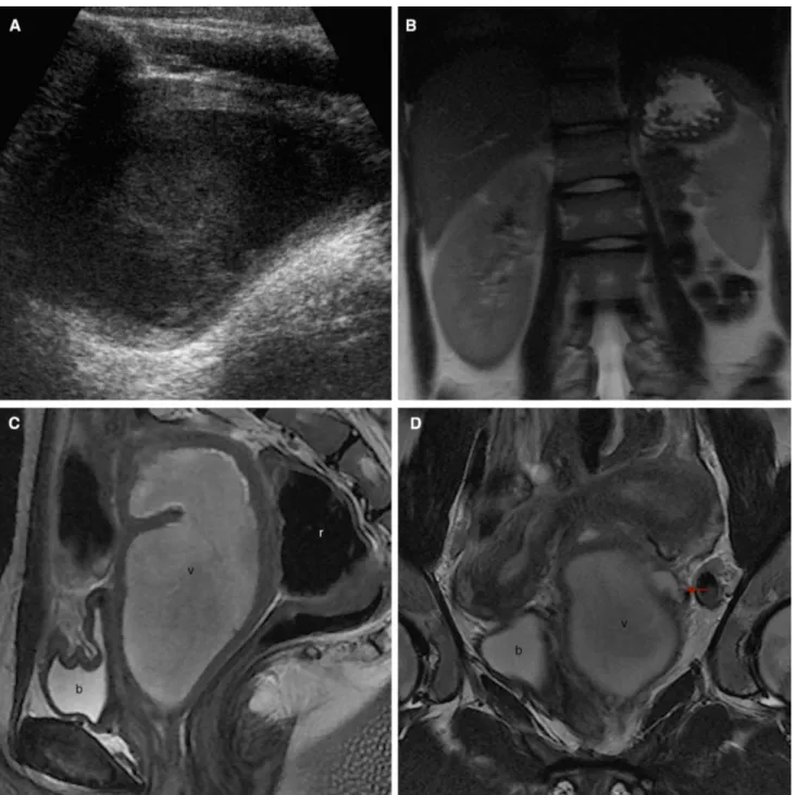
Fig. 2. Images in a 13-year-old girl who presented to the emergency department with a 3-month history of dysmenor-rhea (A–C US; D axial T1WI; E axial T2WI; F, G coronal T2WI; H sagittal T2WI). US revealed the absence of the right kidney (A), two uterine horns (B), and a dilated round cavity with homogenous, hypoechogenic content, and posterior en-hancement (C). MRI was performed, showing a dilated right hemivagina with hematic content (v), hyperintense on T1WI (D) and hypointense on T2WI (E–H). Two uterine horns (F arrows) and two cervices (G dashed arrow) are clearly de-picted. These findings are compatible with uterus didelphys
and, in combination with right hematocolpos and ipsilateral renal agenesis, correspond to the HWWS. A schematic rep-resentation (I) shows the septum position and the obstructed right hemivagina (pink). Patient underwent a corrective sep-tectomy 3 months later. On the post-surgery MRI (1 year la-ter), uterus didelphys is easily identified, with two non-dilated uterine cavities (J) and two independent cervices (K dashed arrow). No vaginal obstruction is found. A schematic repre-sentation (L) reveals a post-surgical non-septate single vagina. b bladder; r rectum; v obstructed hemivagina.
mass that may mimic ischiorectal swelling on initial ex-amination, as reported by Asha and Manila [24].
Pelvic pain is generally intermittent and worsens during menses. Hematocolpos and hematometra are more prevalent at the right side and result from retained menstrual flow. These manifestations are more painful when the vaginal septum fuses with the vaginal wall, leading to a complete obstruction [1,2,20,25]. If there is not a complete obstruction, symptoms may delay or be only mild [12].
When no fusion occurred between the septum and the vaginal wall, the former extends either completely or partially from the cervices to the vaginal introitus. In these cases, two vaginal openings are present and pa-tients are frequently asymptomatic and only incidentally detected [25]. These vaginal duplications are commonly mistaken for HWWS, but actually do not belong to the typical presentation [13].
HWWS is often misdiagnosed even after menarche. Several reasons may justify the delayed symptoms: first,
due to its distention proprieties, the vagina can accom-modate a large volume of blood; second, some of the blood is absorbed between menses [23]; third, anti-in-flammatory drugs and oral contraceptives are usually prescribed to relieve dysmenorrhea [2, 26]; and finally, some patients presenting with isolated vaginal discharge are erroneously treated with long-term antibiotics. At the emergency department, the diagnosis is also challenging and frequently simulates inflammatory and infectious disorders like tubo-ovarian abscess [25].
Only a few cases of prepubertal presentation are re-ported. Pansini et al. [27] reported the case of a 5-month-old infant with an acute urinary retention due to didel-phys uterus associated with an obstructed hemivagina. In these early cases, the initial manifestations result from collected secretions within the obstructed hemivagina by the effects of maternal hormones [9]. In another report, Hansen and DeWitt [28] described the case of a 5-year-old female presenting with an 8-month history of recur-rent, infectious vaginal discharge secondary to uterus
Fig. 2. continued
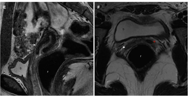
Fig. 3. Images in a 16-year-old girl who presented to the emergency department with a 2-month history of severe pel-vic pain and anomalous uterine hemorrhage (A US; B coronal T2WI; C sagittal T2WI; D, E coronal T2WI; G coronal T2WI; H axial T2WI). US revealed the absence of the left kidney (not shown) and a pelvic, thick-walled cystic structure with ho-mogeneous, low-level internal echoes, and posterior en-hancement (A). MRI showed the left renal agenesis (B), a huge left hematocolpos (C–E v), and a uterus didelphys (D). The non-obstructed right hemivagina (E white arrow) may be also seen. A dilated left ureteral stump (D, E red arrows) is
also identified, coursing lateral, and posterior to the hemato-colpos. This triad—uterus didelphys, obstructed hemivagina, and ipsilateral renal agenesis—characterizes the HWWS. A schematic representation (F) shows the septum position and the obstructed right hemivagina (pink), as well as the dilated left ureteral stump (gray line). Patient underwent a corrective septectomy 4 months later. On the post-surgery MRI (1 year later), uterus didelphys is clearly depicted (G), as well as a single non-obstructed vagina (G, H arrow). A schematic rep-resentation (I) better illustrates the post-surgical anatomy. b bladder; r rectum; v obstructed hemivagina.
didelphys with partial hemivagina obstruction. Never-theless, prepubertal cases are not always symptom-rich. Actually, the normal external genitalia appearance and age-appropriate development may mask asymptomatic
abnormalities of the internal reproductive organs [13]. So, the prepubertal diagnosis of the HWWS generally implies a high index of suspicion, which may come from prenatal or neonatal renal US. If renal agenesis is found
Fig. 3. continued
in prenatal or neonatal US, particular attention should be provided to the external genitalia and, even in the absence of a pelvic or vulvar mass, the risk of further genital anomaly should be highlighted [19].
Complications
Pyohematocolpos, pyosalpinx, and pelviperitonitis may appear as acute complications, while endometriosis, pelvic adhesions, infertility, and obstetric complications constitute potential long-term problems [2,13].
In some cases, the hemivaginas communicate through a partially fenestrated septum or the two cervices com-municate through a fistula, which may lead to pyocolpos due to infection of the retained menstrual blood. These patients tend to present with purulent vaginal discharge [1,25]. Peritonitis is a rare acute complication and occurs when hematocolpos progress to hematosalpinx and rupture [12].
Pelvic endometriosis is a well-known long-term complication of the HWWS. Tong et al. [1] reported a prevalence of 17.1% to endometriosis among 70 patients with HWWS. Actually, endometriosis is not more com-mon in Mu¨llerian anomalies as a whole, but its incidence increases when outflow obstruction, hematosalpinx, he-matometra, or hematocolpos are present. The onset of pelvic endometriosis in patients with HWWS often oc-curs in adolescence, and some authors believe that long-term continual reverse menstrual flow due to hemivagina obstruction is the main pathological contributor [8,29]. In another study of Tong et al. [8], all of the 14 ovarian endometriotic cysts found among 94 women with HWWS were ipsilateral to the vaginal septum.
Besides relieving symptoms, early diagnosis and treatment of endometriosis may avoid major anatomical
distortion due to pelvic adhesions therefore preventing infertility. Ovarian endometriomas should be removed, especially if large, in order to preserve healthy ovarian tissue and reduce recurrence of endometriosis. On the other hand, surgical correction of the obstructive anomaly usually leads to significant regression of en-dometriosis [8]. However, Silveira and Laufer [29] re-ported the cases of five patients in whom endometriosis developed or persisted after surgical correction. The au-thors advocate that persistence of prior peritoneal im-plants, coelomic metaplasia, metastases, immunologic deficiency, and genetic predisposition may explain these non-successful cases.
The risk of infertility has constituted matter of con-cern in patients with HWWS. Nonetheless, some studies like that of Tong et al. [1] showed that the incidence of primary infertility is not increased among women with HWWS. Actually, infertility is a potential complication of HWWS when the diagnosis is delayed and treatment is inappropriate, and appears to be more related to en-dometriosis and infectious complications than to uterus didelphys [20,23,29].
The risk of abortion, preterm delivery, and neonatal morbidity is higher in patients who conceived before sur-gical treatment [12,21,30]. However, women who carry a pregnancy to term often have no obstetric difficulties [13]. Watanabe et al. [31] reported two cases of lower genital tract adenocarcinomas in patients with HWWS, one endometrioid adenocarcinoma within the obstructed cervix and one primary clear cell carcinoma within the obstructed upper vagina. Although malignancy is not a well-established complication yet, the authors recom-mended periodic imaging evaluation of the blind side of the vagina and uterus.
Diagnostic procedures
US and MRI are the most commonly used imaging modalities for pre-surgical diagnosis of the HWWS [1]. US is a readily available and non-invasive technique that does not use contrast material or radiation. Transab-dominal US is unreliable and particularly limited in the presence of insufficient bladder distension, bowel inter-position, uterine retroflexion, and leiomyomas [32]. On the other hand, transvaginal US improves anatomical depiction and better assesses contour anomalies like fundal clefts. According to Pellerito et al. [32], transvaginal US may suffice to make a correct diagnosis. However, this is not a valid option in the pediatric set before the beginning of sexual life.
Three-dimensional (3D) US has been developed over recent years. It may be used with transabdominal or transvaginal probes and allows the radiologist to view
Fig. 4. Images in a 12-year-old girl who presented to the emergency department with a 1-month history of severe dys-menorrhea (A US; B axial T1WI; C coronal T2WI; D, E sagittal T2WI; G axial T2WI; H sagittal T2WI). US revealed the ab-sence of the left kidney (not shown) and a pelvic, thick-walled tubular structure with heterogeneous, mixed echogenicity, and posterior enhancement (A). MRI showed a uterus didelphys with a non-dilated right horn (C red arrow) and left hemato-colpos (v), hematocervix (c), hematometra (u), and hematos-alpinx (t). Blood appeared hyperintense on T1WI (B) and slightly less intense on T2WI (C–E). The hematocolpos essentially involved the upper vagina and was heterogeneous and lobulated, suggesting co-infection. Both ovaries were
identified (C white arrows), the left one encased in a dilated Fallopian tube (t). A schematic representation (F) shows the septum position, the obstructed right hemivagina and blood regurgitation to the ipsilateral Fallopian tube (pink). This case exemplifies an acute complication with hematosalpinx and probable pyocolpos. Ten days later, the patient underwent a left hemihysterectomy with salpingectomy and preservation of the left ovary. On the post-surgery MRI (1 year later), single uterus (G red arrow) and vagina (H white arrow) are seen, as well as a remnant hemorrhagic cystic structure (G blue arrow). A schematic representation (I) better illustrates the post-sur-gical anatomy. b bladder, c cervix, r rectum, t Fallopian tube, u uterine body, v obstructed hemivagina.
images of the three orthogonal planes simultaneously. 3D US appears to provide high-quality images similar to those yielded by MRI and achieve high-accuracy levels in the diagnosis of congenital uterine anomalies. Moreover, its lower cost and superior tolerance by patients consti-tute relevant advantages [11].
According to our experience, transabdominal US should be performed prior to the transvaginal approach even in sexually active patients. The former enables a global evaluation of the pelvis, which is particularly useful in patients with uterus didelphys since the uterine
horns may diverge and be laterally placed within the pelvis. In these cases, an isolated transvaginal approach may not suffice if the field of view is too limited. At our institution, we perform transabdominal US with a con-vex probe with a frequency bandwidth ranging from 6 to 2 MHz (Siemens Acuson Antares). When indicated, transvaginal US is performed using an endocavitary probe with a frequency bandwidth ranging from 4 to 9 MHz (GE Voluson E8).
US usually identifies two hemiuteri compatible with uterus didelphys and dilatation of the proximal vagina
(Figs.2, 3, 4). The content of the dilated reservoir is hemorrhagic, ranging from anechoic fluid to heteroge-neous collections with mixed echogenicity. In the set of infection, some echogenic foci representing gas bubbles may be found. In some cases, hydrometra or he-matometra may be found as a result of regurgitation of the vaginal content to the endometrial cavity. The dis-tinction between an obstructed vagina and an adnexal cystic lesion like endometriomas or other complex ovarian cysts may be difficult [9].
MRI is safe, non-invasive, and free of ionizing ra-diation. It better depicts pelvic anatomy and documents uterine anomalies, thus being useful in the management of surgical treatment options [9, 23]. MRI is more ac-curate than hysterosalpingography (HSG) or US not only in the evaluation of the uterine contours, the shape of the endometrial cavity and the course of the vaginal septum, but also in the detection of acute and chronic complications [9]. High-resolution T2WI should be ob-tained in three different planes, one of them parallel to the long axis of the uterus, in order to better understand the uterine morphology and the features of the sep-tum [9]. Sometimes, the non-obstructed hemivagina is
flattened by the hematocolpos and difficult to recognize (Fig.3). T1-weighted images (T1WI) are generally ob-tained in the axial plane, showing the high signal inten-sity of the hemorrhagic fluid within the obstructed hemivagina (Figs.2, 4). In some cases, this fluid looses signal intensity from T1WI to T2-weighted images (T2W1), due to high concentration of protein and iron from recurrent hemorrhage (Fig.2). This T2 shortening is known as shading sign and was initially described as an MRI sign of ovarian endometriomas. In the set of the HWWS, it is not uncommon to find endometriomas and post-obstructive hematosalpinx, both potentially hyper-intense on T1WI. If any doubt persists, a complementary fat-saturated T1WI sequence should be performed in order to exclude a fat-containing mass. Intravenous contrast material should not be routinely used but may be helpful in the setting of infectious complications or incidental findings. At our institution, MRI is performed on a 3.0 Tesla (T) magnet (Signa HDxt, GE Healthcare). Specific parameters are provided in Table1.
HSG provides valuable information regarding the interior cavity of the uterus. However, this technique fails in the characterization of some uterine subtypes since only patent cavities are demonstrated and the ex-ternal contour and rudimentary non-communicating horns are not visualized. Moreover, it implies contrast material and radiation exposure. Thus, HSG is not cur-rently considered a first-line modality when Mu¨llerian duct anomalies are suspected [11,32].
Computerized tomography (CT) does not easily de-pict pelvic anatomy and is not recommended in the dif-ferential diagnosis of congenital uterine anomalies [9]. The exposure to ionizing radiation and the eventual in-travenous administration of iodinated contrast also constitute important disadvantages in the pediatric con-text. However, CT may be required in the set of acute abdomen when US is not conclusive. A spontaneously hyperdense cystic lesion may be found, corresponding to an obstructed, hemorrhagic, or infected hemivagina [26]. Hysteroscopy allows direct visualization of the in-trauterine cavity and ostia but does not evaluate the external contour of the uterus. Thus, it is frequently in-conclusive and implies further investigation. Moreover, this procedure is invasive, usually needs anesthesia, and is less appropriate in pediatrics [11].
Fig. 4. continued
Laparoscopy and laparotomy may also be used for diagnostic purposes but are expensive and invasive pro-cedures with anesthesia- and surgery-related risks [32]. However, some authors like Zurawin et al. [26] advocate that laparoscopy should be the gold standard for the
complete evaluation of congenital anomalies of the fe-male reproductive tract. Despite referring that MRI may correctly anticipate the diagnosis, the authors consider that laparoscopic evaluation better identifies en-dometriosis, pelvic infection, and adhesions.
Fig. 5. A–F Images in a 17-year-old girl who complained with a 6-year history of severe dysmenorrhea (A–C coronal T2WI; D, E sagittal T2WI; F axial T2WI). MRI showed a left renal agenesis (A) and a uterus didelphys. Two independent uterine horns and two non-communicating cervices are rec-ognized. The right uterine horn (B–D white arrow) and cervix (C–E white dashed arrow) have a normal appearance. The left uterine horn is smaller (B, C red arrow), and the ipsilateral
cervix appears thinner and tortuous (C red dashed arrow). Two independent hemivaginas seem to be found at a lower level (F white and red arrow-heads). Despite the absence of obstruction, these findings appear to be related to the HWWS. Long-term oral contraceptives may explain the lack of he-matocolpos. The existence of incomplete vaginal septa or longitudinal non-fused septa may also be the cause. b blad-der, r rectum.
Treatment
The main purposes of the surgical treatment in HWWS are to relieve symptoms and guarantee successful re-productive outcomes [13].
The treatment of choice for patients with HWWS is the surgical excision and marsupialization of the longi-tudinal vaginal septum (Figs.2, 3) [25]. Some surgeons prefer to perform the vaginal septum resection only after a large hematocolpos develops. It distends and thins the septal tissue consequently becomes easier to excise [26,
33]. Care must be taken during the dissection in order to preserve the hymen and avoid damage to the bladder or the rectum [26].
Some asymptomatic patients with HWWS are suspected after the identification of a renal agenesis at prenatal or neonatal US (Figs.5,6). Some surgeons decide to closely follow these patients and ensure that surgical correction is performed before hormonal impregnation at puberty. In young adolescents, postponement of surgery may be re-quired; in these cases, gonadotropin-releasing hormone analogs may constitute a valid option for maintaining amenorrhea, particularly if cervical atresia coexists [1].
Some morphological features increase the complexity of the surgery and the post-surgical risk of contracture, stenosis, infection, and reoperation need. These include thick septum, narrowed area of resection, collapsing
sidewalls, and high septum location. Vaginal packing, vaginal dilators, and vascular stents are reported in the literature as useful to maintain the vaginal patency in these complicated cases [33].
The use of laparoscopy at the time of vaginal sep-tectomy has been used to assess the uterine anatomy. However, the impact of this approach on definitive treatment remains unclear, and some authors believe that a timely diagnosis avoids unnecessary laparoscopies [13]. Endoscopic ablation of the vaginal septum has been re-ported as an alternative to the conventional open surgical excision. This is a minimally invasive technique that lacks major morbidity and appears to achieve equivalent results to those of open surgical excision, as reported by Roth et al. [34]. Hemihysterectomy with or without salpingo-oophorectomy should be avoided in order to preserve fertility. It is rarely indicated when resecting the vaginal septum is not enough to relieve the hematometra (Fig.4) [13, 23]. Metroplasty, a reconstructive uterine surgery, may also be indicated in some variants. Nabeshima et al. [18] reported the case of a 12-year-old girl with an obstructed non-communicating uterus didelphys without vaginal septum in whom a laparo-scopic metro plastic surgery was successfully performed. Women with a uterine septum and recurrent pregnancy loss may also benefit from metroplasty [21].
Fig. 5. continued
Conclusion
The triad of uterus didelphys, obstructed hemivagina, and ipsilateral renal agenesis characterizes the HWWS. This syndrome usually presents with non-specific symptoms after menarche and is frequently mistaken for inflammatory and infectious pelvic disorders.
Anti-inflammatory drugs, oral contraceptives and/or antibiotics are therefore prescribed, relieving symptoms and delaying the diagnosis. Before puberty, the diag-nosis is even more challenging, unless early acute complications occur, leading to emergent imaging evaluation. If renal agenesis is found at prenatal or
Fig. 6. US images in a 3-year-old patient with prenatal di-agnosis of right multicystic dysplastic kidney. Postnatal right kidney resorption occurred (A). The right ureteral stump is dilated (B, C white arrows) and ends in a tubular structure coursing posterior to the bladder (b). These findings suggest the presence of an obstructed hemivagina in association to an
ectopic ureter. The uterus is not easily identified due to its immature morphology. This case probably corresponds to a variant of the HWWS. A rigorous follow-up should be per-formed in order to characterize the uterine anatomy and guide the patient toward timely and appropriate surgical correction. b bladder, v hemivagina.
neonatal US, the risk of further genital anomaly should be considered.
Pediatricians, radiologists, gynecologists, and pedi-atric surgeons should become familiar with this syn-drome and other congenital anomalies of the female genital tract. Despite its rarity, an accurate preoperative diagnosis can be achieved and depends on a high clinical suspicion. Prognosis may be excellent if a timely cor-rective surgery is performed, thus preventing long-term complications and preserving future fertility.
Acknowledgments.The authors sincerely thank Cristina Borges, MD, Filomena Sousa, MD, and Pedro Ferreira, RT, for their valuable contributions.
References
1. Tong J, Zhu L, Lang J (2013) Clinical characteristics of 70 patients with Herlyn–Werner–Wunderlich syndrome. Int J Gynecol Obstet 121:173–175. doi:10.1016/j.ijgo.2012.11.023
2. Del Vescovo R, Battisti S, Di Paola V, et al. (2012) Herlyn-Werner-Wunderlich syndrome: MRI findings, radiological guide (two cases and literature review), and differential diagnosis. BMC Med Imaging 12:4. doi:10.1186/1471-2342-12-4
3. Purslow C (1922) A case of unilateral haematokolpos, haematometra and haematosalpinx. J Obstet Gynecol Br Emp 29:643. doi:
10.1111/j.1471-0528.1922.tb16100.x
4. Herlyn U, Werner H (1971) Simultaneous occurrence of an open Gartner-duct cyst, a homolateral aplasia of the kidney and a double uterus as a typical syndrome of abnormalities. Geburtshilfe Frauenheilkd 31:340–347
5. Wunderlich M (1976) Unusual form of genital malformation with aplasia of the right kidney. Zentralbl Gynakol 98:559–562 6. Mandava A, Prabhakar R, Smitha S (2012) OHVIRA syndrome
(obstructed hemivagina and ipsilateral renal anomaly) with uterus didelphys, an unusual presentation. J Pediatr Adolesc Gynecol 25:e23–e25. doi:10.1016/j.jpag.2011.11.004
7. Ugurlucan FG, Bastu E, Gulsen G, Eken MK, Akhan SA (2014) OHVIRA syndrome presenting with acute abdomen: a case report and review of the literature. J Clin Imaging 38:357–359. doi:
10.1016/j.clinimag.2013.12.011
8. Tong J, Zhu L, Chen N, Lang J (2014) Endometriosis in association with Herlyn-Werner-Wunderlich syndrome. Fertil Steril 102(3): 790–794. doi:10.1016/j.fertnstert.2014.05.025
9. Yavuz A, Bora A, Kurdog˘lu M, et al. (2014) Herlyn-Werner-Wunderlich syndrome: merits of sonographic and magnetic re-sonance imaging for accurate diagnosis and patient management in 13 cases. J Pediatr Adolesc Gynecol 28:1–5. doi:10.1016/j.jpag. 2014.03.004
10. Sajjad Y (2010) Development of the genital ducts and external genitalia in the early human embryo. J Obstet Gynaecol Res 36(5):929–937. doi:10.1111/j.1447-0756.2010.01272.x
11. Saravelos SH, Cocksedge KA, Li T-C (2008) Prevalence and di-agnosis of congenital uterine anomalies in women with reproduc-tive failure: a critical appraisal. Hum Reprod Update 14(5):415– 429. doi:10.1093/humupd/dmn018
12. Beer WM, Carstairs SD (2013) Herlyn-Werner-Wunderlich syn-drome: an unusual presentation of acute vaginal pain. J Emerg Med 45(4):541–543. doi:10.1016/j.jemermed.2013.03.035
13. Gholoum S, Puligandla PS, Hui T, et al. (2006) Management and outcome of patients with combined vaginal septum, bifid uterus, and ipsilateral renal agenesis (Herlyn-Werner-Wunderlich syndrome). J Pediatr Surg 41:987–992. doi:10.1016/j.jpedsurg.2006.01.021
14. Rastogi M, Revannasiddaiah S, Thakur P, et al. (2013) Mu¨llerian duct anomalies and their effect on the radiotherapeutic manage-ment of cervical cancer. Chin J Cancer 32(8):434–440. doi:
10.5732/cjc.012.10222
15. Grimbizis GF, Gordts S, Di Spiezio Sardo A, et al. (2013) The ESHRE/ESGE consensus on the classification of female genital tract congenital anomalies. Hum Reprod 28(8):2032–2044. doi:
10.1093/humrep/det098
16. Wang J, Zhu L, Lang J, et al. (2014) Clinical characteristics and treatment of Herlyn-Werner-Wunderlich syndrome. Arch Gynecol Obstet 290(5):947–950. doi:10.1007/s00404-014-3286-5
17. Dorais J, Milroy C, Hammoud A, et al. (2011) Conservative treatment of a Herlyn-Werner-Wunderlich mullerian anomaly variant, non-communicating hemiuterus with gartner duct pseudocyst. J Minim Invasive Gynecol 18:262–266. doi:10.1016/j.jmig.2010.12.006
18. Nabeshima H, Nishimoto M, Shiga N, Utsunomiya H, Yaegashi N (2013) Laparoscopic strassman metroplasty in a postmenarcheal adolescent girl with Herlyn-Werner-Wunderlich mullerian anomaly variant, obstructed noncommunicating didelphic uterus without gartner duct pseudocyst. J Minim Invasive Gynecol 20(2):255–258. doi:10.1016/j.jmig.2012.10.016
19. Wu TH, Wu TT, Ng YY, et al. (2012) Herlyn-Werner-Wunderlich syndrome consisting of uterine didelphys, obstructed hemivagina and ipsilateral renal agenesis in a newborn. Pediatr Neonatol 53(1):68–71. doi:10.1016/j.pedneo.2011.11.014
20. Sanghvi Y, Shastri P, Mane SB, Dhende NP (2011) Prepubertal presentation of Herlyn-Werner-Wunderlich syndrome: a case report. J Pediatr Surg 46:1277–1280. doi:10.1016/j.jpedsurg.2011.02.067
21. Patton PE, Novy MJ, Lee DM, Hickok LR (2004) The diagnosis and reproductive outcome after surgical treatment of the complete septate uterus, duplicated cervix and vaginal septum. Am J Obstet Gynecol 190:1669–1675. doi:10.1016/j.ajog.2004.02.046
22. Nawfal AK, Blacker CM, Strickler RC, Eisenstein D (2011) La-paroscopic management of pregnancy in a patient with uterus didelphys, obstructed hemivagina, and ipsilateral renal agenesis. J Minim Invasive Gynecol 18(3):381–385. doi:10.1016/j.jmig.2011. 01.005
23. Gu¨du¨cu¨ N, Go¨nenc¸ G, Is¸c¸i H, Yig˘iter AB, Du¨nder I (2012) Herlyn-Werner-Wunderlich syndrome—timely diagnosis is important to preserve fertility. J Pediatr Adolesc Gynecol 25:e111–e112. doi:
10.1016/j.jpag.2012.05.013
24. Asha B, Manila K (2008) An unusual presentation of uterus didelphys with obstructed hemivagina with ipsilateral renal agene-sis. Fertil Steril 90(3):e9–e10. doi:10.1016/j.fertnstert.2007.08.003
Table 1. MRI protocol
Axial T2WI Coronal T2WI Sagittal T2WI Axial T1WI Axial FS T1WI Dynamic CE
FS T1WIa CoronalT2WIb
Sequence type frFSE frFSE frFSE FSE FSFSE GRE SSFSE
FOV (cm) 18 20 22 32 32 34 48
Matrix 352 9 256 384 9 256 384 9 256 512 9 320 384 9 256 320 9 256 512 9 320 Slice thickness (mm)/spacing (mm) 3.5/0.3 3/0.3 4/0.4 5/0.5 5/0.5 3/0 6/0.6
TR 4860 5160 3440 760 680 3.5 1800
TE 120 120 100 Minimum full Minimum full 1.7 100
Flip angle (°) 90 90 90 90 90 12 90
a
Phases at 50, 100, 150 and 200 s after contrast administration bUpper abdomen
FS, fat saturation; CE, contrast-enhanced; frFSE, fast relaxation fast spin-echo; FSE, fast spin-echo; GRE, gradient echo; SSFSE, single-shot fast spin-echo; FOV, field of view; TR, time to repetition; TE, echo time
25. Wozniakowska E, Torres A, Milart P, et al. (2014) Delayed diag-nosis of Herlyn-Werner-Wunderlich syndrome due to microperfo-ration and pyocolpos in obstructed vaginal canal. J Pediatr Adolesc Gynecol 27:e79–e81. doi:10.1016/j.jpag.2013.07.009
26. Zurawin RK, Dietrich JE, Heard MJ, Edwards CL (2004) Didel-phic uterus and obstructed hemivagina with renal agenesis: case report and review of the literature. J Pediatr Adolesc Gynecol 17:137–141. doi:10.1016/j.jpag.2004.01.016
27. Pansini L, Torricelli M, Gomarasca A, et al. (1988) Acute urinary retention due to didelphys uterus associated with an obstructed hemivagina in a 5-month-old infant. J Pediatr Surg 23(10):984–985 28. Hansen KA, DeWitt J (2005) Premenarchal, recurrent vaginal discharge associated with an incomplete obstructing longitudinal vaginal septum. J Pediatr Adolesc Gynecol 18:423–426. doi:
10.1016/j.jpag.2005.09.008
29. Silveira SA, Laufer MR (2013) Persistence of endometriosis after correction of an obstructed reproductive tract anomaly. J Pediatr Adolesc Gynecol 26:e93–e94. doi:10.1016/j.jpag.2013.01.002
30. Stassart J, Nagel T, Prem K, Phipps W (1992) Uterus didelphys, obstructed hemivagina, and ipsilateral renal agenesis: the Univer-sity of Minnesota experience. Fertil Steril 57(4):756–761
31. Watanabe Y, Etoh T, Nakai H (2012) Adenocarcinoma of the lower female genital tract in patients with Herlyn-Werner-Wun-derlich syndrome. Am J Obstet 207(6):e5–e6. doi:10.1016/j.ajog. 2012.09.009
32. Pellerito JS, McCarthy S, Doyle M, Glickman M, DeCherney AH (1992) Diagnosis of uterine anomalies: relative accuracy of MR imaging, endovaginal sonography, and hysterosalpingography. Radiology 183:795–800
33. Cooper AR, Merritt DF (2010) Novel use of a tracheobronchial stent in a patient with uterine didelphys and obstructed hemivagina. Fertil Steril 93(3):900–903. doi:10.1016/j.fertnstert.2008.10.022
34. Roth M, Mingin G, Dharamsi N, Psooy K, Koyle M (2010) En-doscopic ablation of longitudinal vaginal septa in prepubertal girls: a minimally invasive alternative to open resection. J Pediatr Urol 6:464–468. doi:10.1016/j.jpurol.2009.12.009