83
DOI:
10.1590/0004-282X20150155
IMAGES IN NEUROLOGY
Sneddon syndrome – imaging findings
Síndrome de Sneddon – achados de imagem
Pedro Henrique Teixeira Junqueira
1,2, Paulo Puglia Jr.
1, Lázaro Luís Faria do Amaral
1,2, Mauricio Hoshino
1he Sneddon syndrome is a rare clinical syndrome that
associates strokes and livedo, etiologically related with the
antiphospholipid antibody syndrome (Figure 1)
1,2,3,4,5.
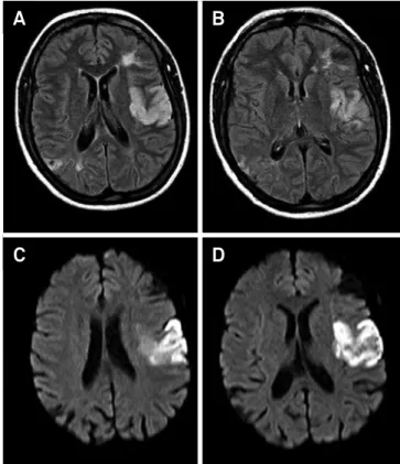
his 42 year-old woman with headache and right
hemipa-resis had a MRI that showed acute/subacute ischemia in part
of the territory of the left middle cerebral artery (MCA) and
bi-hemispheric gliosis/encephalomalacia (Figure 2). MR
angiog-raphy showed normal cervical carotid and vertebral circulation,
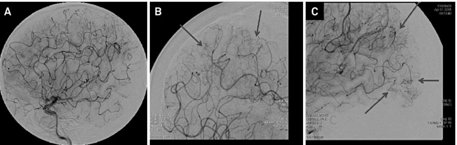
illing defect in M2 segment of the left MCA. Catheter
angiog-raphy disclosed difuse cortical occlusive arteriopathy associated
with a rich collateral cortical network. he M2 occlusion was
re-perfused, with infarct sign in its territory (Figures 3 and 4).
1Hospital Santa Catarina, Sao Paulo SP, Brazil;
2Hospital Beneicência Portuguesa de São Paulo, MedImagem, Sao Paulo SP, Brazil.
Correspondence: Pedro Henrique Teixeira Junqueira; Med Imagem, Hospital Beneicência Portuguesa de São Paulo; Rua Maestro Cardim, 407; 01323-000 São Paulo SP, Brasil; E-mail: [email protected]
Conflicts of interest: There is no conlict of interest to declare. Received 11 July 2015; Accepted 04 August 2015.
Figure 1.
Reticular livedo on limbs.
Figure 3.
Angiography early arterial phase: (A) right side
apparently normal; (B) slow illing area (red circle) and luxury
perfusion (arrow).
A
B
Figure 2.
AXIAL FLAIR (A e B) showing areas corresponding
to bi-hemispheric gliosis /encephalomalacia and area of
acute/subacute ischemic stroke in part of the territory of the
left MCA. AXIAL DIFFUSION (C and D) proves acute /subacute
schemic stroke area.
A
B
84
Arq Neuropsiquiatr 2016;74(1):83-84References
1. Maamar M, Rahmani M, Aidi S, Benabdeljlil M, El HassaniMy R, Jiddane M et al. [Sneddon’s syndrome: 15 cases with cerebral angiography]. Rev Neurol (Paris). 2007;163(8-9):809-16. French. doi:10.1016/S0035-3787(07)91463-0
2. Marinho JL, Piovesan EJ, Leite Neto MP, Kotze LR, Noronha L, Twardowschy CA et al. Clinical, neurovascular and neuropathological features in Sneddon’s syndrome. ArqNeuropsiquiatr.
2007;65(2B):390-5. doi:10.1590/S0004-282X2007000300005 3. Boesch SM, Plörer AL, Auer AJ, Poewe W, Aichner FT, Felber SR
et al. The natural course of Sneddon syndrome: clinical and
magnetic resonance imaging indings in a prospective six year observation study. J Neurol Neurosug Psychiatry. 2003;74(4):542-4. doi:10.1136/jnnp.74.4.542
4. Stockhammer G, Felber SR, Zelger B, Sepp N, Birbamer GG, Fritsch POet al. Sneddon’s syndrome: diagnosis by skin biopsy and MRI in 17 patients. Stroke.1993;24(5):685-90. doi:10.1161/01.STR.24.5.685
5. Uthman, I.W., Khamashta, M.A. Livedo racemosa: a striking dermatological sign for the antiphospholipid syndrome [Editorial]. J Rheumatol. 2006;33(12):2379-82.