Faculdade de Medicina de Lisboa
RESPOSTA DOS LINFÓCITOS T
RECRUTAMENTO, RECONHECIMENTO E FUNÇÕES
Prof
Doutoramento em Ciências Biomédicas
Faculdade de Medicina de Lisboa
RESPOSTA DOS LINFÓCITOS T
γγγγδδδδ
A TUMOUR
RECRUTAMENTO, RECONHECIMENTO E FUNÇÕES
Telma Lança
Tese orientada pelo
Professor Doutor Bruno Silva-Santos
Doutoramento em Ciências Biomédicas
Especialidade em Imunologia
2012
Faculdade de Medicina de Lisboa
TUMOURES:
RECRUTAMENTO, RECONHECIMENTO E FUNÇÕES
Prof. Doutor Bruno Silva-Santos, PhD
Instituto de Medicina Molecular
Faculdade de Medicina
Universidade de Lisboa
A presente dissertação foi realizada na Unidade de Imunologia Molecular do Instituto de Medicina Molecular, e foi apresentada à Faculdade de Medicina
da Universidade de Lisboa, para obtenção do grau de Doutor em Imunologia
Todas as afirmações efectuadas no presente documento são da exclusiva
responsabilidade do seu autor, não cabendo qualquer responsabilidade à Faculdade de Medicina de Lisboa pelos conteúdos nele apresentados.
A impressão desta dissertação foi aprovada em reunião do Conselho Científico da Faculdade de Medicina de Lisboa da Universidade de Lisboa em 20 deNovembro de 2012.
European
Fundação para a Ciência e Tecnologia
O trabalho aqui apresentado foi financiado
European Molecular Biology Organization- Young Investigator Programme European Research Council (StG_26052) Fundação Calouste Gulbenkian Fundação para a Ciência e Tecnologia e Bolsa de Doutoramento SFRH/BD47342/2008 O trabalho aqui apresentado foi financiado por:
Investigator Programme European Research Council (StG_26052) Fundação Calouste Gulbenkian outoramento SFRH/BD47342/2008
i
Acknowledgments / Agradecimentos ... vii
Abbreviations ... ix
Resumo ... xiii
Summary ... xvi
General introduction 1.1 Immune response to tumours ... 1
1.1.1 Tumour immunosurveillance theory ... 1
1.1.2 Cancer immunoediting hypothesis ... 2
1.1.3 Tumour microenvironment ... 3
1.1.3.1 Tumour-infiltrating leukocytes ... 3
1.1.3.1.1 The traditional players: NK, CD8+ T and Th1 cells lymphocytes . 3 1.1.3.1.2 “New” effectors : γδ T, NKT and Th17 lymphocytes ... 5
1.1.3.1.3 The inflammatory phagocytes: TAMs and TANs ... 6
1.1.3.1.4 Immunosuppressive leukocytes: Treg and MDSCs ... 7
1.1.3.2 Other cells (non-leukocytes) ... 8
1.1.3.3 Key molecular players: Cytokines and Chemokines ... 9
1.1.4 TILs as prognostic factors ... 12
1.1.5. Principles of cancer immunotherapy ... 13
1.2 Role of γδγδγδγδ T cells in tumour immunity ... 17
1.2.1 Thymic development of γδ T cells ... 17
1.2.2 TCRγδ repertoires and functions ... 18
1.2.2.1 Mouse γδ T cell subsets ... 19
1.2.2.2 Human γδ T cell subsets ... 20
1.2.3.γδ T cell activation:TCRγδ ligands ... 21
1.2.4 NKG2D and its ligands ... 23
1.2.4.1. NKG2D receptor ... 23
1.2.4.2 NKG2D ligands ... 24
1.2.5 Other important receptors in γδ T cell activation ... 27
1.2.6 γδ T cell response to tumours ... 28
ii 1.3 Objectives of the Thesis ... 33
Tumour cell recognition by γδγδγδγδ T cells
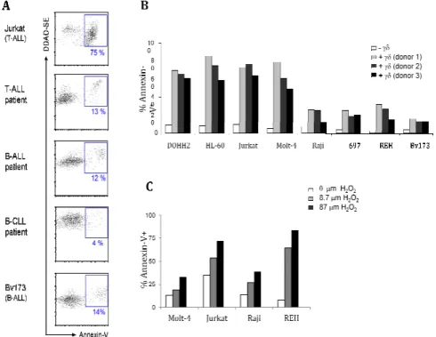
2.1 Abstract ... 37 2.2 Introduction ... 37 2.3 Results... 38 2.3.1 Expanded and activated γδ-PBL efficiently kill leukaemia cells (Molt-4) in
vitro and in vivo ... 38
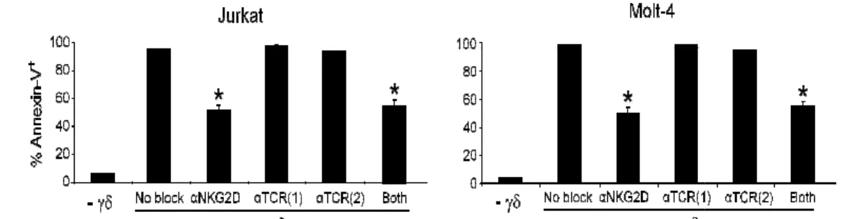
2.3.2 Differential susceptibility of leukaemia and lymphoma cells to γδ−PBL cytotoxicity ... 39 2.3.3 NKG2D mediates Vγ9Vδ2 T-cell recognition of haematopoietic tumours that endogenously express ULBP1 and MICA ... 41 2.3.4 ULBP1 is required for Vγ9Vδ2 T cell-recognition of leukaemia/
lymphoma ... 42 2.3.5 ULBP1 displays a highly heterogeneous expression in cancer patients . 44 2.4. Discussion ... 45 2.5 Materials and Methods ... 47
Recruitment of γδγδγδγδ tumour-infiltrating lymphocytes
3.1 Abstract ... 53 3.2 Introduction ... 53 3.3 Results and Discussion ... 54 3.3.1 Murine cytotoxic γδ T cells play non-redundant anti-tumour role in B16 lesions ... 54 3.3.2 CCR2/CCL2 pathway is required for γδ T cell recruitment to B16 tumours
in vivo ... 56
3.3.3 Human CCR2+Vδ1 T cells migrate towards CCL2 which is deregulated in multiple cancer types ... 56 3.4 Material and Methods ... 60
iii
4.1 Abstract ... 69
4.2 Introduction ... 69
4.3 Results... 70
4.3.1 γδ T cells play opposite roles in tumour progression in two distinct tumour models ... 70
4.3.2 Distinct γδ T cell functions in ID8 and B16 models are not accounted by cytotoxicity of IFN-γ production ... 71
4.3.3 Accumulation of suppressive MDSCs during B16 tumour progression .. 74
4.3.4 Accumulation of IL-17+ producing γδ T cells in the peritoneal cavity of ID8 tumour bearing mice may promote tumour progression ... 75
4.5 Material and methods ... 79
General discussion and future perspectives 5.1 General discussion ... 83
5.1.1 Tumour cell recognition by γδ T cells ... 83
5.1.2 Recruitment of γδ tumour-infiltrating lymphocytes... 86
5.1.3 Promotion of tumour growth by γδ T cells ... 90
5.2 Future perspectives ... 92
References: ... 97
iv General Introduction
Figure 1 - Tumour immunosurveillance theory and Cancer immunoediting
hypothesis. ... 1
Figure 2 - Chemokine-mediated interaction between tumour cells and stromal cells ... 11
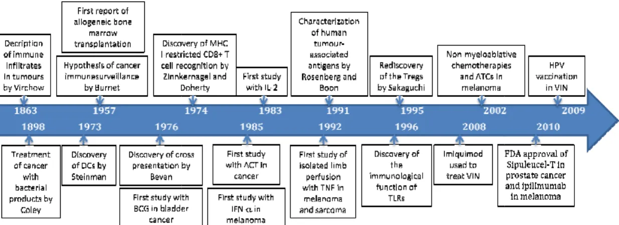
Figure 3 - Timeline: the history of cancer immunotherapy. ... 14
Figure 4 - Mouse γδ cell generation is developmentally programmed ... 18
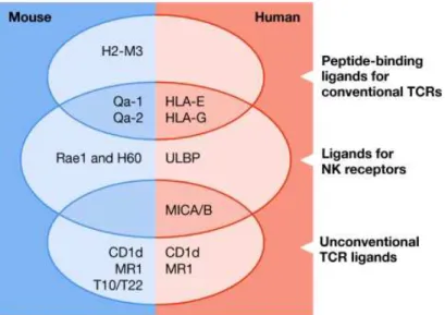
Figure 5 – Non-classical MHC molecules and their receptors.. ... 25
Tumour cell recognition by γδ γδ γδ γδ T cells Figure 1 - Expanded and activated γδ-PBL efficiently kill Molt-4 acute leukaemia cells ... 38
Figure 2 - Differential susceptibility of leukaemia and lymphoma cells to γδ−PBL cytotoxicity ... 40
Figure 3 - NKG2D (but not TCR) mediates Vγ9Vδ2 T-cell leukaemia line tumour cell recognition. ... 41
Figure 4 - NKG2D mediates Vγ9Vδ2 T-cell recognition of haematopoietic tumours that endogenously express ULBP1 and MICA . ... 42
Figure 5 - ULBP1 is required for Vγ9Vδ2 T cell-recognition of leukaemia/ lymphoma cells.. ... 43
Figure 6 - ULBP1 is required for Vγ9Vδ2 T cell-recognition of leukaemia/ lymphoma cells.. ... 44
Figure 7 - ULBP1 displays a highly heterogeneous expression in cancer patients. . 45
Recruitment of γδ γδ γδ γδ tumour-infiltrating lymphocytes Figure 1 - Murine cytotoxic γδ T-cells play non-redundant anti-tumour role in B16 lesions.. ... 55
Figure 2 - CCR2/CCL2 pathway is required for γδ T-cell recruitment to B16 tumours in vivo. ... 57
Figure 3 - Human CCR2+ Vδ1 T-cells migrate towards CCL2 that is deregulated in multiple cancer types. ... 59
Supplemental Figure 1 - IFN−γ production by T-cell subsets from tumour-draining lymph nodes. ... 63
Supplemental Figure 2 - Chemokine concentrations in protein extracts from day 14-tumours from WT or Tcrd-/- mice, as measured by Quantibody Chemokine Mouse Array.. ... 63
Supplemental Figure 3 - Gating strategies for identification of tumour-infiltrating leukocyte subsets by flow cytometry.. ... 64
v ... 65 Supplemental Figure 5 -- In vitro responses of human γδ T-cells to recombinant CCL2... 65
Promotion of tumour growth by γδ γδ γδ γδ T cells
Figure 1 – TCRδ-deficient mice show decreased survival upon B16 tumour challenge and increased survival upon ID8 tumour challenge. ... 71 Figure 2 – Activated γδ T cells show equivalent in vitro cytotoxicity toward B16 and ID8 tumour cell lines. ... 72 Figure 3 – Peritoneal γδ T cells expand upon B16 and ID8 tumour transplantation.. 72 Figure 4 - IFN-γ+ γδ T cells accumulate in the peritoneal cavity of ID8 tumour bearing mice.. ... 73 Figure 5 - ID8 tumour growth in CD27-deficient mice. ... 74 Figure 6 - MDSCs accumulate in the peritoneal cavity of B16 tumour bearing mice and inhibit γδ T cell proliferation in vitro. ... 75 Figure 7 - IL-17+ γδ T cells accumulate in the peritoneal cavity of ID8 tumour bearing mice.. ... 76 Figure 8 - γδ T cells are a major source of IL-17 in the peritoneal cavity of ID8 tumour bearing mice... 76
vi General Introduction
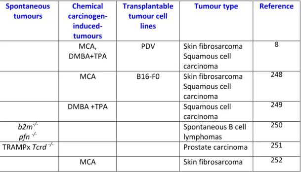
Table 1 - Tumour-infiltrating leukocytes associated with prognosis for cancer patients ... 12 Table 2- Frequency, distribution and repertoires of γδ T cells ... 19 Table 3 - Expression of NKG2D by immune cells ... 23 Table 4 - Percentage of amino acid identities between human NKG2D MICA/B and ULBPs ... 26 Table 5 - Mouse tumour models implicating γδ T cells in tumour immunosurveillance ... 29 Table 6 – Clinical trials involving γδ T cells ... 32
Tumour cell recognition by γδ γδ γδ γδ T cells
vii
Acknowledgments / Agradecimentos
Ao escrever estes agradecimentos é com muito prazer e saudade que olho para trás e revejo os momentos que fizeram parte dos quase 5 anos que passei na Unimol. Queria agradecer em primeiro lugar ao Prof. Dr Bruno Silva Santos, por todo o apoio que me deu, por ter acreditado e confiado em mim desde o início; por ter puxado por mim até ao limite pois isso fez-me crescer enquanto cientista e consequentemente enquanto pessoa. Por me ter criticado quando eu merecia e por me ter elogiado quando merecia. Por ser uma pessoa justa, entusiasta, e no fundo por se uma inspiração para qualquer cientista. Por desafiar a minha segurança. Por me ter dado a oportunidade de ir a vários congressos internacionais (o que considero uma das partes mais estimulantes da minha estadia da Unimol) que me permitiu conhecer muitas pessoas de diferentes nacionalidades e portanto com outra cultura e outra perspectiva da ciência ( e do mundo).
Dentro da minha unidade queria fazer um agradecimento especial ao Daniel Correia, Anita Gomes, Natacha Sousa e Margarida Rei porque participaram directamente nos projectos em que estive envolvida. Um obrigado pela ajuda técnica e pelas discussões científicas. Sem vocês nada do que consegui seria possível.
Um outro obrigado muito especial à Julie Ribot que por me ter ensinado muitas das técnicas que aprendi no laboratório, por me ter ajudado muitas vezes a interpretar os meus resultados e pelas longas conversas (científicas e pessoais). Obrigada à Ana de Barros e à Ana Pena pelos divertidos momentos que passámos (nomeadamente à volta do Wall of Fame); esses momentos foram tão importantes como os momentos que passámos a trabalhar. Obrigada às restantes pessoas da unidade que sempre estiveram presentes quando precisei: Karine Serre, Ana Pamplona, Rita Neres, Francisco Caiado, Hakaan Norell, Eva Rolo, Nina Schmolka, Sérgio Ribeiro e Andreia Carneiro. Todos os membros da Unidade são mais que colegas, são amigos. Obrigada pelos momentos que passámos a trabalhar mas também a diverti-nos, nomeadamente no dia-a dia, nos retiros e nos congressos.
Obrigada aos nosso colaboradores: Prof. Doutor Daniel Pennington, obrigada por sempre ter acreditado em mim; Prof Doutoura Carmen Penido, por me ter acolhido no seu laboratório no Rio de Janeiro. Obrigada também à Dr Maria Fernanda Sousa-Costa, pela realização de algumas experiências.
Obrigada Dra Ana Rita Grosso pela ajuda bio-informatica e ao Dr José Ramalho pelo fornecimento de materiais. Obrigada à Dra Maria Gomes da Silva pela cedência de amostras de doentes.
Obrigada toda a UIB (Unidade de ImunBiologia) nomeadamente ao Prof. Doutor Henrique Veiga-Fernandes. Obrigada aos meus, não só “vizinhos” mas também amigos, Diogo Pereira, Sílvia Madeira, Rita Domingues, Inês Barbosa (ex-membro), Afonso Almeida. Obrigada pela ajuda técnica, pela cedência de reagentes, pelos lanches, almoços e longas noites no FACS.
viii ajuda técnica, pela cedência de reagentes, pelos bons momentos, pelas longas conversas da sala de cultura.
Obrigada ao Prof. Doutor João Barata da UBCA (Unidade da Biologia do Cancro). Obrigada Leila Martins e ao Bruno Cardoso pela ajuda técnica e cedência de reagentes.
Obrigada ao Prof. Doutor João Eurico Fonseca, da UIR (Unidade de Investigação em Reumatologia). Obrigada Rita Cascão pela ajuda técnica e cedência de reagentes. Obrigada à Pamela Weinmann pelas colheitas de sangue.
Obrigada aos membros do meu Comité de tese , Prof. Doutor Henrique Veiga-Fernandes, Prof. Doutor João Eurico Fonseca e Prof. Doutor João Ferreira.
Obrigada aos membros (actuais e passados) da UCF (Unidade de Citometria de Fluxo) Dra Maria Soares, Ana Luísa Caetano, Ana Isabel Pinto, Ana Vieira. Obrigada por terem mantido funcional a Unidade na qual passei mais tempo. Obrigada por me terem ajudado sempre que precisei e por partilharem alguns do melhores momentos de “descoberta”.
Obrigada aos a todos os membros do Biotério.
Obrigada à Sónia Pereira e Catarina Silveira da Genomed pelas colheitas de sangue. Obrigada a todos os dadores de sangue do IPS (Instituto Português do Sangue).
Obrigada à Dra Fernanda Kyle pela amizade e por me ter ajudado na minha estadia no Rio de Janeiro.
Obrigada a todos os meus colegas da Comissão dos Estudantes de Doutoramento, nomeadamente : Margarida Rodrigues, Henri Franquelim, Carolina Góis, Joana Xavier, Jorge Santos , Alice Melão, Daniel Silva e Joana Tato. Um obrigada especial à Inês Crisóstomo (directora do Programa Doutoral do CAML).
Queria também agradecer à Ana Teixeira , Nuno Carinhas e Marlene Carmo, meus antigos colegas do ITQB pela amizade e por me terem ajudado a desenvolver o espírito cientifico. Obrigada às minhas professoras da Faculdade de Ciências e Tecnologia, Universidade Nova de Lisboa, Prof Dr Alice Pereira, Prof Dr Teresa Moura, Prof Dr Ilda Sanches e Prof Dr Madalena Ludovice. Obrigada às minhas amigas e companheiras de trabalho e de estudo Joana Costa, Débora Tavares e Ana Teresa Avelar.
Esta importante fase da minha vida não teria decorrido do mesmo modo sem o apoio, amizade, e carinho dos meus amigos Ana Barbosa, Cristina Ferreira, Rita Pombo, Bruna Macedo, Pedro Mira, Sandro Costa, Marta Leal, Hélder Aires, Susana Paulino, Gustavo Bastos e Patrícia Brás.
Por fim um agradecimento muito especial aos meus pais Nélia e Carlos e irmã Ângela pelo apoio incondicional.
ix
Abbreviations
ACT Adoptive cell transfer
ADCC Antibody-dependent cell cytotoxicity AIDS Acquired immunodeficiency syndrome ALL Acute lymphoblastic leukaemia
AML Acute myeloid leukaemia APC(s) Antigen presenting cell (s) AVC(s) Angiogenic vascular cell(s)
β2m β2-microglobulin
BCG Bacilli Calmette-Guérin
BrHPP Bromohydrin pyrophosphate,
CAF(s) Cancer-associated fibroblast(s) CAR Chimeric antigen receptor CD Cluster of differentiation CLL Chronic lymphocytic leukaemia
CMV Cytomegalovirus
CRC Colorectal carcinoma
CTL(s) Cytotoxic T lymphocyte(s)
CTLA-4 Cytotoxic T-lymphocyte antigen 4
CTX Cyclophosphamide
CVID Common variable immunodeficiency
DC(s) Dendritic cells
DETC(s) Dendritic epidermal T cells DLBCL Diffuse large B cell lymphoma
DMBA Dimethylbenanthracene
DNAM-1 DNAX accessory molecule-1
EPCR Endothelial protein C receptor FACS Fluoresce-assisted cell sorting FCS Foetal calf serum
FDA Food and drug administration FITC Fluorescein
FL Follicular lymphoma FoxP3 Forkhead box P3
GITR Glucocorticoid-induced TNFR family related gene GM-CSF Granulocyte-macrophage colony stimulating factor H60 Histocompatibility antigen 60
HDMA-PP Hydroxy-dimethyl-allyl-pyrophosphate
HMB-PP 4-hydroxy-3-methyl-but-2 enyl-pyrophosphate
HCMV Human cytomegalovirus
x IL Interleukin
IPP Isopentenyl pyrophosphate
KIR(s) Killer-cell immunoglobulin-like receptor(s) M1 “classical” activated macrophage
M2 “alternatively” activated macrophage MACS Magnetic-activated cell sorting MCA Methylcholanthrene
MDSC(s) Myeloid-derived suppressor cell(s) MFI Mean fluorescence intensity MHC Major histocompatibility complex
MICA MHC class I polypeptide-related sequence A MICB MHC class I polypeptide-related sequence B MMP Matrix metalloproteinase
mRNA Messenger RNA
MULT1 Murine UL16-binding protein-like transcript 1 MVA Mevalonate
N1 “classical” activated neutrophil N2 “alternatively” activated neutrophil NCR Natural cytotoxicity triggering receptor NHL Non-Hodgkin´s lymphoma
NK Natural killer
NKG2A Natural killer group 2 member A NKG2D Natural killer group 2 member D
NKR(s) Natural killer cell-associated receptor(s) NKT Natural killer T cell
NOD Non-obese diabetic NSCLC Non-small cell lung cancer OKT3 Anti-CD3 antibody, clone OKT3 PBL(s) Peripheral blood lymphocyte (s) PBMC(s) Peripheral blood mononuclear cells (s) PBS Phosphate buffered saline
PCR Polymerase chain reaction PD-1 Programmed cell death protein 1 PE Phycoerythrin
Pen/Strep Penicillin Streptomycin PerCP Peridinn chlorophyll PerCP-Cy5.5 Peridinn chlorophyll-Cy5.5 Pfn Perforin
PHA Phytohemagglutinin
PMA Phorbol 12-myristate 13-acetate Rae1 Retinoic acid early transcript RAG Recombination activating gene
xi RPMI Roswell park memorial institute cell culture medium RT-qPCR Real-time-quantitative polymerase chain reaction SCID Severe combined immunodeficiency
SCT Stem cell transplantation
STAT Signal transducer and activator of transcription T10 Thymus leukaemia antigen 10
T22 Thymus leukaemia antigen 22 TAA(s) Tumour-associated antigens TAM(s) Tumour-associated macrophage(s) TAN(s) Tumour-associated neutrophil(s) TCR(s) T cell receptor(s)
TME Tumour microenvironment TGF-β Transforming growth factor beta TIL(s) Tumour-infiltrating lymphocytes Th1 T helper cell type 1
Th2 T helper cell type 2 Th17 T helper cell type 17 TLR(s) Toll-like receptor(s) TNF Tumour necrosis factor
TNFR Tumour necrosis factor receptor TPA 12-0-tetra-decanoylphorbol
TRAIL Tumour necrosis factor related apoptosis inducing ligand TRAMP Trangenic adenocarcinoma mouse prostate cancer Treg Regulatory T cell
ULBP(s) UL-16 binding proteins
VEGF Vascular endothelial growth factor Zol Zoledronate
xiii A teoria da imunovigilância do cancro postula que as células do sistema imunitário são capazes de eliminar células transformadas, da mesma forma que combatem patogénios e células infectadas por patogénios. Esta teoria constitui a base da imunoterapia do cancro, a qual explora as propriedades anti-tumorais do sistema imunitário (tanto do sistema inato como do adaptativo) no tratamento de doenças malignas.
As células T γδ são linfócitos que constitutem uma pequena percentagem (1-10%) dos linfócitos periféricos do sangue humano, mas que representam a maioria das células T em tecidos epiteliais. Estas células apresentam algumas propriedades que as tornam uma boa aposta para protocolos de imunoterapia para o cancro, como por exemplo, citotoxicidade independente da apresentação de antigénios por moléculas do complexo major de histocompatibilidade (MHC) e reactividade selectiva a fosfoantigénios que podem ser sintetizados em larga escala. Contudo, apesar do entusiasmo inicial, o sucesso clínico do uso de células T γδ tem sido limitado dadas as baixas percentagens de resposta clínica objectiva obtidas. Estes resultados revelam a necessidade de mais investigação acerca dos mecanismos que determinam uma interação produtiva entre células T γδ e células tumorais.
Uma das maiores lacunas na biologia das células T γδ são os mecanismos que controlam o reconhecimento de células tumorais. Por esse motivo decidimos analisar quais as moléculas envolvidas na detecção de tumores hematológicos por células Vγ9Vδ2, as quais constituem a maior fracção das células T γδ no sangue periférico humano. Observámos uma grande variabilidade de susceptibilidade de linhas celulares de leucemia e linfoma à citotoxicidade mediada por células Vγ9Vδ2. Verificámos que o receptor NKG2D é necessário, enquanto que o receptor de células T (TCR) é dispensável, para a eliminação de linhas tumorais susceptíveis. Analisámos a expressão de ligandos de NKG2D nas linhas celulares tumorais resistentes e susceptíveis, e observámos que a expressão de ULBP1 (tanto ao nível do mRNA como ao nível da proteína) está associada a tumores susceptíveis. Realizámos ensaios de perda e ganho de função in vitro e concluímos que o ULBP1 é um determinante não redundante do reconhecimento de leucemias e linfomas por parte das células T γδ humanas. De realçar que observámos uma grande heterogeneidade de expressão de ULBP1 em amostras primárias de doentes de leucemia e linfoma, o que poderá contribuir para a variabilidade de respostas observadas em ensaios clínicos hematológicos baseados em células T γδ.
Outro aspeto limitante para o sucesso da utilização de células T γδ na imunoterapia do cancro, é a falta de conhecimento dos fatores que controlam a migração e infiltração tumoral das células T γδ . Dado o papel fulcral das quimiocinas e dos seus receptores na migração de leucócitos, investigámos o seu envolvimento o recrutamento tumoral das células T γδ. Usámos o modelo murino pré-clinico de melanoma, baseado na injecção da linha tumoral B16, e comparámos a composição de quimiocinas em extratos proteicos de tumores provenientes de murganhos suficientes (“selvagens”) ou deficientes em linfócitos T γδ (Tcrd-/-), tendo observado
xiv apresentavam tumores maiores do que os murganhos selvagens. Analisámos a composição leucocitária e concluímos que os tumores de murganhos Ccr2-/- e também de murganhos Ccl2-/- continham significativamente menos células T γδ infiltrantes do que os murganhos selvagens. Verificámos também que a migração de outras populações de linfócitos T, nomeadamente células CD4+ ou CD8+, não foram afetadas. Analisámos as populações mielóides, incluindo macrófagos, neutrófilos e “myeloid-derived suppressor cells” (MDSCs), e observámos (como previsto na literatura) que estas populações leucocitárias se encontravam reduzidas em tumores originários de murganhos Ccr2
-/-. Considerando que a infiltração de
tumores por células mielóides está geralmente associada com mau prognóstico, é notável que os murganhos Ccr2
apresentem tumores maiores em comparação com
murganhos selvagens. Adicionalmente, como os murganhos Tcrd-/- também apresentaram tumores aumentados (em comparação com murganhos selvagens), os nossos dados sugerem uma nova função protetora da via inflamatória CCR2/CCL2 através do recrutamento de células T γδ citototóxicas.
Dadas as diferenças significativas entre células T γδ de murganho e humanas, e no sentido de aplicar estas descobertas à medicina, investigámos se as células T γδ humanas dependiam de CCR2/CCL2 para a sua migração. Verificámos que a sub-população Vδ1 expressa CCR2 e migra para CCL2 in vitro; pelo contrário, a sub-população Vδ2 não expressa CCR2 (mesmo após ativação)e não responde a CCL2. Adicionalmente, observámos uma grande variabilidade de níveis de expressão de CCL2 em vários tipos de tumores humanos; alguns tipos sobrexpressam, enquanto que outros subexpressam CCL2 comparativamente com os respetivos tecidos saudáveis. Estes dados salientam a importância de correlacionar a infiltração de células T Vδ1 com a expressão de CCL2 in situ em ensaios clínicos futuros.
Apesar das potentes propriedades anti-tumourais das células T γδ estarem bem estabelecidas, alguns estudos recentes reportaram uma atividade pro-tumoral das células T γδ. Consequentemente, proposemo-nos investigar as condições que determinam as propriedades anti- ou pro-tumorais das células T γδ. Através da comparação de dois modelos murinos de melanoma (linha B16) e de carcinoma do ovário (linha ID8), mostrámos que o efeito pro-tumoral das células T γδ se associa a elevada produção de produção de interleucina-17 (IL-17). Verificámos que murganhos Tcrd-/- apresentam sobrevivência reduzida (comparado com murganhos selvagens) após transplante do tumor B16, mas sobrevivência aumentada após transplante do tumor ID8. A acumulação de células T γδ produtoras de IL-17 exclusivamente na cavidade peritoneal de murganhos transplantados com tumores ID8, levou-nos a propôr um efeito pro-tumoral dependente de IL-17. É importante notar que a maior fonte de IL-17 nos tumores ID8 foram as células T γδ, e estas que estas expressaram níveis mais elevados de IL-17 (ao nível de cada célula) em comparação com as células T CD4+.
Resumindo, nesta tese caracterizámos vários aspetos-chave da resposta das células T γδ a tumores , nomeadamente o reconhecimento molecular de linfomas e
xv propriedades pro-tumorais. Acreditamos que estas descobertas contribuem significativamente para o conhecimento da biologia das células T γδ, e esperamos que possam melhorar os protocolos atuais de imunoterapia do cancro baseados da ativação de células T γδ.
Palavras-chave: Células T γδ; ULBP1; NKG2D; leucemia; linfoma; imunoterapia do cancro; CCR2; CCL2; IL-17
xvi Tumour immunosurveillance postulates that immune cells are able to eliminate transformed cells, as much as they eliminate pathogens or pathogen-infected cells. This theory constitutes the basis of cancer immunotherapy which explores anti-tumour properties of the immune system (both innate and adaptive) for the treatment of human malignancies.
γδ T cells are innate-like lymphocytes that account for a small percentage (1-10%) of human peripheral blood lymphocytes, but represent the majority of T cells in epithelial tissues. Several properties make γδ T cells attractive targets for cancer immunotherapy, namely their MHC-unrestricted cytotoxicity and unique responsiveness to clinical grade available (phospho) agonists. Despite the promise of
γδ T cells in cancer immunotherapy, the clinical success has been limited by low percentages of objective responses. This urges more research on the mechanisms that govern the interactions between γδ T cells and tumours.
One of the major gaps in the γδ T cell field is the lack of mechanistic knowledge on tumour cell recognition. We decided to analyze which molecules determine haematological tumour recognition by Vγ9Vδ2 T cells, the major γδ T cell subsets in human peripheral blood. We observed widely variable susceptibility of leukaemia and lymphoma cell lines to Vγ9Vδ2 T cell-mediated cytotoxicity. For those tumours that were efficiently targeted by Vγ9Vδ2 T cells, we found that this required the NK receptor NKG2D, but not the signature T cell receptor (TCR). We then analyzed the expression of NKG2D ligands in susceptible or resistant tumour cells lines, and observed that ULBP1 expression (both at the mRNA level and protein level) segregated with susceptible targets. Through a series of loss- and gain-of-function assays, we demonstrated that ULBP1 constitutes a non-redundant determinant of leukaemia and lymphoma cell recognition by human γδ T cells. Importantly, we observed a dramatic heterogeneity of ULBP1 expression in primary samples obtained from leukaemia and lymphoma patients, which can thus contribute to the highly variable outcomes of γδ T cell-based clinical trials in haematological tumours.
Another important limitation to the modulation of γδ T cells in cancer immunotherapy is the lack of molecular cues that direct γδ T cell migration to tumours. Given the pivotal role played by chemokines and their receptors in leukocyte migration, we investigated which chemokines could determine γδ T cell recruitment to tumours. We used the “gold standard” pre-clinical transplantable B16 melanoma model and compared chemokine composition of tumour extracts from WT and TCRδ-deficient mice, and observed an accumulation of the CCR2 ligands, CCL2 and CCL12, in tumour extracts from TCRδ-deficient animals. Interestingly, the comparison of WT and CCR2-deficient hosts revealed increased tumour growth in CCR2-/- mice. Critically, we showed that tumours from CCR2-/- (as well as CCL2-/-) mice contained significantly less infiltrating γδ T cells compared to WT tumours, whereas other T cell populations, particularly CD4+ and CD8+ T cells, were not affected. We also analysed myeloid populations, namely macrophages, neutrophils and
myeloid-xvii literature, these leukocyte populations were reduced in tumours from CCR2 mice. Considering that myeloid cell infiltration into tumours is usually associated with poor prognosis, it is noticeable that CCR2-/- mice displayed increased tumour growth when compared to WT mice. Furthermore, as TCRδ-deficient mice also showed increased tumour burden (compared to WT mice), our data suggests a novel protective role for the CCR2/ CCL2 chemokine pathway through the recruitment of cytotoxic γδ T cells.
Considering the significant differences between murine and human γδ T cells, and trying to translate these findings into the human setting, we also investigated if human γδ T cells relied on CCR2 for migration. We observed that the Vδ1 subset expresses CCR2 and migrates towards CCL2 in vitro; by contrast with the Vδ2 population that lacks CCR2 expression, even after activation. Moreover, we observed a dramatic variety of CCL2 levels in several cancer types, with some overexpressing and other downmodulating CCL2 when compared to healthy tissue controls. These data collectively highlight the importance of correlating Vδ1 T cell infiltration with CCL2 expression in situ in future cancer clinical studies.
While the potent anti-tumour properties of γδ T cells have been widely demonstrated, some recent reports have implied a pro-tumour role for γδ T cells. We therefore set out to investigate which conditions determine the anti- versus pro-tumour properties of γδ T cells. By comparing two different murine tumour models, B16 melanoma and ID8 ovarian carcinoma, we proposed that the pro-tumour role of
γδ T cells may be determined by IL-17 production. TCRδ-deficient mice showed reduced survival (compared to WT) when challenged with B16 tumours, but enhanced survival when challenged with ID8 tumours. We demonstrated an accumulation of IL-17-producing γδ T cells in the peritoneal cavity of ID8 tumour bearing mice that seemed to dominate over anti-tumour IFN-γ production. Importantly, the major source of IL-17 in the ID8 tumour model were γδ T cells, and these expressed higher levels of IL-17 (on a per cell basis) than their CD4+ T cell counterparts.
In summary, in this thesis we have characterized some key features of γδ T cells in tumour immunosurveillance, namely leukaemia/ lymphoma cell recognition; migration and tumour infiltration; and putative pro-tumour properties. We believe these findings make an important contribution to our understanding of γδ T cell function, and may help to improve current cancer immunotherapy protocols based on γδ T cell activation.
Keywords: γδ T cells; ULBP1; NKG2D; leukaemia; lymphoma; tumour
CHAPTER I:
General Introduction
1
1.1 Immune response to tumours
1.1.1 Tumour immunosurveillance theory
Cell transformation arises as a consequence of accumulating genetic alterations affecting intrinsic cellular programs, for example, cell cycle check point control, programmed cell death, differentiation or metabolism. However, not all transformed cells lead to cancer because tumour cell growth and dissemination is highly dependent upon reciprocal interactions between genetically modified cells and the dynamic microenvironment that surrounds them.
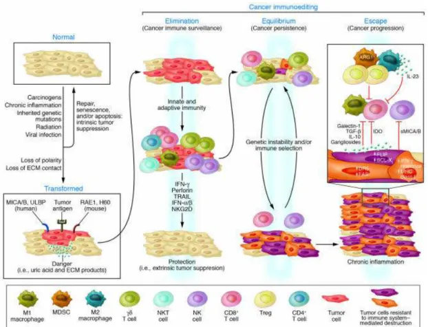
The idea that the immune system was part of the tumour microenvironment, and could potentially control tumour cells, was postulated by Paul Ehrlich in the early 1900s. He suggested that cells of the immune system are able to eliminate tumours, much like they destroy pathogens or pathogen-infected cells. However, so little was known about the composition and function of the immune system at that time, that it was not possible to assess the validity of its prediction. It took nearly 50 years for MacFarlane Burnet and Lewis Thomas to conceive their tumour immunosurveillance theory (Figure 1), which proposed that adaptive immunity was responsible for preventing cancer development in immunocompetent hosts, by recognizing and eliminating continuously arising, transformed cells 1.
Figure 1 - Tumour immunosurveillance theory and Cancer immunoediting hypothesis. Adapted from Swann& Smyth 20072
2 However, the first in vivo evidence for tumour immunosurveillance was only demonstrated in the 1990´s and derived from the observation that transplanted tumours grew more robustly in mice treated with neutralizing monoclonal antibodies specific for interferon-γ (IFN-γ)3. By that time with the advent of genetically modified immunodeficient mice, the relative contribution of the several components of the immune system for tumour immunosurveillance was examined4 . For example, lymphocyte-deficient mice, such as: recombination activating gene
Rag1-/- and Rag2-/-, severe combined immunedeficient (SCID), Tcrb-/-, Tcrd-/- and nude mice all display an increased susceptibility to tumour induction after methylcholanthere (MCA) treatment5-9. Moreover, immunodeficient mice show increased spontaneous tumour development with age. One striking example is the incidence of immunogenic B cell lymphomas in aged mice (> 1 year) on either C57BL/6 or BALB/c backgrounds, which increases from 0-6% in wild-type mice, to 40-60% in perforin-deficient mice10, 11. The genetic absence of other lymphocyte cytotoxic pathways, such as TRAIL or FasL, also increases the susceptibility of mice to spontaneous lymphomas12, 13. All together, these data provided strong evidence that lymphocytes (mainly due to critical cytotoxic molecules they produce) protect hosts from tumour development4.
In humans, the importance of immune surveillance against tumour emergence and progression was reinforced with the observation that immune deficiency states, including iatrogenic immune suppression, severe combined immunodeficiency (SCID), common variable immunodeficiency (CVID), and acquired immunodeficiency syndrome (AIDS), greatly increased the susceptibility of patients to many types of malignancies14.
Collectively, these were the foundations for the development of tumour immunotherapy as a “discipline”, and cancer immunotherapy as its clinical application.
1.1.2 Cancer immunoediting hypothesis
A hallmark study in 2001 showing that the immune system not only protects the host against tumour formation, but also shapes tumour immunogenicity7 prompted a major revision of the tumour immunosurveillance theory. Robert Schreiber postulated the cancer immunoediting hypothesis, which stresses the dual host-protective and tumour-promoting actions of the immune system in developing tumours.
The cancer immunoediting hypothesis15 states that tumour development is a dynamic process composed of three sequential phases: elimination, equilibrium and escape (Figure 1). In the elimination phase, innate and adaptive immunity destroy developing tumours before they become clinically apparent. If this phase goes to completion, the host remains free of cancer, and elimination thus represent the full extent of the process. If, however, a cancer cell variant is not destroyed in the elimination phase, it may then enter the equilibrium phase, in which tumour cells
3 are maintained in a state of functional dormancy. This phase is dependent on the adaptive immunity and it´s when editing of tumour immunogenicity occurs. Equilibrium may also represent an end stage of the cancer immunoediting process. However, as a consequence of constant immune selection pressure placed on genetically unstable tumour cells held in equilibrium, tumour cell variants may emerge that (i) are no longer recognized by adaptive immunity (antigen loss variants or tumour cells that develop defects in antigen processing or presentation), (ii) become insensitive to immune effector mechanisms, or (iii) induce an immunosuppressive state within the tumour microenvironment. These tumours may then enter the escape phase, in which their outgrowth is no longer blocked by immunity. These tumours emerge to cause clinically apparent disease.
1.1.3 Tumour microenvironment
Tumour immunosurveillance relies on the presence of leukocytes, most notably lymphocytes, in the tumour microenvironment. There, tumour growth, invasiveness, and metastasis are dynamic processes that involve the interaction of cancer cells with the extracellular matrix, the vasculature, and various types of non-cancerous host cells that form the tumour stroma. Commonly, the tumour microenvironment (TME) is constituted by: angiogenic vascular cells (AVCs), cancer-associated fibroblastic cells (CAFs) and infiltrating leukocytes16.
1.1.3.1 Tumour-infiltrating leukocytes
Within leukocytes, lymphocytes have gathered most attention given the phenotypes of lymphocyte-deficient mice and cancer patients14, and the prognosis value of their infiltration into tumours (see ahead 1.1.4.). However, it has become clear that a wide variety of leukocyte subtypes is present in the tumour microenvironment and play important anti-tumour roles, but also sometimes pro-tumour roles therein.
1.1.3.1.1 The traditional players: NK, CD8
+T and Th1 cells lymphocytes
It has been known for three decades that NK cells and CD8+ T lymphocytes, including those extracted from tumour biopsies, can efficiently kill transformed cells. Collectively, these killer lymphocytes recognize two important types of tumour antigens (among others): processed peptides presented by MHC class Ia proteins via TCRαβ; and non-classical (class Ib) MHC proteins via NKG2D17. The latter, which is expressed on NK, CD8+ and also γδ T cells, has been recently shown to be a key genetic determinant of cancer immunosurveillance18.
NK and CD8+ cells provide highly complementary anti-tumour strategies. Indeed, as demonstrated by the seminal work of Kärre and Kiessling, the downregulation of MHC class Ia, which is a common mechanism of evasion against CD8+ cells, renders tumours more susceptible to NK cell-mediated lysis. This “missing
4 self” recognition by NK cells is based on a set of MHC class Ia-specific inhibitory receptors that include killer cell immunoglobulin-like receptors (KIRs) in humans, lectin-like Ly49 molecules in mice, and CD94/NKG2A heterodimers in both species; in fact, NK cells express a complex repertoire of inhibitory and activating receptors that calibrate this anti-tumour function, while ensuring self-tolerance19, 20. In result, NK cells eliminate tumours that lack MHC class Ia expression; or that overexpress ligands for activating NK receptors like NKG2D or the natural cytotoxicity receptors NKp30, NKp44 and NKp4620. Furthermore, NK cells express high levels of low-affinity Fc receptor for IgG (CD16), which allows them to mediate antibody-dependent cell-mediated cytotoxicity (ADCC)21.
NK cells have been described to infiltrate various types of tumours in the skin, lung, gut and kidney18. Recent data on human NK cells infiltrating highly aggressive non-small cell lung cancers (NSCLC) showed a profound alteration of their phenotype, with decreased ability to degranulate and to produce IFN-γ, when compared with NK cells from distal lung tissues or blood from the same patients or from healthy donors22. This functional impairment of NK-TILs correlated with decreased expression of NKp30, NKp80, DNAM-1, CD16 and ILT2 receptors. Interestingly, among these, NKp30 has been shown to affect the prognosis of gastrointestinal stromal tumours through a specific pattern of alternative splicing23.
Various immunotherapeutic strategies have been proposed to tackle the common defects of NK cell activity in cancer patients20: activation of endogenous NK cells (with cytokines like IL-2, IL-15 and IL-18), cell adoptive immunotherapy, NK-cell-based donor lymphocyte infusions and allogenic stem cell transplantation (SCT)21. Although globally the objective responses have been disappointing, some data from allogenic and, more recently, haploidentical hematopoietic SCT have shown clinical (in the absence of adverse) effects mediated by NK cells20. This inspires further translational studies aimed at enhancing NK cell recruitment to tumours and their functional activity in situ.
With regard to CD8+ T cell-based immunotherapy, many recent efforts have focused in activating and expanding CD8+ tumour-infiltrating lymphocytes (TILs) ex
vivo and then re-infusing them into the cancer patients - adoptive cell therapy (ACT).
ACT of CD8+ TILs into lymphodepleted metastatic melanoma patients has shown very high objective response rates, ranging from 50% up to 81%24. In fact, TIL-ACT (combined with high doses of IL-2) has mediated cancer regression in 49-72% of melanoma patients, and durable complete responses, beyond 3-7 years, are currently ongoing in 40% of the patients25.
In pre-clinical models, adoptively transferred naïve CD8+ cells were shown to infiltrate melanoma lesions, be activated in situ and differentiate into functional cytotoxic T lymphocytes (CTLs)26. The naïve status of the infused population appeared to be an important parameter, as the differentiation stage of CTLs inversely correlated with their anti-tumour efficacy in vivo27. The enhanced anti-tumour function of naïve T cells was related to sustained effector cell development, prolonged cytokine production, and increased expansion in vivo.
Transduction of tumour antigen-specific TCRs28 or chimeric antigen receptors (CARs)29, 30 represent exciting prospects to increase the efficacy of cytotoxic ACT.
5 These strategies have thus far enabled cancer regression in patients with metastatic melanoma, synovial sarcoma, neuroblastoma and refractory lymphoma or leukaemia25.
In addition to cytotoxicity, IFN-γ secretion is a key anti-tumour function of CD8+ and NK cells, who share this property with various other lymphocyte populations, most notably “helper type 1” (Th1) CD4+ cells. These were first described 25 years ago in the context of the “Th1/ Th2” paradigm of immunity to infection, and since then clearly implicated in promoting anti-tumour responses: Th1 cells enhance the cytotoxic functions of NK and CD8+ cells, upregulate MHC class I expression in tumour cells (a direct effect of IFN-γ), and support CD8+ cell proliferation through the secretion of IL-231. Moreover, Th1 cells condition the antigen-presenting capacity of DCs and macrophages, thus shaping the CTL response. In fact, the combination of Th1 cell therapy with local radiation therapy augmented the generation of tumour-specific CTL at the tumour site and induced a complete regression of subcutaneous tumours32.
1.1.3.1.2 “New” effectors :
γδ
γδ
γδ
γδ
T, NKT and Th17 lymphocytes
The immune response to tumours by γδ T cells is the main focus of this thesis, so it will be discussed in the next chapter.
The “Th1/ Th2” paradigm for CD4+ T cell differentiation has been recently revised with the addition of Th17 cells, characterized by the production of interleukin-17 (IL-17). IL-17-deficient mice were shown to be more susceptible (than wild type animals) to tumour growth and lung metastasis33, 34. Adoptive transfer studies from the Restifo lab showed that in vitro generated Th17 cells were more efficient at eradicating tumours than Th1 cells35, and this was recently associated with stem cell-like properties of Th17 cells36. Importantly, adoptively transferred Th17 cells gave rise in vivo to Th1-like effector cell progeny36, and IFN-γ was actually necessary for the protective effects of adoptively transferred Th17 cells35. These data suggest that acquisition of Th1-like properties is required for an anti-tumour function by Th17 cells.
In stark contrast to the previous studies, IL-17-deficient mice presented reduced tumour growth in other models such as B16 melanoma and MB49 bladder carcinoma37, DMBA/TPA-induced skin carcinoma38, or in a spontaneous intestinal tumour model (driven by a mutation in the tumour suppressor gene APC)39.
The pro-tumour functions of IL-17 have been tightly linked to angiogenesis: IL-17 has been shown to act on endothelial, stromal and tumour cells to induce the expression of pro-angiogenic factors like VEGF, Angiotensins, PGE2 and IL-8, and thus promote tumour vascularization40. The precise conditions that determine pro- versus anti-tumour functions of Th17 TILs remain unclear and require further investigation.
Although Th17 cells are important providers of IL-17, this cytokine can be abundantly produced by other tumour-infiltrating leukocyte populations. Namely, murine γδ T cells can be the major source of IL-17, not only in homeostatic
6 conditions41, but also upon infection or tumour challenge42, 43. Like for Th17 cells, the role of IL-17 produced by γδ cells within the tumour microenvironment is controversial: it has been associated both with angiogenesis and promotion of tumour growth42, 44; and with CD8+ T cell recruitment and the therapeutic effects of chemotherapy against several subcutaneous tumour lines43, 44.
NKT cells also employ NK receptors, as well as CD1d-restricted TCRs to recognize tumour targets. The vast majority of these T cells are canonical or invariant NKT (type I NKT) cells that possess a specific TCRα rearrangement (Vα14Jα18 in mice; Vα24Jα18 in humans), associated with Vβ chains of limited diversity. All the other NKT cells that are CD1d-restricted and do not express this invariant TCR are called type II NKT cells45, 46. Although CD1d-deficient mice showed increased susceptibility to MCA-induced sarcomas47, there is evidence of functional heterogeneity also within NKT cells: while type I NKT cells seem to be protective, type II NKT cells mostly suppress tumour immunity46, 48.
In terms of cytokine production, activated NKT cells are potent providers of IFN-γ and IL-4 (and, to lesser extent, of IL-17). In the B16 metastatic melanoma model, a dual role of NKT cells was linked to immune suppressive IL-4 production by the thymus-derived subpopulation; and protective IFN-γ production by liver-derived type I NKT cells49.
Based on the pre-clinical evidence for an anti-tumour role of type I NKT cells, and the availability of a specific TCR agonist, α-Gal-Cer, several clinical trials have attempted to activate endogenous iNKT cells, or – more promising given by relative rarity of NKT cells in humans – perform ACT with (ex vivo expanded) type I NKT cells. However, the clinical effects of a-Gal-Cer or NKT ACT have been very limited46, thus illustrating the difficulty in translating findings from animal models of cancer into improved immunotherapies.
1.1.3.1.3 The inflammatory phagocytes: TAMs and TANs
Macrophages and neutrophils are important myeloid cells of the innate immune system and major drivers of inflammatory responses. Given the long-established association between cancer and inflammation, it is not surprising that tumour-associated macrophages (TAMs) and neutrophils (TANs) can have great impact on the course of tumour progression. While most studies have associated TAM and TAN infiltration with promotion of tumour cell growth, some other reports have proposed some anti-tumour roles. Once again, these opposing behaviors may be explained by heterogeneous TAM and TAN phenotypes, with distinct intra-tumour dynamics in various models.
Mirroring Th1/ Th2 polarization of CD4+ T cells, two distinct subsets of macrophages have been recognized: the “classical” activated (M1) macrophage phenotype and the “alternatively” activated (M2) macrophage phenotype50. IFN-γ drives the polarization towards M1 macrophages, which are characterized by abundant production of TNF-α, IL-12 and IL-23, CXCL9 and CXCL10, reactive nitrogen
7 and oxygen species; and by high expression of MHC class II and costimulatory molecules (making them efficient antigen-presenting cells)51. Conversely, IL-4 polarizes macrophages towards the M2 phenotype, which is associated with low levels of IL-12 but high levels of IL-10, IL-1RA and IL-1 decoy receptor. M2 cells also produce CCL17, CCL22 and CCL24, which results in the recruitment of Tregs and Th2 cells, eosinophils and basophils51.
The balance between M1 and M2 phenotypes seems to be controlled by NFkB signaling. Thus, NFkB targeting switched macrophages from an M2 to an M1 phenotype and led to ovarian tumour regression in vivo52. Nonetheless, the most frequent TAM phenotype seems to be M250. Consistent with this, TAM depletion was associated with improved anti-tumour immunity in models of metastatic breast, colon and non-small lung cancers53. The pro-tumour roles of M2 macrophages derive from various molecular mechanisms, including the production of the pro-angiogenic mediator semaphoring 4D54 and the invasive proteases cathepsins B and S55.
In the case of neutrophils, besides secreting cytokines and chemokines (such as IL-1b, IL-8, and IL-12), they produce large amounts of proteinases that remodel the extracellular matrix and promote the release of pro-angiogenic VEGF, thus supporting tumour cell growth and invasiveness56. Particularly important neutrophil proteinases are elastase57 and matrix metalloproteinases MMP-8 and MMP-958.
Despite being widely accepted as pro-tumour mediators based on multiple pre-clinical and clinical studies57, a dual nature of tumour-infiltrating neutrophils has also been suggested recently59, 60. Thus, anti-tumour N1 and pro-tumour N2 subsets were described and modulated within tumours by TGF-β59or IFN-β61. Consistent with such a complex neutrophil activity within the tumour microenvironment, the concentration of reactive oxygen species also seems to determine either pro-tumour (genotoxicity at modest concentrations) or anti-tumour (cytotoxicity at high concentrations) effects57. Consequently, the depletion of total neutrophils can lead to either reduced59 or increased62 tumour burden, further illustrating the globally paradoxical roles of tumour-infiltrating leukocytes.
1.1.3.1.4 Immunosuppressive leukocytes: Treg and MDSCs
Myeloid-derived suppressor cells (MDSCs) represent a heterogeneous population of myeloid progenitors and precursors of macrophages, granulocytes and dendritic cells, which are better characterized by their strong capacity to inhibit both innate and acquired immunity63 particularly T cell responses64. Murine MDSCs can be identified by the expression of Gr1 (includes Ly6C and Ly6G, macrophage and neutrophil markers, respectively) and CD11b (characteristic of macrophages). In humans, MDSCs are characterized by a CD11b+ CD33+ CD34+ CD14- HLA-DR -phenotype. Tumours produce various factors that promote MDSC expansion, such as IL-6, VEGF or GM-CSF, whereas they get further activated by local IFN-γ, IL-1β or Toll-like receptor (TLR) signal64.
MDSCs use a diversity of mechanisms to suppress T cell function, including the uptake of arginine and cysteine (essential amino acid for T cell activation) and
8 the nitration of the TCR63. In addition, MDSCs have been recently shown to directly support tumour growth by promoting the epithelial-to-mesenchymal transition in melanocytes65.
The possibility of improving anti-tumour immune responses by targeting MDSCs has been explored in pre-clinical models. One of the chemical drugs that seem to be more effective for MDSC depletion was 5-fluorouracil (5-FU). In a model of thymoma EL4 cells transplanted subcutaneously, tumour-bearing mice treated with 5-FU showed reduced number of MDSC in tumour lesions. This associated with prolonged mouse survival and enhanced intratumoral CD8+ T cell antigen-specific capacity to produce IFN-γ66. Interestingly, combination therapy with an agent (cyclophosphamide, CTX) that reduces Tregs led to a synergistic protective effect. Consistent with this, another study showed that inhibition of MDSC and Treg function within B16 melanomas using blocking antibodies to CTLA-4 (already in clinical use - ipilimumab - in late-stage melanoma) and to PD-1 reduced tumour development and increased mouse survival67.
Foxp3+ Tregs are well known to suppress the activation, proliferation and effector functions (such as cytokine production) of a wide range of immune cells, including αβ and γδ T cells, NK and NKT cells, B cells, macrophages and DCs. Suppressive functions displayed by Tregs include contact-dependent mechanisms, such as those that involve CTLA-4, PD-1 and GITR; and cytokine-mediated mechanisms such as TGF-β, IL-10 and IL-3568. TGF-β is particularly critical since, besides being strongly immunosuppressive, creates a potent positive feedback mechanism by instructing the differentiation of “inducible” Tregs69.
Experimental Treg depletion has been usually accomplished using anti-CD25 monoclonal antibodies, since there is a good correlation between CD25 and Foxp3 expression within CD4+ T cells (although activated effector cells also upregulate CD25). Prophylactic Treg depletion in renal cell carcinoma and MCA carcinoma was shown to reduce tumour growth, with protection being dependent on CD8+ and NK cells70, 71.
While most studies have concentrated on the immunosuppressive function of Tregs, two recent reports have shown that they can also act by directly promoting tumour growth and dissemination. Thus, Treg TILs in ovarian cancer they secrete VEGF that promotes endothelial cell proliferation72; and in breast cancer they produce RANKL, which associates with lung metastasis73. Importantly, the latter study is one of many that demonstrates that Treg accumulation within tumours is a marker for poor clinical outcome74.
1.1.3.2 Other cells (non-leukocytes)
Historically, tumour angiogenesis was thought to be regulated by cancer cells expressing proangiogenic factors, which is indeed one mechanism; however there is now abundant evidence that stromal cells in the tumour microenvironment are instrumental in switching on and sustaining chronic angiogenesis in many tumour
9 types16. The angiogenic switch was reported to be accompanied by reduced apoptosis and increased proliferation of cancer cells; the vascularization of tumours serves to attenuate cell death that would otherwise result from hypoxia and lack of serum-derived nutrients and survival factors75, 76.
Recently, angiogenic vascular cells have been implicated in local supply of growth-promoting trophic factors that are expressed by endothelial cells, potentially acting to stimulate, in a paracrine way, proliferation of tumour cells77. To hamper neovascularisation to tumours, potent angiogenic inhibitors have been developed, mainly acting at the vascular endothelial growth factor (VEGF) and other proangiogenic signalling pathways. This notwithstanding, the clinical responses are typically transitory, and survival benefit limited to duration of the treatments16.
A variety of cancer-associated fibroblasts can be recruited and/or be activated at the tumour microenvironment to contribute to tumour progression. For example, cancer-associated fibroblasts can produce mitogenic epithelial growth factors and also orchestrate the epithelial-to-mesenchymal transition (EMT) via secretion of TGF-β78. Moreover, cancer-associated fibroblasts in different tumour microenvironments can produce a number of proangiogenic factors, namely VEGF and IL-8/CXCL879.
Interestingly, platelets have been also shown to be able to promote tumour progression. By physically associating with cancer cells, platelets secrete TGF-β and induce transitory epithelial-to-mesenchymal transition, facilitating extravasation and seeding of metastases80.
1.1.3.3 Key molecular players: Cytokines and Chemokines
The cells that compose the tumour microenvironment produce a number of cytokines that may modulate the immune response and tumour progression. The hallmark cytokines that show potent anti-tumour effect are IFN-γ and TNF-α. IFN-γ is mainly produced by lymphocytic cytotoxic cells, including CD8+ and γδ T cells, NK cells, and as well as by Th1 CD4+ T cells. The most well characterized function of
IFN-γ is the upregulation of MHC class I molecules to aid the priming and presentation of antigens in APCs81. Moreover, IFN-γ regulates all aspects of Th1-mediated immune responses and activates macrophages82. IFN-γ has been used in the clinical management of a variety of malignancies, including bladder carcinoma, colorectal cancer, ovarian cancer, and adult T-cell leukaemia83.
TNF-α is primarily produced by macrophages, but also by a variety of other cells, including NK cells, T lymphocytes, smooth muscle cells, fibroblasts. The biological effect of TNF-α binding depends on the type of receptor activated (TNFR1 or TNFR2) and the cellular stage during activation, but the late stage event in TNF-α stimulation is cell apoptosis84. The potential use of TNF-α as a therapeutic agent was intensively studied in vitro and in vivo studies. These studies highlighted its possible role as an anticancer agent and prompted support for the numerous phase I and
10 phase II studies that followed85. Nevertheless, systemic TNF-α has not yet been translated to a patient therapy mainly due to elevated toxicity and lack of efficacy.
Although the anti-tumoral roles of IFN-γ (type II interferon) are well described, type I interferons (IFN-α and IFN-β) also play a role in tumour immunosurveillance. These are ubiquitously expressed and signal through the interferon type I receptor. Preclinical mechanistic experiments revealed that host type I IFN signalling was necessary upstream for spontaneous CD8+ T cell response against tumour antigens in mice86. The mechanism of this effect was predominantly through the action of host type I IFNs on CD8α+ dendritic cells (DCs), the subset involved in cross-presentation of antigens to CD8+ T cells. The therapeutic effect of IFN-α was explored in several cancer clinical trials, most importantly in melanoma87. A meta-analysis of the available published data from randomized clinical trials reported an event-free survival and overall survival in patients with high-risk melanoma treated with IFN-α adjuvant therapy88. To date, the mechanism of the therapeutic effects of IFN-α is not completely known but the examination of the nodal tumour taken before and after treatment, revealed increased infiltration of the tumour tissue by dendritic cells and T cells; and a striking ablation of STAT3 expression that is typically constitutively active in melanoma89.
TGF-β is a potent cytokine that regulates cell proliferation, differentiation and apoptosis. During tumourigenesis, two distinct roles have been reported for this cytokine: it has been reported as a tumour suppressor at early stages of the disease, and as a tumour promoter at later stages. These dual effects may be determined by the cell type where TGF-β acts as well as by the tumour microenvironment90. Another important cytokine that modulates immune responses at the tumour microenvironment is IL-10. The biological role of IL-10 in cancer is quite complex; however its role in promoting tumour progression is evidenced by the presence of IL-10 in advanced metastases and by the correlation of serum IL-10 levels to progression of disease91. Several suppressive cells, including TAMs, MDSCs and Tregs are important sources of TGF-β and IL-10.
The control of leukocyte trafficking is determined by a complex network of interactions involving tissue-specific integrins and chemokines, with selectins and chemokine receptors expressed on the leukocytes. It is now widely accepted that leukocytes are localized both in the tumour-supporting stroma and the tumour areas (and actually may account for up to 50% if the tumour mass) and that their migration from lymphoid tissues to tumours is determined by chemokines (Fig 2).
11 Figure 2 - Chemokine-mediated interaction between tumour cells and stromal cells. Adapted from Mukaida et al. 201292
Several reports support a critical role for CXCR3, a receptor for CXCL9/10/11 chemokines, in T cell recruitment to tumours. In mouse and human melanoma, CXCR3 ligands and CCL5 synergize to promote T-cell infiltration into cutaneous lesions93. In hepatocarcinoma, inflammatory cytokines such as TNF-α and IFN-γ, or TLR ligands, induce the production of CXCL10 and CCL5 in tumour epithelial cells or tumour-infiltrating immune cells94. Importantly, CXCL9/10 were associated with better survival for colorectal cancer patients95 and positive outcome in ovarian cancer patients96.
By contrast to CXCR3 ligands, usually correlated with good prognosis for cancer patients, some chemokines predict a poor outcome mostly due to the attraction of potentially suppressive cells. Several lines of evidence indicate that CCL2 plays an important role in TAMs and MDSCs recruitment. For example, CCL2 recruitment of TAMs to breast, prostate and colon carcinoma correlated with negative prognostic value for patients97. Also, CCL2 seems to recruit MDSCs in several types of mouse tumour models, including Lewis lung carcinoma, methA sarcoma, melanoma and lymphoma98.
A large number of Treg cells often infiltrate into tumours and systemic removal of Treg cells enhances natural as well as vaccine-induced anti-tumour T cell immunity. Tregs usually express CCR4, and its ligand, CCL22, regulates intratumoral Treg infiltration into various tumours99.
Overall, chemokines are thought to be promising candidates for immunomodulatory strategies in cancer, and several clinical trials are underway100.
12
1.1.4 TILs as prognostic factors
Although a favorable association of high numbers of TILs in primary tumours had been generally reported for decades in many human cancers, TILs had never reached the level of recognized prognostic marker (or proof for tumour immunosurveillance) probably due to their phenotypic and functional heterogeneity101. The recent observations that specific immune parameters have better prognostic value than standard staging systems, highlights the importance of the endogenous immune response in determining the clinical outcome. This may help to modify current classifications, and - most importantly - to identify the patients who would benefit the most from adjuvant immunotherapy (Table 1). Considering the data reviewed above, it is tempting to assume that good prognosis associates (for example) with CD8+ and NK cells, whereas bad prognosis is linked to the accumulation of Tregs and MDSCs. Moreover, given the functional heterogeneity within many leukocyte populations, clearly distinct outcomes could be expected from Th1 versus Th2 or Th17 CD4+ subsets, M1 versus M2 macrophages, N1 versus N2 neutrophils. This level of refinement is obviously incompatible with traditional immunohistochemistry of cancer patient samples, thus requiring additional techniques like flow cytometry and molecular biology to provide an adequate characterization of tumour-infiltrating leukocytes. Furthermore, detailed imaging may also be important as to define the localization of TILs within the tumour mass. For example, in a pre-clinical model, CD8+ T cells were recently shown to be trapped in the stroma and thus excluded from the core of tumour due to post-translational modifications (nitration) of the chemokine CCL2102. Interestingly, novel drugs that inhibited CCL2 nitration facilitated CD8+ T cell infiltration and tumour regression
Table 1 - Tumour-infiltrating leukocytes associated with prognosis for cancer patients. Adapted from Lança & Silva-Santos 2012103
Good prognosis Bad prognosis
TIL Cancer type Reference Cancer type Reference
CD8+ Colorectal cancer Hepatocellular carcinoma Esophageal carcinoma Breast cancer 104105106 94107108 105, 106, 109 Th1 (CD4+) Colorectal cancer Hepatocellular carcinoma Breast cancer 110 94 111 Th2 (CD4+) Pancreatic cancer 112
Th17 (CD4+) Esophageal carcinoma 109 Colorectal cancer Hepatocellular carcinoma
Prostate cancer
110 113 114
Tregs (CD4+) Colorectal cancer
Hepatocellular carcinoma Ovarian carcinoma Breast Cancer 115 107 116 117 γδ γδ γδ
γδ T cells Ovarian carcinoma 118 Breast Cancer 119
B cells Breast cancer 120
NK cells Esophageal carcinoma Hepatocellular carcinoma
109 94
MDSCs Esophageal, pancreatic and gastric cancer 121
Macrophages Breast cancer 122