IN ST IT U T O D E C IÊ N C IA S B IO M ÉD IC A S A B EL S A LA Z A R FA C U LD A D E D E CI ÊN CI A S FA C U LD A DE DE M ED IC IN A
R
aq
ue
l R
ea
l. H
um
an i
PS
Cs a
nd d
ise
ase mo
de
llin
g in
vivo
C
ont
rib
ut
io
n o
f h
um
an i
PS
Cs t
o t
he s
tu
dy
of n
eu
ro
n d
ev
elo
pm
ent a
nd d
is
ea
se m
od
elli
ng
in
viv
o
Ra
qu
el M
aria
Coe
lho
R
ea
l
Contribution of human iPSCs to
the study of neuron development
and disease modelling in vivo
Raquel Real
D
2019D
.ICB
AS
2019
SE D E A D MIN IST RA TIVRAQUEL MARIA COELHO REAL
Contribution of human iPSCs to the study of neuron development and disease
modelling in vivo
Tese de Candidatura ao grau de Doutor em
Biologia Básica e Aplicada submetida ao Instituto
de Ciências Biomédicas Abel Salazar da
Universidade do Porto
Orientador – Doutor Vincenzo De Paola
Categoria – Group Leader
Afiliação – Institute of Clinical Sciences, Faculty
of Medicine, Imperial College London, UK
Coorientador – Doutor João Carlos Bettencourt
de Medeiros Relvas
Categoria – Investigador Principal
Afiliação – Instituto de Biologia Molecular e
Celular / Instituto de Inovação e Investigação em
Saúde, Universidade do Porto
Raquel Real was supported by a PhD fellowship (PD/BD/52198/2013) awarded by Fundação para a Ciência e Tecnologia (FCT). This work was funded by the Medical
Acknowledgments
I would like to thank:
My supervisor Vincenzo De Paola, for receiving me in the Synaptic Plasticity and Repair Group and giving me the opportunity to work on this project.
My co-supervisor João Relvas, for the wise advice and encouragement. Rick Livesey and Manuel Peter, for enthusiastically entering this collaboration. My mentor Richard Festenstein, for the very helpful career talks.
My lab mates Shabana Khan and Maria Tortora, for their precious help in the final stages of this project and their friendship.
Joana Dopp, for being the best research assistant ever.
Cher Bass, for the helpful discussions on calcium imaging analysis.
Marta and Theresa, the best flatmates anyone could wish for. Thank you for the night-long conversations and the amazing trips together!
The amazing members of the GABBA team, Rita, Delfim and Elsa, for their infinite support. The GABBA Programme and FCT, for believing in me and giving me the opportunity to pursue my dreams.
The Rosetrees Trust, for the additional funding to support this project. Helen Figueira, for the talent to solve admin and logistics nightmares. And finally, my family, who always stood by me.
Contributions
Manuel Peter (The Gurdon Institute, University of Cambridge, UK) conducted the human
iPSC-derived neuron differentiation protocol, lentiviral vector preparation and transductions.
Ayiba Momoh (The Gurdon Institute, University of Cambridge, UK) developed the new Ts21
iPSC line used in this study. Samuel J. Barnes (Dementia Research Institute, Imperial College London, UK) developed customised MATLAB software and analysed in vivo calcium time series images. Mark A. Smith (Institute of Clinical Sciences, Imperial College London, UK) performed electrophysiological recordings in acute brain slice preparations. Chad
Whilding (Microscopy Facility, MRC London Institute of Medical Sciences, UK) developed a
customised ImageJ (NIH) script for confocal microscopy image analysis. Graham Knott (BioEM Facility, EPFL, Switzerland) performed the electron microscopy. Alessio Strano (The Gurdon Institute, University of Cambridge, UK) performed the gene expression experiment, the short tandem repeat microarray and the genome-wide SNP array (with Illumina, Inc.).
Emanuela Volpi (University of Westminster, UK) analysed fluorescent in situ hybridization
signals. Prof Gordon Stamp (The London Clinic, UK) analysed the histology material stained with Haematoxylin-Eosin for the presence of teratoma. Shabana Khan and Maria Tortora (Institute of Clinical Sciences, Imperial College London, UK) assisted with the analysis of a subset of confocal immunohistochemistry images. Joana Dopp (Institute of Clinical Sciences, Imperial College London, UK) assisted with hNCAM stainings. Antonio Trabalza (Institute of Clinical Sciences, Imperial College London, UK) helped with the initial in vivo imaging and surgeries.
Table of contents
RESUMO ... 4 ABSTRACT ... 6 LIST OF PUBLICATIONS ... 8 LIST OF FIGURES ... 9 LIST OF TABLES ... 11 LIST OF ABBREVIATIONS ... 12 CHAPTER 1. INTRODUCTION ... 141.1 Human induced pluripotent stem cells as model systems for central nervous system development and disease ... 14
1.2 Neural induction of hiPSCs recapitulates human neurogenesis ... 15
1.3 hiPSCs generate functional neurons and neural networks ... 16
1.4 In vivo transplantation of human PSC-derived neurons ... 17
1.5 Neurons derived from hPSC as model systems of disease ... 19
1.6 Human iPSCs and Down syndrome ... 20
1.7 Down syndrome brain phenotypes ... 21
1.8 Insights from animal models of Down syndrome ... 22
1.9 iPSCs in the study of Down syndrome ... 26
CHAPTER 2. METHODS ... 31
2.1 Generation of a new Ts21 iPSC line ... 31
2.2 Animals ... 31
2.3 Surgical procedures ... 31
2.4 Live imaging ... 32
2.5 Two-photon image analysis ... 33
2.6 Electrophysiology ... 35
2.7 Immunohistochemistry ... 35
2.8 Histology ... 36 2.9 Retrograde trans-synaptic tracing ... 37
2.10 In situ hybridization ... 37
2.11 Electron microscopy ... 38
2.12 Gene expression analysis ... 38
2.13 Copy number variation detection ... 39
2.14 Short tandem repeat assay ... 39
2.15 Statistics and data presentation ... 39
CHAPTER 3. IN VIVO MODELLING OF HUMAN NEURON DYNAMICS ... 42
3.1 Cellular characterization of transplanted human grafts ... 42
3.2 In vivo characterization of human neuron structural development ... 49
3.3 Functional characterization of transplanted human grafts connectivity ... 57
3.4 In vivo development of neural network activity in transplanted human grafts ... 61
CHAPTER 4. IMAGING HUMAN NEURON STRUCTURAL AND FUNCTIONAL
DYNAMICS IN DOWN SYNDROME ... 63
4.1 In vivo modelling of Down syndrome diseases with hiPSC-derived neurons ... 63 4.2 Ts21 neurons transplanted in vivo recapitulate aspects of the cortical
neurogenesis defect in Down syndrome ... 65 4.3 Ts21 neurons exhibit normal developmental neurite refinement ... 68
4.4 Ts21 neurons have increased structural synaptic stability ... 71 4.5 Ts21 neurons show no evidence of major functional defects ... 75
4.6 In vivo calcium imaging reveals impaired network synchronization in Ts21
neurons ... 80
CHAPTER 5. DISCUSSION ... 83
5.1 Transplanted human iPSC-derived neurons as a model system for the study of
5.2 Transplanted human iPSC-derived neurons as a model system for human
neurodevelopmental disorders ... 87
5.3 Concluding remarks ... 89
REFERENCES ... 92
APPENDIX ... 105
Resumo
O estudo dos mecanismos celulares que regulam o desenvolvimento neuronal humano esteve até recentemente praticamente inacessível, mas a descoberta na década passada que células somáticas de dadores podem ser reprogramadas in vitro para células estaminais pluripotentes induzidas (iPSCs) permitiu a criação de diversos tipos celulares, incluindo várias classes de neurónios e células da glia. Estas células têm a vantagem de serem facilmente acessíveis e de não estarem sujeitas aos mesmos constrangimentos éticos que o uso de células derivadas de células estaminais embrionárias humanas. Estudos in vitro com recurso a neurónios corticais humanos derivados de iPSCs permitiram aprofundar de forma inédita o conhecimento dos processos fundamentais do neurodesenvolvimento humano. No entanto, a longevidade e o potencial de maturação neuronal in vitro são limitados, presumivelmente devido à ausência de interações celulares complexas em culturas celulares em monocamada e à ausência de vascularização em culturas tridimensionais de organóides cerebrais. Consequentemente, os sistemas in vitro não permitem uma caracterização plena da sequência de eventos que ocorre durante o desenvolvimento neuronal humano. Tal conhecimento é fundamental para compreender as especificidades do desenvolvimento do cérebro humano, e que o distinguem dos cérebros de outros mamíferos. Adicionalmente, o conhecimento detalhado dos processos celulares que caracterizam o normal desenvolvimento neuronal em humanos é fundamental para compreender as múltiplas doenças que afetam o desenvolvimento cerebral em fases precoces da vida, geralmente com consequências devastadoras. No sentido de ultrapassar as limitações dos sistemas in vitro, células neuroprogenitoras e neurónios excitatórios prosencefálicos derivados de iPSCs foram transplantados no córtex somato-sensitivo de ratos adultos, nos quais uma janela craniana foi implantada em simultâneo para permitir microscopia multi-fotão intravital. Esta estratégia permitiu efetuar o estudo dinâmico da neuritogénese e sinaptogénese, assim como o desenvolvimento de redes neurais, ao longo de vários meses, no contexto de um microambiente multicelular complexo e vascularizado. Estes estudos revelaram que neurónios humanos em fases precoces do desenvolvimento adquirem plasticidade sináptica estrutural e conectividade, com um padrão de actividade neural de carácter oscilatório e dinâmica espaço-temporal complexa.
A abordagem acima delineada foi ainda utilizada no estudo da síndrome de Down, um distúrbio do neurodesenvolvimento comum e uma causa importante de deficiência intelectual congénita. O progresso no conhecimento dos mecanismos celulares e moleculares
subjacentes à síndrome de Down tem sido dificultado pela disponibilidade limitada de modelos que recapitulem esta complexa condição cromossómica. A compreensão do espectro de fenótipos celulares que caracterizam a síndrome de Down requer um modelo que interrogue todas as fases do desenvolvimento neural, o que torna os estudos em tecidos cerebrais humanos post mortem necessariamente incompletos, uma vez que estes apenas permitem observar fenótipos de forma estática. As células estaminais embrionárias humanas, por outro lado, são relativamente inacessíveis e o seu uso limitado por considerações éticas. Os modelos murinos permitem recapitular todas as fases do desenvolvimento neural e dissecar os mecanismos patogénicos da síndrome de Down. Consistentemente, estes modelos têm sido extremamente úteis para aprofundar o conhecimento da patogénese da síndrome de Down, mas existem limitações importantes que importa reconhecer. Em primeiro lugar, na maioria dos modelos murinos nem todos os genes do cromossoma 21 (Hsa21) estão presentes em triplicado. Em segundo lugar, a homologia entre os cromossomas murinos e o Hsa21 é apenas parcial. Finalmente, a regulação genética do Hsa21 é diferente da regulação genética dos cromossomas ortológos em células murinas. Consequentemente, a reprogramação de células somáticas de indivíduos com síndrome de Down em células estaminais pluripotentes induzidas constitui uma preciosa ferramenta para dissecar
mecanismos patogénicos nesta síndrome cromossómica complexa. Células
neuroprogenitoras, neurónios e astrócitos derivados de iPSCs humanas portadoras de trissomia 21 (Ts21) podem recapitular as fases iniciais do desenvolvimento neural de modo longitudinal. Adicionalmente, a sua transplantação no cérebro murino tem o potencial de revelar fenótipos inéditos, que não são evidentes nos sistemas in vitro. De facto, imagiologia multi-fotão de neurónios portadores de trissomia 21 transplantados, provenientes de dois dadores independentes com síndrome de Down, revelou que as estruturas sinápticas são menos dinâmicas nestes neurónios, e que os circuitos neurais nos transplantes com trissomia 21 são muito menos síncronos que os transplantes euploides. Ambos estes fatores têm o potencial de contribuir para os defeitos cognitivos características da síndrome de Down, o que ilustra a potencialidade das técnicas de microscopia in vivo de neurónios derivados de doentes para o estudo de distúrbios do neurodesenvolvimento.
Abstract
The study of cellular mechanism of human neuron development has until recently been largely inaccessible, but the discovery in the past decade that somatic donor cells can be reprogrammed in vitro to induced pluripotent stem cells (iPSCs) has enabled the generation of large pools of diverse cell types, including several classes of neurons and glial cells that are readily accessible and not subject to the same ethic constraints as the use of cells derived from human embryonic stem cells. In vitro studies using human iPSC-derived cortical neurons as a model system have advanced our understanding of basic neurodevelopmental processes in a remarkable way. However, there are limitations to the longevity and maturation potential of neurons in vitro, either due to a lack of more complex cell-cell interactions in monolayer neuronal cultures or of inner vascularization in three-dimensional brain organoid cultures. Consequently, in vitro systems are unable to fully characterize the sequence of events that occurs during the development of human neurons. Such knowledge is fundamental to understand the specificities of human brain development that set it apart from other mammalian brains. In addition, an in-depth understanding of the cellular processes that characterize normal neural development in human cells is necessary to comprehend the multiple disorders that affect early brain development, often with devastating consequences. To overcome the limitation of in vitro systems, human cortical neural progenitor cells (NPCs) and excitatory neurons of forebrain identity derived from iPSCs were transplanted into the somatosensory cortex of adult mice, in which a cranial window was simultaneously implanted for longitudinal multiphoton intravital imaging. This strategy has allowed the dynamic study of neuritogenesis, synaptogenesis and the development of neural network activity over several months, in a multicellular and highly vascularized complex microenvironment, highlighting that human cortical neurons in early stages of development undergo structural synaptic rewiring and acquire circuit connectivity with an oscillatory pattern of neural activity and complex spatiotemporal dynamics.
Down syndrome (DS) is a common neurodevelopmental disorder and a major cause of congenital intellectual disability. Advances in our understanding of the underlying cellular and molecular mechanisms of DS have been hampered by the limited availability of model systems that recapitulate this complex chromosomal condition. Elucidation of the full spectrum of cellular phenotypes of DS requires a model system that interrogates all stages of neural development, which precludes the usefulness of human post-mortem and foetal brain tissue that provide only discrete snapshots of the disease phenotypes. Human embryonic stem cells
are relatively inaccessible and there are often ethical limitations to their use. Animal models can recapitulate all stages of neural development and enable the dissection of the pathogenic mechanisms of DS. These have been extremely useful to our understanding of disease pathogenesis, but there are important caveats. First, triplication of chromosome 21 (Hsa21) genes is incomplete in most DS mouse models. Second, there is only partial mouse-human homology of Hsa21 regions. Finally, Hsa21 genes are differentially regulated in a mouse cellular context and in human cells. Therefore, iPSCs reprogrammed from somatic cells of DS individuals represent a valuable tool to dissect disease mechanisms in this complex disorder. Human iPSC-derived NPCs, neurons and astrocytes that harbour trisomy of chromosome 21 (Ts21) can longitudinally recapitulate the early stages of neural development in DS, and its transplantation into the mouse brain has the potential to uncover new disease phenotypes not evident in in vitro systems. Indeed, longitudinal multiphoton imaging of transplanted Ts21 neurons derived from two independent individuals with DS has uncovered that synaptic structures are less dynamic in Ts21 neurons and that Ts21 neural circuits are much less synchronous. Both these factors could contribute to the cognitive deficits characteristic of DS and illustrate the potential of in vivo imaging of patient-derived neurons for disease modelling.
List of Publications
The work described in this dissertation is included in the following peer-reviewed publication:
Raquel Real*, Manuel Peter*, Antonio Trabalza*, Shabana Khan, Mark A. Smith, Joana Dopp,
Samuel J. Barnes, Ayiba Momoh, Alessio Strano, Emanuela Volpi, Graham Knott, Frederick J. Livesey and Vincenzo De Paola. In vivo modeling of human neuron dynamics and Down syndrome (2018). Science 362, eaau1810. doi: 10.1126/science.aau1810.
List of Figures
Figure 1.1 Human corticogenesis………...…….……….……… 16
Figure 1.2 Human chromosome 21 and mouse models of Down syndrome …………. 25
Figure 1.3 Strategies to obtain isogenic disomic Ts21-iPSC lines ………...… 30
Figure 2.1 Schematic of experimental procedures and timeline ………... 34
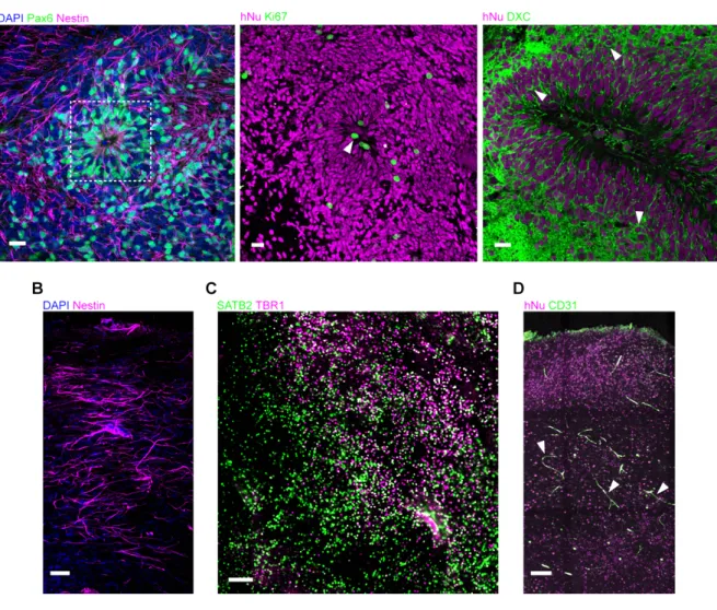
Figure 3.1 Ongoing neurogenesis in transplanted human grafts ………...…. 44
Figure 3.2 Vascularization and complex cytoarchitecture of the human graft ……... 45
Figure 3.3 Host cell identity within the human graft ………...…. 46
Figure 3.4 Minimal recruitment of host-derived microglia to the human graft ……….… 47
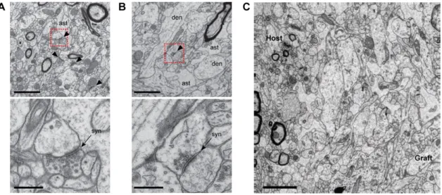
Figure 3.5 Ultrastructure of human grafts reveals immature neural tissue ……….……. 48
Figure 3.6 No teratomas developed in the human grafts ………... 48
Figure 3.7 In vivo two-photon imaging of human cortical grafts reveals cellular mechanisms of pruning ……….……….... 51
Figure 3.8 Human neurons project to known target areas of the somatosensory cortex ……….……….…. 52
Figure 3.9 Developing dendritic synaptic structures are characterized by highly dynamic restructuring and balanced rates of gains and losses ……….……. 54
Figure 3.10 Developing axonal synaptic structures mirror the events in dendritic synaptic structures ………... 56
Figure 3.11 Human neurons are excitable and have functional synaptic input …………. 58
Figure 3.12 Synaptic input to the human graft is mainly from other human neurons … 60 Figure 3.13 Synchronized spontaneous neural activity emerges early and has a defined spatiotemporal order ……….……… 62
Figure 4.1 Fluorescent in situ hybridization assay ……….……….… 64
Figure 4.2 Neurogenesis in Ts21 human cortical grafts ………... 67
Figure 4.4 In vivo neurite development is normal in Ts21 neurons ……….…… 70 Figure 4.5 Ts21 neurons show decreased dendritic spine plasticity ……… 73 Figure 4.6 Axonal synaptic structures in Ts21 neurons have enhanced stability ……... 74 Figure 4.7 Ex vivo electrophysiological characterisation of Ts21 and control neurons 77 Figure 4.8 Transplanted Ts21 and control neurons have similar synaptic input ………. 78
Figure 4.9 Reduced Ih activation in Ts21 neurons ………. 79
Figure 4.10 Ts21 neurons exhibit decreased burst activity ………. 81 Figure 4.11 Reproducibility of calcium dynamics in an independent control line …... 82
List of Tables
Table 1 Lentiviral vectors used for cell transduction ….…….………...……. 40 Table 2 Cell lines used to generate transplanted cortical neurons ………....….………. 40
Table 3 Antibodies used for immunofluorescence ……….………… 41
Table 4 Biophysical properties of WT and Ts21 neurons ………... 76
List of Abbreviations
2P Two-photon
AD Alzheimer’s disease
AMPA α-amino-3-hydroxy-5-methyl-4-isoxazolepropionic acid
APP Amyloid precursor protein
Ab Amyloid-b
CD31 Cluster of differentiation 31
ChR2 Channelrhodopsin-2
CNS Central nervous system
CTIP2 COUP-TF-interacting protein 2
DAPI 4',6-Diamidino-2-phenylindole dihydrochloride
DNA Deoxyribonucleic acid
DS Down syndrome
DSCR Down syndrome critical region
DXC Doublecortin
EEG Electroencephalogram
EM Electron microscopy
EnvA Envelope glycoprotein of avian sarcoma/leukosis virus subtype A EPB En passant bouton
FISH Fluorescent in situ hybridization
GABA Gamma-aminobutyric acid
GAD67 Glutamate decarboxylase 1
GECI Genetically encoded calcium indicator GFAP Glial fibrillary acidic protein
GFP Green fluorescent protein
GW Gigaohm
hESCs Human embryonic stem cells
hiPSCs Human induced pluripotent stem cells
hNu Human nuclei
Hsa21 Human chromosome 21
hSYN Human synapsin
Ih Hyperpolarization-activated cation current IBA1 Ionized calcium binding adaptor molecule 1
mEPSC Miniature excitatory postsynaptic current mIPSC Miniature inhibitory postsynaptic current
Mmu10 Mouse chromosome 10
Mmu16 Mouse chromosome 16
Mmu17 Mouse chromosome 17
MRI Magnetic resonance imaging
mV Millivolt
NBQX 2,3-Dioxo-6-nitro-1,2,3,4-tetrahydrobenzo[f]quinoxaline-7-sulfonamide NCAM Neural cell adhesion molecule
NPC Neural progenitor cell
NSG NOD scid gamma
NuMA Nuclear mitotic aparatus
OLIG2 Oligodendrocyte transcription factor
PAX6 Paired Box 6
PBS Phosphate buffered saline
PDGFRα Platelet-derived growth factor receptor α
pF Picofarad
PFA Paraformaldehyde
ROI Region of interest
ROS Reactive oxygen species
RV Rabies virus
RV-∆G Glycoprotein-deleted rabies virus
SATB2 Special AT-rich sequence-binding protein 2 SNAP Synaptosomal nerve-associated protein
SNP Single nucleotide polymorphism
SD Standard deviation
SEM Standard error of the mean
SSC Saline sodium citrate
TBR1 T-box, brain 1
TOR Turnover ratio
Ts21 Trisomy of chromosome 21
TVA Tumour virus receptor A
VGLUT1 Vesicular glutamate transporter 1
Chapter 1. Introduction
1.1 Human induced pluripotent stem cells as model systems for central
nervous system development and disease
Animal models have been the preferred model system to study the development of the central nervous system and its many disorders. Due to the array of sophisticated genetic engineering tools currently available for mice, this species has been the most widely used to model human brain disorders. However, not only is the neurodevelopmental process much faster in rodents than in human, rodents also lack the structural complexity of the human brain, particularly in the cortex. Cortical areas associated with higher-order cognitive functions, such as prefrontal and temporal cortices, are particularly well developed in the human brain compared to mice. These interspecies differences account for the limited validity of rodent models to recapitulate human brain development and diseases (Ardhanareeswaran et al., 2017). Alternatives include post-mortem human brain tissue, which lack a temporal dimension, primary human brain cells, which are difficult to access and are not expandable, and human embryonic stem cells (hESCs), which have the advantage of being pluripotent and can be differentiated into brain cells in vitro, making them the ideal model to study neurodevelopmental and disease mechanisms at distinct developmental stages. However, the use of hESCs in research is hampered by ethical and accessibility issues.
In 2007, two independent groups reported the generation of induced pluripotent stem cells (iPSCs) from adult human somatic cells, by the ectopic introduction of a defined set of pluripotency-related transcription factors into somatic cells (Takahashi et al., 2007; Yu et al., 2007). The most commonly used combination of transcription factors includes the so-called Yamanaka factors: Oct3/4, Sox2, Klf4 and c-Myc. These cells closely resemble human embryonic stem cells, as they express hESC surface markers, are capable of self-renewal and are pluripotent (i.e., they have the potential to differentiate into cell types from the three germinal layers). It is known that hiPSCs partially retain the epigenetic memory of the somatic cells they originate from, which leads to a differentiation bias towards the original cell type (Ohi et al., 2011). In addition, the study of gene expression profiles of hESCs and iPSCs revealed unique gene expression and epigenetic signatures in iPSCs (Chin et al., 2009; Ruiz et al., 2012). Extended in vitro culture partially corrected the differences in gene expression between hiPSCs and hESCs (Chin et al., 2009), but whether these residual molecular differences have any physiological significance is still a matter of debate. Nonetheless, hiPSCs are widely considered to be hESC-like cells (Choi et al., 2015; Guenther et al., 2010). In addition to
transcriptomics comparing the two types of pluripotent stem cells, multiple studies have generated different subtypes of neuronal cells from hESCs and hiPSCs in parallel, without distinction in differentiation potential or efficiency, developmental time course, or morphological and functional properties of the mature neurons (Kim et al., 2011; Shi et al., 2012a; Song et al., 2013). However, unlike hESCs, the generation of hiPSCs from somatic cells for research is not subject to the same ethical concerns and legal restrictions. Additionally, hiPSCs are more readily available in large numbers and from a wider pool of individuals than hESCs, which is particularly relevant when studying complex and multigenic disorders. Therefore, hiPSCs have emerged in the neuroscience field as an essential research tool for understanding human brain development, identification of disease mechanisms and drug discovery (Liang et al., 2018).
1.2 Neural induction of hiPSCs recapitulates human neurogenesis
During neural induction of hiPSCs, exposure to appropriate concentrations and gradients of specific morphogens that are normally expressed during brain development leads to the generation of different subtypes of neurons. Protocols have been developed for the in vitro generation of cortical glutamatergic excitatory neurons (Shi et al., 2012b), GABAergic inhibitory interneurons (Nicholas et al., 2013), midbrain dopaminergic neurons (Chambers et al., 2009; Soldner et al., 2009), hippocampal granule neurons (Yu et al., 2014) and spinal motor neurons (Chambers et al., 2009; Dimos et al., 2008). Importantly, in vitro differentiation of hiPSCs into cortical neurons has been shown to recapitulate many aspects of in vivo human cortical development. The human cortex is composed of six layers of neurons that contain a majority of glutamatergic excitatory neurons generated in the ventricular zone, and approximately 20% of GABAergic interneurons that are formed in the medial ganglionic eminence of the ventral telencephalon and migrate into the cortex during development (Molyneaux et al., 2007). The glutamatergic neurons destined for each cortical layer are born in a defined temporal order over several weeks, with deep layer neurons generated earlier than upper layer neurons, followed by the late production of astrocytes. It has been revealed that the generation of cortical neurons from both hESC- and hiPSC-derived neural progenitor cells (NPCs) follows the same temporal pattern (i.e., deeper layer neurons form earlier than upper layer neurons) (Espuny-Camacho et al., 2013; Shi et al., 2012a). Additionally, the extended time-course of human neurogenesis and neuronal maturation is preserved in vitro, as hiPSC-derived neurons require prolonged periods in culture to achieve morphological and electrophysiological patterns of maturity (Shi et al., 2012a) (Figure 1.1). Interestingly, transplantation of hESCs into the rodent brain did not accelerate the timing of neurogenesis and neuronal maturation (Espuny-Camacho et al., 2013), which suggests that developmental
processes are intrinsically regulated in a species-specific manner (van den Ameele et al., 2014). Similar observations of a protracted timeline akin to human embryonic development were made in the differentiation and maturation of forebrain GABAergic interneurons in vitro (Nicholas et al., 2013).
1.3 hiPSCs originate functional neurons and neural networks
Several studies have addressed the fundamental question of whether hiPSC-derived neurons are able to achieve a state of functional maturation analogous to human neurons in vivo. Neural progenitor cells cultured in vitro have been shown to progressively develop passive and active membrane properties resembling mature human neurons (Niclis et al., 2017; Prè et al., 2014; Song et al., 2013). Whole-cell patch-clamp recordings of forebrain neurons derived from hiPSC performed over time reveal the appearance of voltage-dependent sodium and potassium currents and a progressive increase in the proportion of cells that fire trains of action potentials in response to current injection, indicating the gradual addition of ionic channels to the membrane and the intrinsic excitability of these neurons (Prè et al., 2014; Shi et al., 2012a). Moreover, the characteristics of the action potentials evolve over time, from relatively immature spiking phenotypes to more mature firing properties, including an increase in spike amplitudes, faster spikes and lower thresholds for action potential generation. Also, neurons derived from hiPSCs in vitro eventually develop spontaneous miniature excitatory and inhibitory postsynaptic currents (mEPSCs and mIPSCs, respectively) that increase in frequency and amplitude over time, a hallmark of functional synapses. Importantly, the temporal profile of functional maturation and synaptogenesis in hiPSC-derived neurons is protracted relative to rodent neurons, suggesting neural progenitor cells derived from hiPSCs follow an intrinsic species-specific developmental programme lasting several months (Avaliani et al., 2014; Niclis et al., 2017).
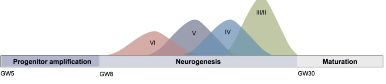
Figure 1.1 Human cortical neurogenesis.
In vitro generation and maturation of cortical neurons from hiPSCs recapitulates the time-course and temporal patterning of in vivo cortical neurogenesis, with deeper layer neurons being generated before upper layer neurons over several months. GW, gestational week. Adapted from van den Ameele et al., 2014.
Spontaneous neural electrical activity has been documented in several mammalian species during development and has been linked to many essential developmental processes, including neuronal differentiation and migration, synaptogenesis and synaptic plasticity (Khazipov and Luhmann, 2006). In addition, spontaneous oscillatory patterns of synchronized electrical activity have been shown to play a key role in early neural network development. Importantly, hESC- and hiPSC in vitro also exhibit spontaneous activity transients that mature over time, in parallel to increased morphological complexity and progressive maturation of membrane properties (Heikkilä et al., 2009; Kirwan et al., 2015; Prè et al., 2014). Given that calcium activity reflects neuronal activity and the size of a calcium transient directly correlates with the number of repetitive action potentials fired (Smetters et al., 1999), Kirwan and colleagues have monitored the development of calcium transients longitudinally to ascertain whether hiPSC-derived cortical neurons can form neuronal networks in vitro (Kirwan et al., 2015). Indeed, hiPSC-derived neurons in vitro are not only functional and synaptically connected, but they also develop glutamatergic-dependent calcium transients that mature over time in a stereotypical manner reminiscent of cortical neural network development in vivo (Khazipov and Luhmann, 2006). Thus, neurons derived from hiPSCs constitute a very useful tool to model human neuron and neural network development in vitro.
1.4 In vivo transplantation of human PSC-derived neurons
hPSC-based in vitro systems can provide significant insights into human-specific features of brain development. However, monolayer neuronal cultures lack the spatial organisation and complex cytoarchitecture of the human brain, which limit its usefulness to model the full complexity of neurodevelopmental processes. In addition, cultures of ESC/iPSC-derived neurons are limited by short-term survival and incomplete neuronal maturation (Korecka et al., 2016). Under specific culturing conditions, hiPSC-derived neural progenitors and neurons in vitro have the intrinsic ability to self-organise into three-dimensional (3D) multi-layered cortical structures (Mariani et al., 2012). These 3D cortical structures exhibit a gene expression profile typical of the early embryonic telencephalon, recapitulating the transcriptional programs that are active during human cortical development. The generation of 3D multicellular culture systems (or organoids) has been an important development in the field of hiPSCs. Lancaster and colleagues were the first to develop a hiPSC-derived brain organoid, a structure that contains several identifiable brain regions and cortex-like layered structures (Lancaster et al., 2013). The cellular composition, gene expression profiles and epigenomic signatures of cerebral organoids are remarkably similar to those of the human foetal tissue (Camp et al., 2015; Luo et al., 2016). Therefore, brain organoids constitute an important tool for modelling human neurodevelopment, in a multicellular context that more
closely mimics human physiology. The multicellular environment of organoids enables cells to achieve a functionally more mature state than in 2D cultures, and the interaction between different cell types within the organoid allows for the study of non-cell autonomous aspects of disease pathology. Despite the obvious advantages, the organoid technology still has limitations, the most significant of which is the lack of vascularisation that limits growth and maturation. To circumvent this issue, brain organoids generated in vitro from hESCs have recently been transplanted into the brains of mice, which resulted in organoid vascularisation in vivo and prolonged survival (Mansour et al., 2018). The transplantation of dissociated hESC/iPSC-derived neurons in the central nervous system of rodents has also been shown to enhance the survival and accelerate the functional maturation of neurons (Espuny-Camacho et al., 2013; Niclis et al., 2017), and substantial efforts are under way to fully elucidate if hESC/iPSC-derived neurons can form synapses and functionally integrate into the host neural circuitry. This is significant, due to the prospect of cell replacement therapy using patient-specific iPSC-derived neurons following injury or in the context of disease. In a landmark study, mouse embryonic neurons transplanted in the primary visual cortex of adult mice after a lesion in the same cortical area extended axons and received input from host neurons in a target-specific manner (Falkner et al., 2016). More importantly, by labelling the transplanted neurons with a genetically encoded calcium indicator and performing in vivo imaging of stimulus-evoked responses, it was demonstrated that transplanted neurons respond to visual stimuli, indicating full integration within the host visual circuitry. It is well established that neurons derived from both hESCs and hiPSCs are also able to grow long-range axonal projections along white matter tracts of the host brain (Espuny-Camacho et al., 2013; Niclis et al., 2017; Steinbeck et al., 2012). Importantly, the innervation patterns of grafted hESC-derived neurons have been shown to be target-specific and to mimic the endogenous host patterns of axonal projections (Espuny-Camacho et al., 2013; Steinbeck et al., 2012). However, whether hESC/iPSC-derived axonal projections into the host brain are functionally connected to the host neural circuitry is less obvious. Several studies have tried to demonstrate host-to-graft functional connectivity through different strategies. Using an optogenetic approach, Avaliani and colleagues demonstrated that graft neurons receive synaptic afferents originating from the host. This study, in which neural progenitors derived from hiPSCs were transplanted into the hippocampus of adult rats after AAV-mediated expression of Channelrhodopsin-2 (ChR2) in host neurons, revealed evoked postsynaptic currents in human neurons upon ChR2 activation of the host hippocampal neurons (Avaliani et al., 2014). An independent study has also shown a small subset of grafted cortical hiPSC-derived neurons responding to light stimulation of host ChR2-expressing thalamic neurons (Tornero et al., 2017). In the same study, hiPSC-derived neurons exhibited electrophysiological responses to physiological sensory stimuli, providing further evidence
that host afferent synaptic inputs to human neurons are functional and that transplanted human neurons integrate into the host neural microcircuitry, in a target-specific manner. Furthermore, motor benefits observed after hESC-derived mesencephalic dopaminergic (mesDA) neurons were transplanted into the striatum of a Parkinson’s disease mouse model, were reversibly abolished by using optogenetics to selectively silence the activity of the transplanted neurons (Steinbeck et al., 2015), which again indicates that transplanted neurons derived from either hESCs or hiPSCs can establish functional connections within the host circuitry. Using a combination of acute brain slice electrophysiology and optogenetics, Steinbeck and colleagues were also able to demonstrate that grafted hESC-derived mesDA neurons modulate glutamatergic synaptic transmission onto the host striatum, an indication that transplanted neurons can influence the activity of endogenous host functional networks. Similarly, light activation of ChR2-expressing hESC-derived neurons induced postsynaptic currents in mouse neurons, both in vitro and after transplantation, suggesting hPSC-derived neurons can integrate in established neural networks (Weick et al., 2011).
1.5 Neurons derived from hPSC as model systems of disease
The finding that hESC/iPSC-derived neurons can integrate in the host neural circuits is an exciting prospect, not only from the perspective of cell replacement therapies, but also because many of the fundamental questions regarding the biology of neural connectivity were until now restricted to non-human models. Animal models of complex neurological diseases often do not recapitulate the full extent of human phenotypes, and have limited value for disease modelling and drug discovery. The generation of chimeric human-rodent models of neurological diseases is thus a step forward, providing a valuable tool to investigate human specific phenotypes and disease mechanisms. A possible approach is to transplant hESC/iPSC-derived neurons from healthy individuals into animal models of disease. In a recent study, the transplantation of non-affected hESC-derived neurons in a well characterized mouse model of Alzheimer’s disease (AD) led to the development of AD pathology in the human graft, including neuronal loss and the appearance of pathological tau species, features that were not observed in the host brain (Espuny-Camacho et al., 2017). An alternative approach is the use of iPSC-derived neurons from patients with specific neurological disorders, which provides insights into the cell autonomous factors contributing to disease phenotypes. The use of chimeric human-rodent models of disease is especially relevant to uncover neuronal maturation defects, as well as synaptic and neural circuitry dysfunction. In this context, the combination of in vivo morphological and calcium imaging, optogenetic stimulation, and electrophysiological recordings of transplanted human neurons can elucidate
cellular and network connectivity phenotypes, helping to understand the mechanisms underlying complex human brain diseases.
1.6 Human iPSCs and Down syndrome
Down syndrome (DS) is the most common genetic cause of congenital intellectual disability, estimated to occur in approximately 1 in 750 live births (Antonarakis et al., 2004). It is caused by trisomy of human chromosome 21 (Hsa21), which results in overexpression of many of the genes encoded on this chromosome (Figure 1.2). The genetic cause of DS was identified 60 years ago (Lejeune et al., 1959), but the precise pathogenic mechanisms are still largely unknown. Several hypotheses have been put forward to explain how an extra copy of Hsa21 contributes to disease-related phenotypes. The ‘gene dosage effect’ hypothesis states that the DS phenotypes are a direct consequence of a gene dosage imbalance resulting in overexpression of specific protein-coding genes (Dierssen, 2012). Furthermore, the identification of individuals with rare segmental duplications of Hsa21 who share a common region of triplicated genes, led to the hypothesis that genes located on this overlap region (Down Syndrome Critical Region, DSCR) were sufficient to produce DS phenotypes when present in three copies (Delabar et al., 1993). However, only some of the Hsa21 genes are likely to be dosage-sensitive and the concept of a DSCR has been discredited by the recognition that partial trisomy of other segments of the Hsa21 not overlapping with the DSCR are also associated with a diagnosis of DS (Korbel et al., 2009; Korenberg et al., 1994). Increasing evidence indicates that the presence of an extra copy of Hsa21 leads to a genome-wide transcriptome dysregulation that affects the expression of genes outside of Hsa21, which will ultimately contribute to the disease phenotype (Letourneau et al., 2014). The proposed mechanisms by which the presence of an extra copy of Hsa21 causes widespread gene expression dysregulation include the overexpression of regulatory coding and non-coding elements in Hsa21, including transcription factors and microRNAs, and chromatin modifications brought about by the overexpression of one or more Hsa21 candidate genes (Dierssen, 2012; Letourneau et al., 2014). While it is possible that some aspects of DS may be due primarily to the effects of single dosage-sensitive Hsa21 genes, the most likely scenario that explains the full complexity of DS phenotypes is one of genomic instability that leads to altered expression of whole chromosomal domains (Gardiner et al., 2010). In addition, the incidence and severity of many of the disease-related phenotypes is highly variable among DS individuals, suggesting phenotypes are modified by the genetic background of each affected individual and by gene-environment interactions (Roper and Reeves, 2006).
1.7 Down syndrome brain phenotypes
Although DS phenotypes are not restricted to the brain, cognitive manifestations of the disorder are the most prevalent and severe. Several imaging and post-mortem anatomical human studies have identified the many structural anomalies present in the brains of individuals with DS from foetal to adult age. A persistent finding is the overall reduction in brain size that can be detected from early developmental stages (Guihard-Costa et al., 2006; Pinter et al., 2001; Raz et al., 1995). While neuronal cell density determined by quantification of neuronal specific enolase in foetal cortical samples appears to be normal during early development (Weitzdoerfer et al., 2001), stereological quantification of total cell numbers reveals that hypocellularity is present in the neocortex (Larsen et al., 2008), hippocampus and parahippocampal gyrus (Guidi et al., 2008), and cerebellum (Guidi et al., 2011) of foetuses aged between 17 and 21 weeks of gestation, as well as postnatally (Wisniewski KE, 1990). Additionally, cortical lamination has been shown to be compromised in the temporal neocortex of foetuses with DS (Golden and Hyman, 1994). Another major anatomical defect in DS is the altered development of dendritic structures. Golgi studies have demonstrated that starting in early infancy, the DS brain exhibits dendritic hypotrophy, with reduced dendritic branching and length, reduced dendritic spine density and alterations in spine morphology in cortical pyramidal neurons (Becker et al., 1986; Takashima et al., 1981, 1994), which predict an impaired synaptic function and information processing within neuronal networks (Dierssen and Ramakers, 2006). Consistent with impaired synaptogenesis, there is significantly reduced expression of synaptic proteins such as drebrin, a neuron-specific F-actin binding protein that regulates spine morphology, and the Synaptosomal Nerve-Associated Proteins aSNAP and SNAP 25, which participate in neurotransmitter release from the presynaptic terminal, in the brain of foetuses with DS (Weitzdoerfer et al., 2001).
In addition to these morphological defects, there is evidence that neurogenesis is severely impaired in numerous regions of the foetal DS brain. The density of proliferating cells was found to be severely reduced in the ventricular germinal matrix (Contestabile et al., 2007), which originates neuronal and glial cells destined to form neocortical layers, and in the neurogenic zones of the hippocampal region (Guidi et al., 2008) of foetal tissue aged between 17 and 21 weeks of gestation. The proportion of proliferating cells was also significantly reduced in the neurogenic regions of the foetal cerebellum (Guidi et al., 2011). These findings point to a widespread reduction in the proliferation potency of precursor cells in neurogenic areas during critical stages of brain development, which is likely to be an essential determinant of the generalized hypocellularity that characterizes the DS brain from early life. An impairment in cell proliferation was also confirmed in vitro by a nucleotide analogue incorporation assay
in DS foetal fibroblasts (Gimeno et al., 2014) and in neural progenitors obtained from the cortical ventricular zone of 18 weeks old foetuses (Lu et al., 2012). Of note, apoptosis markers have not been found to be consistently elevated across all regions of the foetal brain (Guidi et al., 2011), suggesting a defect in neurogenesis plays a more crucial role in the reduction of brain size and hypocellularity observed in individuals with DS. In contrast to reduced neurogenesis, the number of astrocytes has been shown to be greatly increased both in developing and adult DS brains (Griffin et al., 1998; Guidi et al., 2008; Zdaniuk et al., 2011). Astrocytes form the largest population of cells in the mammalian brain and its many functions include the maintenance of the blood-brain barrier, regulation of neurotransmitter concentrations in the extracellular fluid, ion homeostasis, promotion of myelination, and regulation of synapse formation and plasticity (Lee et al., 2016). While the exact consequences of the increased number of astrocytes in the DS brain are still unclear, this finding suggests that a bias towards the acquisition of an astrocytic phenotype could underlie a reduction of neuronal precursor specification, which could further justify the reduced neuronal densities observed in the DS brain (Stagni et al., 2018).
Furthermore, trisomy of chromosome 21 (Ts21) is also a significant risk factor for neurodegeneration later in life, as all individuals with Down syndrome develop neuropathological features consistent with Alzheimer’s disease (AD) between the third and fourth decades of life, and approximately 60% will develop AD-like dementia by the age of 65 (Ballard et al., 2016).
1.8 Insights from animal models of Down syndrome
Anatomical studies of foetal and post-mortem human brain tissue, as well as structural and functional imaging studies of individuals with DS, have elucidated many of the brain phenotypes of this disorder. However, these studies do not allow for the dissection of disease mechanisms and the interrogation of gene-phenotype correlations. Murine models have for a long time been the best available tool for dissecting the function of individual Hsa21 genes and for understanding the genetic contributions to DS phenotypes. The major advantages of mouse models are the accessibility to developing tissues and the amenability to genetic engineering, making it possible to directly test the contribution of specific genes to disease phenotypes (Briggs et al., 2013a). The mouse genome contains three large chromosome fragments that are syntenically conserved with most of Hsa21 (Figure 1.2 A). The largest syntenic region is located on mouse chromosome 16 (Mmu16), which contains ~102 orthologous protein-coding genes. The second syntenic region is located on mouse chromosome 17 (Mmu17) and corresponds to a region of ~19 orthologous protein-coding
genes on Hsa21. Finally, the third syntenic region is located on mouse chromosome 10 (Mmu10) and contains ~37 orthologous protein-coding genes (Gupta et al., 2016). In addition, 75 non-coding orthologous genes are distributed across all three mouse chromosomes. The first DS mouse model to be developed was trisomic for the complete Mmu16 (Gropp et al., 1975), but its value to investigate the pathogenesis of DS was limited by the fact that Ts16 mice die in utero and Mmu16 also contains many genes with human orthologues located in other chromosomes other than Hsa21. Several mouse models trisomic for segments of Mmu16 were subsequently developed, the best characterised of which is the Ts65Dn model (Davisson et al., 1990). This mouse model is trisomic for a large telomeric fragment of Mmu16 fused to the pericentromeric region of Mmu17. The duplicated region of Mmu16 is syntenic to most of 21q and contains ~90 protein-coding genes orthologous to Hsa21. Ts65Dn mice recapitulate the human DS phenotype to some extent – they have impaired learning and memory, decreased long-term potentiation and several of the neuroanatomical defects found in DS brains, such as cerebellar hypoplasia, delayed expansion of the neocortex during embryogenesis, reduced cortical cell density, increased number of astrocytes in the hippocampus, atrophy of the dendritic tree, reduced dendritic spine density and aberrant spine morphology (Bartesaghi et al., 2011). However, this mouse model also contains an additional Mmu17 region syntenic to Hsa6 that corresponds to 35 protein-coding orthologous genes (Gupta et al., 2016). The fact that approximately 25% of the trisomic genes in this mouse model are non-Hsa21 orthologues is likely to have confounding phenotypic consequences that impair the interpretation of experimental findings. Additional segmental trisomies have been subsequently generated (Figure 1.2 B). Significantly, a triple trisomy mouse model that spans the entire Hsa21 syntenic regions across Mmu16, Mmu17 and Mmu10, without additional contribution of non-Hsa21 orthologous genes, was developed (Ts1Yey;Ts2Yey;Ts3Yey), which recapitulates most of the phenotypes also found in Ts65Dn mice (Yu et al., 2010). This is the most complete DS mouse model developed so far, but it is still not an accurate representation of the genetic complexity of the human condition. In an attempt to generate a mouse model that more closely resembles the human chromosomal disorder, transchromosomic Tc1 mice were constructed, containing an almost complete copy of Hsa21 (O’Doherty et al., 2005). Despite the theoretical advantage of this model, DNA sequencing of the Hsa21q revealed numerous deletions and rearrangements within the human chromosome, leaving intact only 83% of Hsa21 genes. In addition, Tc1 mice are mosaic for Hsa21 due to random loss of the human chromosome in a subset of cells from all tissues. The degree of mosaicism is variable between animals, which confounds the analysis of phenotypical consequences of the trisomy.
Collectively, murine models have contributed to our understanding of the mechanistic relationship between triplicated genes and DS phenotypes, but there are several important limitations that need to be acknowledged. First, some of the developmental processes that are affected in DS differ significantly between the two species (e.g., neurogenesis), which contributes to the inadequacy of mouse models to fully recapitulate the complexity of the human phenotype. Second, there are differences in the regulation of gene expression between species, which might influence the genome-wide consequences of increased expression of Hsa21 genes (Liu et al., 2011). Third, gene-phenotype correlations depend on the genomic context, and the fact that Hsa21 orthologous genes in mice are distributed across three different chromosomes likely affects functional gene interactions (Gupta et al., 2016). This is relevant because individual gene contributions to a given phenotype might be modest and become apparent only when multiple genes that act on the same pathway or complex are affected. Finally, Gardiner and colleagues have compared the gene content of Hsa21 with orthologous mouse genomic regions and found species-specific genes: 98 human transcripts had no orthologues in the mouse genomic sequence, while 38 mouse transcripts had no identifiable human orthologue (Gardiner et al., 2003). Therefore, no animal model generated so far accurately recapitulates the exact genomic content and functional consequences of Ts21.
Figure 1.2 – Human chromosome 21 and mouse models of Down syndrome.
(A) The human chromosome 21 is the smallest of the somatic chromosomes, with 222 protein coding and 325 non-protein coding genes. The DSCR contains 33 protein coding dosage-sensitive genes. The syntenic regions on the mouse genome are represented, as well as the number of orthologous genes (in brackets).
(B) Mouse models of DS. Tc1 is a chimeric transchromosomic mouse model, containing a copy of Hsa21 with a 4.9 Mb pair deletion comprising approximately 19 genes; it contains appriximately 80% of the Hsa21 encoded genes. All the other models contain duplications of mouse regions that are orthologous to Hsa21. Numbers in brackets indicate the number of conserved protein coding genes that are trisomic. Note that the Ts65Dn model contains a pericentromeric region of Mmu17 that is syntenic to a region on Hsa6 containing 35 protein coding genes. Ts1Cje consists of a mouse model with a shorter fragment of Mmu16, containing only 67% of the orthologous genes that are triplicated in Ts65Dn. The Ts1Rhr model is trisomic for the syntenic region on Mmu16 that contains DSCR orthologous genes. Dp16, Dp17 and Dp10 contain a duplication of the entire Mmu16, Mmu17 and Mmu10 syntenic segments, respectively, without additional contribution of non-Hsa21 orthologous genes. Ts3Yah and Ts1Yah are mouse models with trisomy of partial syntenic segments of Mmu16 and Mmu17, respectively. The triple trisomy mouse model results from the combination of the duplicated regions of Dp16, Dp17 and Dp10, producing a model that is trisomic for all mouse orthologs of Hsa21. Adapted from Gupta et al., 2016.
1.9 iPSCs in the study of Down syndrome
After the development of iPSC technology, it was immediately apparent that the capability to generate disease-specific cells offered immense opportunities to understand disease mechanisms in complex genetic disorders. This is even more relevant in the face of the difficulty in obtaining tissue from human embryos for research purposes that precludes access to human embryonic stem cells. Several studies have reported the generation of trisomy 21 iPSC lines (Ts21-iPSCs) using different reprogramming strategies (Briggs et al., 2013b; Chou et al., 2012; Li et al., 2012; Mou et al., 2012; Park et al., 2008; Weick et al., 2013). Importantly, these studies have recapitulated multiple aspects of Down syndrome pathology, which reiterates the advantage of disease-specific iPSCs as model systems for complex genetic disorders. Ts21-iPSCs have confirmed that the presence of an extra copy of Hsa21 is associated with a global transcriptional deregulation (Briggs et al., 2013b; Hibaoui et al., 2014; Weick et al., 2013). In addition, some of the Ts21-iPSC studies conducted so far have provided evidence for neurodevelopmental phenotypes such as defective neurogenesis (Hibaoui et al., 2014; Jiang et al., 2013) and synaptogenesis (Hibaoui et al., 2014; Weick et al., 2013), a shift from neuronal to astroglial and oligodendroglial phenotypes (Briggs et al., 2013b; Hibaoui et al., 2014), a deficit in the proliferation of neural progenitor cells (Hibaoui et al., 2014; Murray et al., 2015), an increased susceptibility to cell death (Briggs et al., 2013b; Hibaoui et al., 2014; Shi et al., 2012c), and morphological defects such as reduced neurite branching and length (Hibaoui et al., 2014).
Individuals with DS have a very high incidence of early-onset dementia with neuropathological features of Alzheimer’s disease, including the presence of amyloid-b (Ab) plaques. This has been attributed to the triplication of the APP gene that is located in Hsa21. Shi and colleagues were the first to model AD pathology in DS using iPSC-derived neurons (Shi et al., 2012c). In this study, increased production of Ab peptides by Ts21 cortical neurons was observed after only 70 days of in vitro differentiation, and a decline in the Ab40/Ab42 ratio was indicative of a
disproportionate increase in the secretion of the pathogenic Ab42 peptide. In accordance to
this finding, Ab42-containig aggregates (a hallmark of AD pathology) were found intra- and
extracellularly in Ts21 neuronal cultures. Furthermore, there was hyper-phosphorylation of tau protein in aberrant subcellular compartments, compatible with previously well-described AD pathology. An independent study in Ts21 neurons derived from two clones of iPSCs from an individual with DS also showed an increase in Ab-containing aggregates in Ts21 neuronal cultures (Murray et al., 2015), which substantiates the assertion that AD pathogenesis can be reproduced in cortical neurons generated from Ts21-iPSC. Additionally, Shi and colleagues found that the pharmacological inhibition of d-secretase, one of the proteases that processes
APP to generate Ab peptides, significantly reduced the production of Ab40 and Ab42, which
highlighted the potential of specific iPSC-based models to test candidate disease-modifying drugs. In another example, Briggs and colleagues found that the increased sensitivity of Ts21 neural cultures to oxidative stress could be reversed by treatment with N-acetylcysteine, an antioxidant drug (Briggs et al., 2013b), suggesting antioxidants might be of benefit to DS individuals. Finally, a study looking at the interactions between astroglia and neurons revealed that several of the neuronal phenotypes observed in Ts21 neurons are mediated by the secretome of Ts21 astroglia. Importantly, these astroglia-mediated effects (i.e., impaired neurogenesis, shorter neurite length, increase cell death and impaired synapse formation) could be partially corrected by treatment with the clinically available antibiotic minocycline, a drug with antioxidant and anti-inflammatory properties (Chen et al., 2014). In addition to drug screening, one of the aspects that is most appealing in an iPSC-based model system is the possibility of genetic or pharmacological manipulation of disease-specific iPSCs, which has great potential for the study of causative links between individual genetic factors and disease phenotypes. In a remarkable example, Hibauoi and colleagues were able to rescue several neurodevelopmental defects in Ts21 neural progenitors and neurons after either pharmacological or short hairpin RNA (shRNA)-induced inhibition of the DYRK1A gene (Hibaoui et al., 2014). This strongly suggests that overexpression of DYRK1A, a gene located in Hsa21 that encodes a dual serine/threonine and tyrosine kinase which phosphorylates multiple targets that have been implicated in several DS-associated phenotypes (Wiseman et al., 2009), is a main contributor to impaired neurogenesis in DS. Similarly, Huo and colleagues were able to partially rescue a migration deficit in Ts21 GABAergic interneurons in vitro by pharmacologic inhibition of PAK1, a protein involved in the regulation of the actin cytoskeleton that has been shown to be important for neural migration during corticogenesis in mice (Huo et al., 2018). Not surprisingly, PAK1 gene is also located on Hsa21 and its expression is significantly increased in DS. Finally, Chen and colleagues inhibited the expression of S100b in Ts21 astroglia by transfection of small interfering RNA (siRNA), thus normalising the production of reactive oxygen species (ROS) in Ts21 astroglia and partially rescuing the deleterious effects of Ts21 astroglia on neurons (Chen et al., 2014). Again, S100b gene is located on Hsa21 and its expression is significantly increased in Ts21 astroglia. Although previous studies had already established a link between overproduction of S100b and impaired neurogenesis, this study demonstrated that overexpression of the S100b gene in Ts21 astroglia plays an essential role in mediating the toxic interactions between Ts21 astroglia and NPCs/neurons, through non-cell autonomous effects.
It is important to note that there is no absolute agreement between the neurodevelopmental phenotype observed in the different studies mentioned, which highlights that some of these phenotypes might be at least partly dependent on cell culture conditions, the reprogramming process itself and/or the specific genetic background of each DS individual from which the iPS cell lines originate, underlying complex interactions between Ts21 and other genetic and epigenetic factors that vary across individuals. In fact, there is significant clinical phenotypic variability in Down syndrome, which might also be present at the cellular level. In an attempt to counteract the genomic variability between individuals, and even between different iPSC lines reprogrammed from the same somatic cell, that could potentially confound the interpretation of disease-related phenotypes, studies using disease-specific iPSCs normally require multiple cell lines derived from unrelated healthy and age-matched affected individuals. Alternatively, the use of isogenic Ds21-iPSCs and Ts21-iPSCs limits the need to generate several iPSCs lines, by eliminating any theoretical genomic differences between disomic and trisomic lines, apart from the supernumerary copy of Hsa21 (Hibaoui and Feki, 2015). There are several strategies to generate isogenic lines reported in the literature (Figure
1.3). In 1-3% of DS cases, individuals exhibit mosaicism for Ts21, which allows the generation
of both Ds21-iPSCs and Ts21-iPSCs from the same individual (Briggs et al., 2013b; Weick et al., 2013). In very rare cases, monozygotic twins can be discordant for trisomy 21, which constitutes another potential source of isogenic iPSCs (Hibaoui et al., 2014). Occasionally, a subclone of Ts21-iPSCs in culture will spontaneously lose one copy of Hsa21 during passaging, leading to mixed cultures of Ds21-iPSCs and Ts21-iPSCs that can then be isolated by clonal expansion (Chen et al., 2014; Maclean et al., 2012). Alternatively, loss of the extra copy of Hsa21 can be induced by AAV-mediated insertion of the TKNEO transgene into one copy of Hsa21, followed by negative selection against TKNEO-expressing cells (Li et al., 2012). More recently, insertion of an inducible XIST transgene in Ts21-iPSCs using zinc finger nucleases corrected Hsa21 gene expression to near normal disomic levels, reverting a proliferation deficit and a delay in neuronal maturation in vitro (Jiang et al., 2013). XIST is a non-coding RNA that is normally exclusively produced from the inactive X chromosome in mammalian females, and induces heterochromatin modifications that transcriptionally silence the inactive X chromosome. XIST-induced silencing of Hsa21 not only provides a means to accurately dissect the cellular pathologies directly due to Ts21, but also represents the first step towards a potential chromosome therapy for DS. Independently of the strategy, isogenic Ds21-iPSCs and Ts21-iPSCs are now the gold standard to study the effects of human trisomy 21, without the confounding effects of genomic variability.
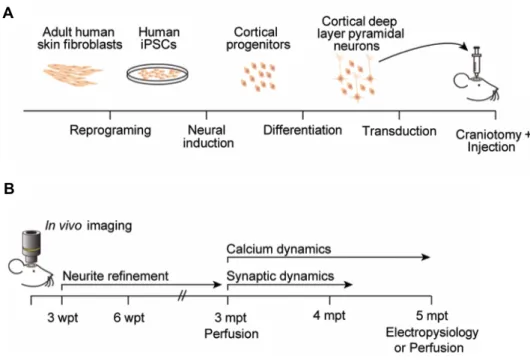
Most of the studies using Ts21 neurons derived from iPSCs have been conducted in vitro. In a recent in vivo study, Huo and colleagues transplanted iPSC-derived GABAergic progenitors
from two DS individuals in the medial septum of adult mice. Both Ds21 and Ts21 GABAergic progenitors generated cortical interneurons with equal efficiency in vivo. However, Ts21 interneurons exhibited reduced soma size, neurite length and branch complexity compared with euploid interneurons. Interestingly, transplanted GABAergic interneurons derived from Ts21-iPSCs also demonstrated a migratory defect from the medial septum to the olfactory bulb and hippocampus. These findings largely confirmed previous in vitro observations, demonstrating that the cellular phenotypes described in vitro were intrinsic to Ts21 GABAergic interneurons and not an artefact of culture conditions (Huo et al., 2018). Similarly, Chen and colleagues transplanted Ts21 astroglia in the lateral ventricles of neonatal mice, to confirm that the negative effects of Ts21 astroglia on neurogenesis and neuronal cell death that had already been detected in vitro could be replicated in the host (Chen et al., 2014). However, it is plausible that neurons in culture cannot capture the full complexity of cellular phenotypes observed in DS and, further to validation of in vitro findings, transplantation of iPSC-derived neural progenitors and neurons could potentially uncover disease phenotypes that are not apparent in vitro. The aim of the present study is therefore to explore the possible contributions of in vivo hiPSC-based studies to the understanding of human neural development and disease modelling. The transplantation into the rodent brain of healthy and disease-specific hiPSC-derived cortical excitatory neurons, followed by subsequent longitudinal intravital imaging, is expected to: (i) enhance the knowledge of dynamic processes such as neuritogenesis, synaptogenesis and neural network formation in human neurons; and (ii) uncover new cellular phenotypes associated with DS that could be relevant to the cognitive impairment characteristic of this condition.
Figure 1.3 Strategies to obtain isogenic disomic Ts21-iPSC lines.
(A) A clone of Ts21-iPSCs can on occasion spontaneously lose one extra copy of Hsa21 in vitro, leading to a mosaic culture. Euploid clones can be isolated to obtain pure Ds21 and Ts21-iPSCs cultures (Weick et al., 2013; Briggs et al., 2013; Huo et al., 2018).
(B) Strategies have been developed to induce the loss or silencing of one extra copy of Hsa21, originating isogenic disomic controls. The introduction of a TKNEO fusion gene into the APP gene of one copy of Hsa21 in Ts21-iPSCs, followed by negative selection against TKNEO, led to a spontaneous chromosome loss in culture (Li et al., 2012). Alternatively, the introduction of the XIST transgene in the DYRK1A locus of one copy of Hsa21 in Ts21-iPSCs led to a chromosome-wide transcriptional silencing of that Hsa21 (Jiang et al., 2013).
(C) Constitutional mosaicism in DS individuals occurs in 1-3% of cases. Isolation and expansion of individual clones from the same tissue generates isogenic iPSCs, identical in every respect except for the extra copy of Hsa21 (Murray et al., 2015).
(D) Isogenic iPSCs have been generated from foetal fibroblasts of monozygotic twins discordant for Ts21 (Hibaoui et al., 2013). Adapted from Hibaoui and Feki, 2015.