Full Terms & Conditions of access and use can be found at
https://www.tandfonline.com/action/journalInformation?journalCode=lspr20
Separation & Purification Reviews
ISSN: 1542-2119 (Print) 1542-2127 (Online) Journal homepage: https://www.tandfonline.com/loi/lspr20
Membrane-based Separation in Flow Analysis for
Environmental and Food Applications
Inês C. Santos, Raquel B. R. Mesquita & António O. S. S. Rangel
To cite this article: Inês C. Santos, Raquel B. R. Mesquita & António O. S. S. Rangel (2020) Membrane-based Separation in Flow Analysis for Environmental and Food Applications, Separation & Purification Reviews, 49:1, 37-54, DOI: 10.1080/15422119.2018.1506810
To link to this article: https://doi.org/10.1080/15422119.2018.1506810
Published online: 20 Aug 2018.
Submit your article to this journal
Article views: 158
View related articles
Membrane-based Separation in Flow Analysis for
Environmental and Food Applications
Inês C. Santos,
1Raquel B. R. Mesquita, and António O. S. S. Rangel
Universidade Católica Portuguesa, CBQF - Centro de Biotecnologia e Química Fina– Laboratório Associado, Escola Superior de Biotecnologia, Rua Arquiteto Lobão Vital, 172, Porto, Portugal
Membrane-based separation techniques have been used as an efficient process for analyte separation or enrichment and matrix removal. By coupling these techniques toflow-based analysis, sample preparation and analyte detection can be automated and miniaturized. Different membrane separation techniques are available but the most used inflow analysis are gas diffusion, dialysis, supported liquid membranes and polymer inclusion membranes. The current state of the art of membrane-based separations hyphenated withflow techniques is presented along with a discussion of the applications to environmental and food analysis. Moreover, a brief description of gas diffusion, dialysis and membrane extraction techniques is also included.
Keywords: Membrane-based separation techniques, flow analysis, gas diffusion, dialysis, supported liquid membranes, polymer inclusion membranes
INTRODUCTION
Sample pre-treatment is one of the bottlenecks of analytical chemistry and because of that it has received special attention. Different sample pre-treatment techniques such as liquid-liquid extraction (LLE), solid-phase extraction (SPE), mem-brane-based separation and digestion have been used (1-3). However, when performed in a batch mode these methods are tedious and time consuming (4). In order to improve the sample throughput and the method precision, sample pre-treat-ment methods have been coupled toflow analysis techniques (5–10).Figure 1shows a timeline of theflow injection tech-niques andFigure 2their representation (11-14).
Thefirst generation of flow analysis was described in 1975 by Ruzicka and Hansen and is based on a continuous flow where the need to achieve chemical and physical equilibrium,
a practice in batch analysis, was discarded. Flow injection analysis (FIA), as it was called, consists in the injection of a well-known volume of sample in aflowing stream of carrier/ reagent. Different reagents can be added by means of con-fluences and the mixing occurs while the sample is propelled toward the detector. This technique provides the analysis of several samples in an automatic way using a simple manifold with high throughput. In spite of its significant advantages, FIA also presents some disadvantages such as the high sample and reagents consumption and the need for physical reconfi-guration in multi-parametric analysis.
To overcome these limitations, sequential injection ana-lysis (SIA) was described in 1990 by Ruzicka and Marshall (12). This technique is based on a programmable and dis-continuous flow, controlled by computer. Sample and reagents are sequentially aspirated and mixing occurs by flow reversal toward the detector. Due to the programmable flow in SIA, reagents are only pumped and consumed when needed, so lower reagents and sample volumes are used, which decrease the consumption values and waste produc-tion. Also, as a multiposition valve is used instead of an injection valve, the coupling of different reagents, detectors and devices is possible without the need for manifold recon-figuration. Therefore, multi-parametric determinations are more easily performed using SIA (15).
Received 22 March 2017, Accepted 3 July 2018.
Address correspondence to António O. S. S. Rangel, CBQF - Centro de Biotecnologia e Química Fina – Escola Superior de Biotecnologia, Universidade Católica Portuguesa, Rua Arquiteto Lobão Vital, 172, Porto 4200-374, Portugal. E-mail:[email protected]
1
Present address: Department of Chemistry and Biochemistry, The University of Texas at Arlington, Arlington TX, USA
Color versions of one or more of thefigures in the article can be found online atwww.tandfonline.com/lspr.
Copyright © Taylor & Francis Group, LLC ISSN: 1542-2119 print / 1542-2127 online
Meanwhile, other flow techniques were also described namely micro-sequential injection lab-on-valve (μSI-LOV), multicommuted flow analysis (MCFA), multisyringe flow injection analysis (MSFIA) and multipumping flow systems (MPFS) (13,14). All theseflow techniques, including FIA and SIA, are based on the same principles: (i) reproducible sample injection; (ii) controlled dispersion of the sample zone and (iii) reproducible timing of its movement from the injection valve to the detection system. Flow techniques are effective tools for analytical chemistry as they can be used for the determination of a wide range of analytes in a fast and automatic way. Moreover, they can also be used for the following purposes: (i) sample introduction into the detector; (ii) automation of
sample pre-treatment; (iii) sample dispersion/dilution and (iv) analyte derivatization (11,15,16). In fact,flow techniques can be coupled with almost any detection device, which allows the detector to be exposed to the sample only for brief periods of time minimizing potential damage or contamination. Furthermore, the conditioning, cleaning and calibration of the detector are effectively performed.
When analyzing complex samples, some challenges may be encountered due to sample matrix interferences and to low analyte concentration (i.e., trace levels). Couplingflow techniques with separation devices enables the collection, enrichment and separation of the analyte from the matrix prior to detection. Therefore, an automation of sample
pre-FIGURE 1. Timeline of the flow analysis techniques.
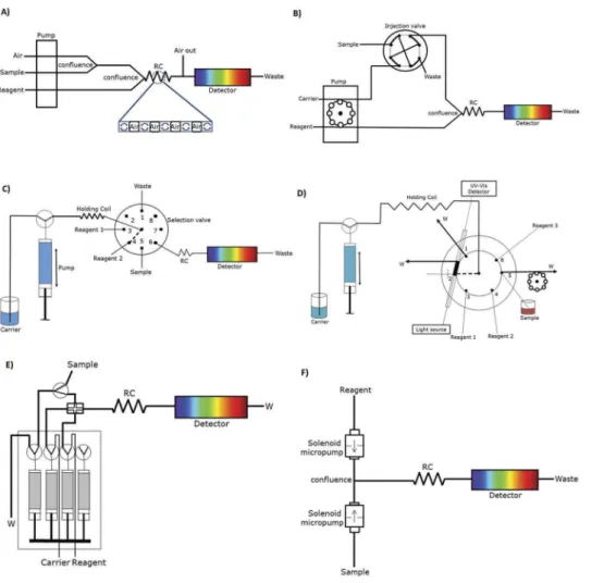
FIGURE 2. Schematic representation of different flow analysis manifolds: (A) segmented flow analysis; (B) flow injection analysis; (C) sequential injection analysis; (D) sequential injection lab-on-valve; (E) multisyringeflow injection analysis; (F) multipumping flow analysis (11–14).
treatment is achieved with a decrease in sample and reagents consumption. As previously shown by Santos et al. (17), SPE is the most used separation method in flow analysis. Nevertheless, membrane-based separations are still pre-ferred to LLE and digestion (18). An overview of the membrane-based separation techniques and their use in flow analysis is presented in the following sections.
MEMBRANE-BASED SEPARATIONS
Membrane-based separation techniques have been used as an alternative to SPE and LLE as they are cheaper and easier to perform (19,20). In membrane-based separation techniques, a membrane (in a solid or liquid state) is used to separate two phases while the components are transferred from a donor in one phase to an acceptor solution in the other phase. This transfer can be achieved by means of different driving forces such as concentration differences, electric potential difference and pressure difference (21). Moreover, the diffusion of the analyte can be of three types: passive, facilitated and active. In passive diffusion, the components diffuse under the influence of a gradient. In facilitated and active diffusion, a carrier is placed in the membrane to increase the membrane permeabil-ity and therefore facilitate the diffusion of the analyte. Nevertheless, in facilitated diffusion, the analyte diffuses from high to low concentration gradients while, in active diffusion, the analyte diffuses against the gradient through a reaction that occurs inside the membrane (22).
Within membrane-based separations and according to the morphology and porosity of the membrane used, different techniques such as gas diffusion (GD), dialysis and liquid membrane extraction can be performed.
Gas Diffusion
GD is used for separation of volatile components that can occur either in the gas phase or in aqueous solutions. A hydrophobic membrane is used to separate the donor from the acceptor phase where the non-selective permeation of gaseous analytes occurs. As only few compounds are volatile at room temperature, GD has a high degree of selectivity since, for example, ionic compounds are excluded. First, the analyte evaporates into the membrane pores and afterward, the volati-lized analytes diffuse through the membrane and are absorbed by theflow of an aqueous solution on the acceptor channel. Chemical species such as dissolved carbon dioxide and dis-solved oxygen can be separated using a GD unit. Furthermore, the applicability of this technique can be increased converting aqueous species into gases by addition of an acid or alkaline solution to the sample in the donor phase. The addition of an acid solution to the donor phase may release gases such as carbon dioxide, hydrogen sulfide and chlorine from carbonate, sulfate and hypochloride, respectively, while the addition of an
alkaline solution to the donor phase can, for example, convert ammonium ions into ammonia gas (20–23).
The driving force in GD membrane separation is the concentration gradient and the diffusion efficiency is mainly governed by the Fick’s law (23). However, the diffusion of a species can also be influenced by the membrane material, membrane path length and area, membrane porosity and membrane thickness. To improve the diffusion efficiency, the membrane path length and area should be as large and as thin as possible, to result in a large contact area easy to cross. However, a compromise between mass-transfer effi-ciency and membrane strength and lifetime may be required. In GD, derivatizing reactions can be carried out in the acceptor stream for two main reasons: to help maintain the concentration gradient thereby increasing the rate of diffu-sion or to form detectable derivatives for detection (23).
Dialysis
Dialysis has been thoroughly described in previous reviews (23–25), which demonstrate its importance in sample pre-treatment. According to the driving forces used, this technique can be referred to as passive dialysis, Donnan dialysis and electrodialysis. The most widely used approach is the passive dialysis, where the analyte is separated from liquid samples by diffusion through a hydrophilic membrane according to its molecular weight. The passive dialysis is a good ultrafiltration molecularfilter as it allows small molecules and ions to pass through the membrane, while macromolecules are held in the donor stream. Due to these reasons, this technique is not considered a selective method for separation and is mainly used to perform sample dilution. In Donnan dialysis or active dialysis, ions are transferred through an ion exchange mem-brane due to the ionic strength gradient while in electrodialy-sis, the analyte is separated by an electricalfield (26).
Dialysis is not usually used as an extraction technique per se due to the lack of discrimination. Therefore, it is usually used as an additional cleanup step. Nevertheless, it has been widely applied to food and biological samples for desalting, buffer exchange, removal of labelling reagents, drug binding, cell growth and feeding, virus purification and blood treatment since macromolecules are excluded from passing through the membrane. Derivatizing reactions can also be carried out in the acceptor stream of the dialysis unit for the same reasons as the ones previously stated in GD (23).
Membrane Extraction
Membrane extraction is a separation technique that uses mainly nonporous membranes, in a liquid or solid state (polymer impregnated with a liquid) that are placed between two liquid phases. Membrane extraction includes different approaches such as supported liquid membrane extraction (SLME), microporous membrane liquid-liquid extraction (MMLLE), polymeric membrane extraction (PME) or
polymer inclusion membranes (PIMs) and membrane extraction with a sorbent interface (MESI) (27,28). Of these approaches, in this manuscript, we will mainly discuss SLME and PIMs for membrane extraction as these are the most used in flow analysis for environmental applications.
Supported Liquid Membranes
Supported liquid membrane (SLM) is the most important liquid membrane technique and was developed to enhance the diffusion through the membrane. As the diffusion coef-ficient is higher in liquids than in solids, the diffusion is enhanced in SLMs when compared to dialysis. The SLM operates with two aqueous phases, the donor and acceptor solutions, separated by an organic phase, the liquid mem-brane, impregnated in the pores of a polymeric support by capillary action. Therefore, in SLM, two LLEs are carried out sequentially: first, the analytes are extracted from the aqueous sample to the organic phase in the membrane and afterward they are extracted from the membrane to the aqueous acceptor phase. In fact, this separation technique is a good alternative to LLE as it has a similar procedure but with significant advantages such as easier automation and operation, and lower organic solvent consumption (28).
In SLMs, reactions in the donor phase can convert the analyte into a non-ionic form therefore favoring mass transfer. Once in the acceptor phase, the non-ionic form must be con-verted back into its ionic form that is irreversibly trapped in this phase. Derivatizing reactions can also be performed in the acceptor stream with the same purposes as for GD and dialysis. The molecules separated by SLM are mainly highly polar ionic or ionizable compounds such as organic acids and bases, charged compounds and metal ions. To improve the analyte permeability and selectivity, the addition of a complexing agent to the liquid membrane to act as carrier can be done to react selectively and reversibly with the analyte. In this way, facilitated or coupled transport processes can be performed.
Polymer Inclusion Membranes
Recently, a new approach, named polymer inclusion membrane (29,30) was developed for the liquid
separation of metal ions and small organic molecules. This approach is a more stable, versatile and selective alternative to SLMs. In PIMs the carrier is entrapped in the membrane matrix while in SLMs the carrier contain-ing solvent is impregnated on a porous polymer film. PIMs consist of a base polymer, usually poly(vinyl chlor-ide) (PVC) or cellulose triacetate (CTA), a carrier for analyte extraction and transport and a plasticizer. The separation procedure occurs in the same way as in SLMs. The main advantage of PIMs is its high selectivity as they are prepared according to the intended analyte.
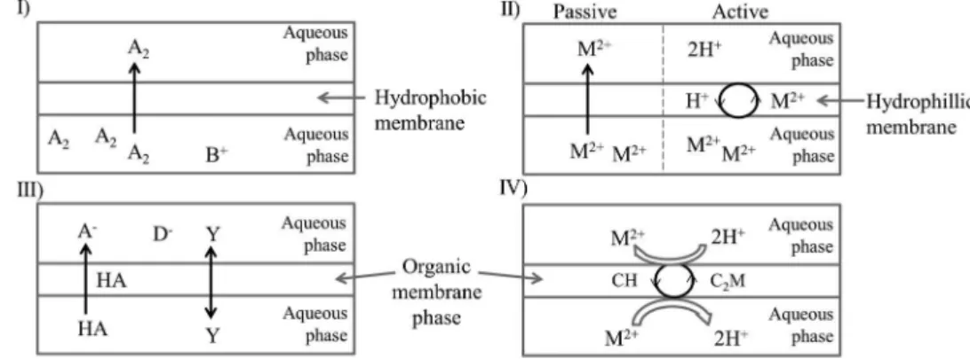
The principles of separation previously described for GD, dialysis, SLM and PIM techniques are schematically shown in Figure 3. A summary of the most important features of each technique is also presented in Table 1.
As previously explained, in GD and dialysis (Figure 3 I and II), the diffusion occurs by concentration gradient. In SLME (Figure 3III), the solute must be in two forms, non-ionic in the donor phase and non-ionic in the acceptor phase, where it is irreversibly trapped. This can be achieved by adjusting the pH in both phases. The separation by SLMs can also be achieved by adding a carrier to the liquid membrane further enhancing the diffusion which resembles the separation with PIMs (Figure 3IV).
Different forms of membrane separation units such as tub-ular (or hollowfiber) and sandwich can be used. Hollow fiber and spiral units increase the surface area and therefore enhance the diffusion of the analyte. Furthermore, when using the sandwich unit, different grooves (spiral, rectangular and wind-ing) can be explored to improve the diffusion efficiency (23).
MEMBRANES – PHYSICOCHEMICAL STRUCTURES AND FEATURES
Different membranes have been used for on-line sample preparation however not yet to their fully extent. Membranes are differentiated based on their pore size, microstructure (homogeneous, microporous,fibrous), physi-cochemical properties (hydrophilic and hydrophobic), reac-tivity and shape.
FIGURE 3. Schematic representation of the membrane extraction processes in: (I) gas diffusion; (II) dialysis; (III) supported liquid membrane and (IV) supported liquid membrane or polymer inclusion membrane (the carrier is represented by the letter C) (23–28).
Microporous membranes are structurally similar to a conventional nanofiltration filter (31) and consist of a solid matrix with defined holes which have diameters from 1–10 nm. Typically, microporous membranes are made of cellophane, cellulose acetate, polycarbonate, polysulfone, polyacrylonitrile, polyamide and others. Separation is achieved solely by a sieving mechanism and so pore dia-meters and molecule sizes are the determining paradia-meters. In this way, a membrane with smaller pores favors the diffusion of small molecules impairing the passage of macromolecules such as colloids. Their structure may be symmetric or asymmetrical (31).
Homogeneous membranes consist of a dense film through which a mixture of molecules is transported by diffusion under the driving force of a pressure, concentra-tion or electrical potential gradient. The separaconcentra-tion of var-ious components of a mixture is related directly to their relative transport rates within the membrane, which are determined by their diffusivity and solubility in the mem-brane phase. Most gas separation, pervaporation and reverse osmosis processes use homogeneous membranes to perform the separation. Homogeneous membranes are usually made of polymers (31).
Electrically charged membranes are highly swollen gels carrying fixed positive (anion-exchange membranes) or negative charges (cation-exchange membranes) where the charged groups arefixed to the polymer. The main applica-tion of ion-exchange membranes is in electrodialysis.
Liquid membranes utilize selective carriers to transport certain molecules at a relatively high rate. These can be made by filling a microporous polymer structure with a liquid membrane phase.
Most membranes are synthetic organic polymers (e.g., polysulfone, cellulose acetate) but can also be prepared from inorganic materials such as ceramics or metals. Polymeric membranes are economical and technologically useful; however, their performance is limited and their permeability
must be sacrificed for selectivity and vice versa. Polymeric porous membranes are widely used in SLMs and polymeric non-porous membranes in PIMs. Most synthetic polymeric membranes are generally hydrophobic, therefore, hydrophi-lic additives such as polyvinylpyrrolidone (PVP) are included to make membranes hydrophilic.
For dialysis, hydrophilic membranes of microporous, homogeneous and ion-exchange types are used, usually made of cellulose acetate or similar materials (e.g., Cuprophane or Cellophane). They need to be hydrated by an overnight stay in water before use. For GD, hydrophobic membranes (e.g., PTFE or PVDF membranes) are used and for SLMs a porous PTFE solvent-filled membrane is used (32). Only a handful of materials are used to make commercial membranes since, in addition to permeability and selectivity, the products must be inexpensive and stable to allow repro-ducible data. Both dialysis and GD membranes are cheap and easy to obtain as they are extensively used in industry. Work is currently being performed to develop new mem-brane materials of higher sensitivity and permeability.
MEMBRANE-BASED SEPARATIONS HYPHENATED TO FLOW TECHNIQUES
Membrane-based separation techniques are fairly easily imple-mented inflow analysis and they have been explored mainly due to advantages such as increased throughput, compatibility with most of the detection techniques, possibility to separate sample matrix and analyte enrichment. In fact, the ability to automate and miniaturize the sample preparation procedure, potentially increasing the precision of the method, is the driv-ing force stimulating the mentioned combination. Additionally, in flow techniques the mass transfer does not need to reach equilibrium as it is required in a batch mode, which allows a faster determination rate. However, due to this reason it is vital to control the operation time.
TABLE 1.
Features of membrane-based separation processes (19) Membrane-based
separation Analyte(s) Driving force Phases (Donor-membrane-acceptor) Membrane material Gas diffusion Volatile compounds Analyte pressure gradient
(concentration gradient)
Aqueous-gas-aqueous Hydrophobic and microporous Dialysis
(passive and active)
Low molecular weight species and ions
Concentration gradient (passive dialysis) Ionic strength gradient (active dialysis)
Aqueous-aqueous (passive dialysis) Aqueous-membrane-aqueous (active dialysis)
Hydrophilic and microporous (passive dialysis)
Ionic and microporous (active dialysis) SLMs Organic acids and bases,
charged compounds and metal ions
Change of partition coefficient
Aqueous-organic-aqueous Hydrophobic and homogeneous PIMs Partition coefficient Aqueous-polymer-aqueous or
organic-polymer-aqueous or aqueous-polymer-organic
Hydrophobic and homogeneous
As previously stated, membrane-based separation pro-cesses inflow analysis can be used in sandwich or tubular modes. Sandwich mode with a flat membrane is the most frequently used, since it is easy to mount and is widely available. The tubular membranes are difficult to mount and the outer chamber usually has a larger dead volume which is considered a drawback. In both approaches the devices can be coupled to different flow techniques such as continuousflow, FIA, and SIA at different locations (23). They can be placed in a pre-valve setup, either before or after the peristaltic pump, inside the loop of the injection valve, between the injection system and the detector or as part of the detector. The latter configuration already exists as a commercial product developed by FIAlab named “sand-wich gas membrane sensor” (33). In this commercial pro-duct, the item includes both separation and detection. The analyte is measured as it is being diffused from the mem-brane and so less dilution occurs and a higher sensitivity can be obtained.
When the membrane device is placed in the injection valve, the acceptor phase is stopped for analyte enrichment and afterward is injected toward the detector. The pre-pump and pre-valve configurations can be an appropriate approach to perform sample dilution or to clean samples containing suspended solids. Nevertheless, the most used configuration is the one that places the membrane unit between the injec-tion system and the detector. The separainjec-tion process can be accomplished by continuously pumping the sample and by keeping the acceptor phase either continuously pumping or stopped. The last strategy may result in increased sensitivity and less analyte dilution as the separation unit can be placed near the detector. Derivatizing reactions in the acceptor channel are also a way to improve the diffusion efficiency as previously explained.
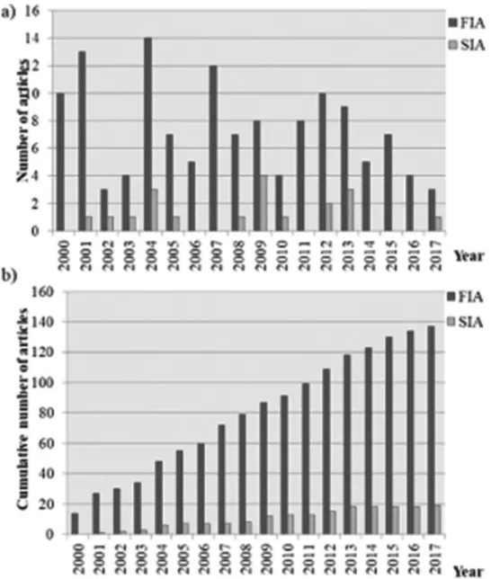
A search for the published papers was made on ISI Web of Knowledge – Web of Science, time span 2000–2016 describing the hyphenation of membrane-based separations with flow techniques. The distribution of the scientific lit-erature in thisfield is shown inFigure 4.
As previously mentioned, membrane separation pro-cesses facilitate analyte enrichment and separation from the matrix minimizing potential interferences. When com-pared to SPE and LLE, membrane-based separation units are an interesting alternative for sample preparation as they are cheaper to obtain and easily coupled toflow techniques with lower organic reagent consumption. However, their utilization in flow analysis has been occasional (Figure 5). Nevertheless, of the membrane-based extraction techniques discussed, GD is the most used inflow analysis. In fact, this technique is more selective than dialysis and simpler to perform than membrane extraction techniques. Few publica-tions exist describing the use of PIMs inflow analysis as it is a fairly recent technique.
The determination of chemical species in both environmen-tal and food matrices presents unique analytical challenges,
namely ultra-low concentrations and variable composition and complexity of the sample matrices. The use offlow methods hyphenated with membrane-based extraction offers many attractive features for addressing these challenges. As pre-viously described, the automation and miniaturization of the sample preparation procedure is possible with higher through-put together with analyte enrichment and matrix interferences minimization. Consequently, although the flow-based mem-brane extraction techniques can be widely applied, the main areas of application are food and environmental analysis (Figure 6). Most of the applications to food analysis have been performed using SIA while the application to environ-mental analysis has been equally performed using FIA and SIA. Combining membrane separation devices with SIA uses the same principles as coupling with FIA but with substantially reduced sample and reagent consumption.
A search of the papers describing the use of membrane-based extraction techniques inflow analysis for environmen-tal (Table 2) (34–60) and food analysis (Table 3) (61–98) since the year of 2000 was made. In environmental analysis, naturally occurring gaseous species (e.g., carbon dioxide, chlorine dioxide and sulfur dioxide) or easily converted spe-cies (e.g., ammonium, dissolved inorganic carbon, sulfite and bromide) are the most targeted using FIA-GD. These deter-minations can be achieved with increased selectivity even in complex environmental samples. Successful applications of this hyphenation can be observed in: the determination of bromide and sulfide in tap and wastewater by spectrophoto-metry (50,55); chlorine dioxide in water byfluorescence (47); and ammonia and inorganic carbon in water with an acid-based indicator by spectrophotometry (42,44), conductimetry (36,38,56,58) and fluorescence (41,49). Due to the high selectivity of GD membranes, most of the methods combine membrane-based separations with non-selective detectors such as spectrophotometric and conductimetric detection where the color change of acid-base indicators or conductiv-ity changes are measured (36,38,40,42,44,48,50,54– 56,58,59). Fluorescence and chemiluminescence detectors are usually chosen when lower detection limits are intended as these are sensitive techniques (37,39,41,43,47,49).
In food analysis, FIA-GDU has also been used for isola-tion of volatile analytes and matrix eliminaisola-tion. Usually, food samples have a complex matrix presenting intrinsic absorption which can interfere with the detection and there-fore it’s elimination is necessary prior to analyte detection. Furthermore, some analytes are expected to be present at high concentration values and a dilution may be needed, which can also be attained with the FIA-GDU technique. Different methodologies using FIA-GDU have been described for the determination of ethanol either by colori-metric determination (77) or based on the schlieren effect (refractive index changes and/or optical variations) (94). In the former work, ethanol is collected into the acceptor stream containing acidic dichromate solution which leads to the formation of green Cr(III), monitored at 600 nm. In
the later work, ethanol diffuses through the membrane and the schlieren effect caused by this compound in the acceptor stream is measured without the need of using reagents. Additionally, GDU can be used to separate volatile com-pounds originated from enzymatic reactions. In the work by Iida et al. (79) an acid urease column was used for the conversion of urea in carbon dioxide that was consequently diffused through a GDU and determined spectrophotome-trically following the color change of a pH indicator.
SIA has also been coupled to GDU (SIA-GDU) for the determination of volatile analytes (37,39,40,48,59,66,70– 72,75,82,96,98) in both food and environmental analysis. In the work by Pais et al. (96), a sequential injection system was developed for ethanol determination using the GDU to perform
in-line dilution. The detection was based on the spectrophoto-metric enzymatic reaction catalyzed by alcohol dehydrogenase in the presence of nicotinamide adenine dinucleotide (NAD+). In the work by Segundo et al. (40), an environmental application of SIA-GDU is described for the determination of ammonium in water samples. Ammonium is converted to ammonia by adding sodium hydroxide in-line. Afterward, ammonia diffuses through the GDU for matrix separation and changes the color of a pH indicator present in the donor channel. This color change is monitored spectrophotometrically.
Dialysis has been more explored for food analysis and therefore fewer works describe environmental applications (52,53). This may be due to the dialysis low selectivity as molecules are separated only by molecular size. In fact,
FIGURE 4. Distribution of the membrane-based flow analysis papers in the timespan 2000–2017; a, count per year; b, cumulative count (search made on ISI Web of Knowledge– Web of Science in October 25th2017).
when using dialysis coupled toflow techniques, a decrease in sensitivity is usually observed (87). Similar to GDU, dialysis is also used in food analysis for matrix removal and sample dilution to fit high concentrations in the linear range of calibration curve therefore increasing the precision of the method. In the works by Silva, Álvares-Ribeiro, Oliveira et al. (67,85) a dialysis unit was introduced in the flow system to attain sample dilution and minimize matrix interferences in wine analysis. Donnan dialysis has been scarcely used in combination with flow techniques in spite of providing high enrichment factors for cations probably due to the requirement of an ionic strength gradient. In the work by Antonia and Allen (86), Donnan dialysis was used for the determination of lead in spiked sweeteners and recoveries higher than 90% were obtained.
In the environmental field, dialysis can be used for the determination of analytes in samples without sample pre-treatment or as an initial step for sample matrix removal prior to analyte pre-concentration removing particles that can clog the analytical column. Ganeshjeevan et al. (53) demonstrated the use of dialysis to separate Cr(VI) from organic matrices based on different molecular sizes. Additionally, dialysis can be used to protect flow-through electrodes and avoid their deterioration by avoiding direct contact of the sample with the active membrane of the sensor (81). Essentially, dialysis coupled toflow techniques is an effective tool for monitoring dynamic systems, a characteristic that could not be achieved using SPE or LLE. Membrane extractions have been used as an alternative to LLE, since SLMs and PIMs use low or insignificant
FIGURE 5. Distribution of the published papers by separation techniques used in hyphenation with: a) FIA and b) SIA (search made on ISI Web of Knowledge– Web of Science in October 25th2017).
amounts of organic reagents and are more easily coupled to flow techniques. When compared to LLE, SLM and PIM enable an easier automation of the sample preparation pro-cedure without the need for large volume ratios between organic and aqueous phase to achieve high enrichment factors as required in LLE. Both SLMs and PIMs have been used for the determination of phenolic compounds, chromium (VI) and orthophosphate by molecular absorption spectroscopy, and for the determination of chlorinated phe-nols by capillary electrophoresis (45,51,57,60) where the authors describe the use of SLMs for analyte enrichment.
Membrane-based separation techniques have been mainly used in FIA manifolds but they can also be employed in SIA. With SIA, a stoppedflow can be more easily performed and the acceptor phase can be kept stopped to improve the diffu-sion through the membrane therefore increasing the diffudiffu-sion efficiency and the pre-concentration capability. In the work by Butwong et al. (37), the acceptor stream was operated in a stopped-flow mode where a 60 s time was adopted to improve the sensitivity of the method. Kolev et al. (99) describe other approaches to improve mass transfer in on-line membrane-based separation in flow analysis by flow manipulation. Approaches such as different fluid structures of the donor stream, stop-flow, oscillating flow and the introduction of air bubbles to separate the sample zone from the donor solution were tested. The authors concluded thatflow manipulation in GD SIA using a second pump for the acceptor stream can improve the sensitivity of the method when compared to conventional GD FIA. This improvement was more pro-nounced for high molecular size analytes. Additionally, an improvement in sensitivity of 7–12.5-fold was obtained when both donor and acceptor streams were oscillated and the sample zone was separated by air bubbles from the donor
solution. Some examples of flow configurations used to per-form membrane-based separation are shown inFigure 7.
Figure 7a illustrates a SIA-SLM approach for in-line hollow-fiber (HF)-assisted three-phase liquid-phase micro-extraction (LPME). The developed method allowed the handling of the donor and acceptor aqueous solutions and of minimal volumes of the organic extracting phase in a programmable flow mode (57). Additionally, the hybrid flow analyzer for HF-LPME with in-line membrane regen-eration allows sample clean-up through the removal of high molecular weight compounds and speciation analysis of Cr (VI) in troublesome harsh environmental samples and high organic load wastewaters.
In Figure 7b, a FIA-PIM approach is shown where the authors used a PVC/Aliquat 336 PIM to extract and pre-concentrate reactive phosphate which was afterward deter-mined by using the well-known molybdenum blue reaction (60). In the described work, the membrane extraction unit was placed in the sample loop of the injection valve which allowed the acceptor solution to be stopped during extrac-tion. The flow system was run in a continuous flow mode unless pre-concentration was attended and, in this case, a 2-min stopped period of time was adopted.
In SIA manifolds, the diffusion unit can be connected to the flow system in two different approaches: (a) the donor and the acceptor channels are connected to two different ports of the selection valve (Figure 7a, andc); (b) the donor channel connected to one port of the selection valve and the acceptor channel connected to a pump in a hybrid FIA-SIA manifold (Figure 7d).
The SIA-GD system shown in Figure 7c has been pro-posed for the determination of cyanides in tap and mineral water samples. In that work, the analyte is removed from
TABLE 2. Me mbrane -based flow method s for envi ronment al analys is Flow system Analy te(s) Separation pr ocess Me mbran e barri er LOD RSD, % Dete r. Rate (h − 1) Sample Detection Refe rence FIA DIC GD U Hydr ophobic mem brane 70 µM < 3 15 Natural w ater Conduc timetry ( 34 ) FIA CO 2 GD U PTFE mem brane 0.00 1 mmol 2.0 -Soil Molecu lar absorption spectro photo metry ( 35 ) FIA Ammo nia GD U PTFE mem brane 0.10 mg L − 1 4.8 12 Lake water CE ( 36 ) SF A Ammo nium GD U PTFE mem brane 5.5 nM 3.8 n. g. Seawater Molecu lar absorption spectro photo metry ( 37 ) MSFI A Bromide GD U PTFE mem brane 0.5x 10 − 5M 3.1 12 T ap, natural and sea water Molecu lar absorption spectro photo metry ( 38 ) FIA Ammo nium and TIC GD U Hydr ophobic mem brane 50, 0.27 µM < 1 15, 17 Coastal wate rs Conduc timetry ( 39 ) FIA Chrom ium(VI) SLM Polypro pylene hollow fi bre mem brane and A liquat 336 4.6 µg L − 1 < 10 n. g. Domestic waters Molecu lar absorption spectro photo metry ( 40 ) MSFI A Ammo nium GD U Hydr ophobic mem brane 45 µg L − 1 2 32 Pond and seawater Conduc timetry ( 41 ) SIA DIC, DOC, CO 2 ,T C and alka linity GD U Millipore Durapor e® membra ne 0.21 –20 µg L − 1 < 6.47 10, 18 Inland bath ing w ater Molecu lar absorption spectro photo metry ( 42 ) FIA and CF A Ortho phospha te PI M Aliqua t 336 0.5 µg L − 1 < 7.7 10 Natural w ater Molecu lar absorption spectro photo metry ( 43 ) FIA TIC and CO 2 GD U PTFE mem brane 2.9 µmol Kg − 1 10 15, 8 Freshwater and sea water Conduc timetry ( 44 ) SIA Arsenic GD U PTFE mem brane 20, 40 µg L − 1 < 1.2 n. g. Sediment Fluore scence ( 45 ) FIA DIC GD U Hollow fi bre micr oporous mem brane 0.12 mM 0.46 90 Marine and estu arine w aters Conduc timetry ( 46 ) SIA Arsenic GD U PTFE mem brane 0.07 mg L − 1 1.4 n. g. Groundwa ter Fluore scence ( 47 ) SIA Ammo nium GD U Millipore Durapor e® membra ne 27 µg L − 1 < 2 20, 28 T ran sitional and coa stal waters Molecu lar absorption spectro photo metry ( 48 ) FIA Ammo nium GD U Gas-dif fusion membra ne (T ecator -Fos s) 5µ gL − 1 < 3.4 15 Standard reference solutio n for wate r analysis Fluore scence ( 49 ) MCF A Ammo nium GD U Millipore Durapor e® membra ne 42 µg L − 1 < 1.5 20 Surface wate r Molecu lar absorption spectro photo metry ( 50 ) FIA Imidacloprid GD U PTFE mem brane 5.6 pmol 0.4 17 Natural w aters Chemilumin escence ( 51 ) FIA Bicarbonate GD U PTFE mem brane 4.41 mg L − 1 < 1 50 Mineral wate rs Molecu lar absorption spectro photo metry ( 52 ) FIA Phenol ic com pounds SLM PTFE mem brane and tribu tyl-phos phate < 1 µg L − 1 < 4.20 2 Natural w aters Molecu lar absorption spectro photo metry ( 53 ) FIA Chlorine dioxi de GD U T efl on membrane 0.03 mg L − 1 2.6, 1.5 30 T ap, mineral and soda water Fluore scence ( 54 ) SIA Chlorine GD U Millipore Durapor e® membra ne 0.6 mg L − 1 < 2 15, 30 T ap and wastew ater Molecu lar absorption spectro photo metry ( 55 ) FIA Ammo nium GD U PTFE mem brane 7 nM 5.7 30 Seawaters Fluore scence ( 56 ) MSFI A Sul fi de GD U PVDF mem brane 0.03 mg L − 1 < 0.8 13 W aste waters Molecu lar absorption spectro photo metry ( 57 ) CF A Chlorinated phenols SLM T ris, NaH 2 PO 4 and meth anol 6.9, 1.0, 1.7 ng mL − 1 5.7, 2.5, 2.8 n. g. Reservoir and drin king w ater CE ( 58 ) FIA Chloride Dialysis Semi-p ermeable pass ive mem brane 1.2 mg L − 1 < 3.0 25 –40 Soil Potentio metry ( 59 ) FIA Chrom ium(VI) Dialysis Cellulose ac etate mem brane 5 µg L − 1 < 2 3 W aste water Molecu lar absorption spectro photo metry ( 60 ) SF A , segm ented fl ow analysis; FI A, fl ow injection analysis; SI A, sequen tial injection analysis; MS FIA , multisyrin ge fl ow injection analysis; MCF A, multicommute d fl ow analysis; CF A, con tinuo us fl ow analysis; TIC , total inor gan ic carbon ; DIC , disso lved inor ganic ca rbon; DOC, disso lved or ganic; TC, total carbon; GDU , gas dif fusio n unit; SLM , sup p orted liquid membra ne; PIM, polym er inclusion membra ne; PTFE , polyt etra fl uoroeth ylene; PVDF , Poly vinyliden e fl uoride; T ris, 2-Amino-2-hy droxyme thyl-propane-1 ,3-diol; CE, capillary ele ctropho resis absorba nce; n. g., not given.
TABLE 3. Membrane-ba sed flow method s for foo d analys is Flow system Analyte(s) Mem brane sepa ration Membrane barri er LOD RS D, % Deter . Ra te (h − 1) Sample Dete ction Refe rence FIA Free and total sulfu r dioxi de GD U Hydropho bic mem brane 0.5 mg L − 1 7, 1 -W ine Pote ntiometry ( 61 ) SIA V olatile and total acidity GD U Hydropho bic PVDF 0.01 , 0.02 g L − 1 35, 62 W ine Mole cular abso rptio n spectro photome try ( 62 ) FIA Phenol s GD U MMPP T ec hno fi lter 3x10 − 8 M 6, 4 4 Smo ked food Ch emiluminesc ence ( 63 ) FIA CO 2 GD U Hydropho bic PVDF mem brane 83 mg L − 1 < 2 30 W ine and beer Mole cular abso rptio n spectro photome try ( 64 ) FIA Sul fi te GD U T efl on 0.26 mg L − 1 0.4 40 Be verages Bia mperom etry ( 65 ) FIA Ethanol GD U T efl on 0.6% < 4.6 120 A lcoholic bev erage s Mole cular abso rptio n spectro photome try ( 66 ) FIA Sul fi de, sul fi te and ethanol GD U T efl on 0.03 5, 0.2 mg L − 1 , 0.18% (v/v ) 2.18, 2.21 , 2.07 15, 57, 29 W ine and molasses Mole cular abso rptio n spectro photome try ( 67 ) SIA Ethanol GD U Hydropho bic PVDF 0.3% (v/v ) < 1 21 W ine Mole cular abso rptio n spectro photome try ( 68 ) FIA Coppe r GD U n. g. 0.32 µM 1.47 , 3.4 60 D rinking water A mperom etry ( 69 ) SIA Chloride Dialysis Celloph ane mem brane 16 mg L − 1 < 3.62 28, 31 Co dfi sh desalting water Mole cular abso rptio n spectro photome try ( 70 ) FIA Sul fi te GD U PTFE 0.043 mg L − 1 1.5 85 Fr uit juice s A mperom etry ( 71 ) FIA Acesulfame-K, saccharin, caf feine, ben zoic acid and sorb ic acid Dialysis Cellulose mem brane 0.1 –1.8 mg L − 1 < 5 4.3 Sof t drin ks Mole cular abso rptio n spectro photome try ( 72 ) FIA Sul fi te GD U PTFE 3 mg L − 1 < 6 15 W ine V oltam metry ( 73 ) SIA Lactic acid GD U PTFE 0.158 mg L − 1 < 2 22 Y oghurt and fer mented mash Mole cular abso rptio n spectro photome try and potentiom etry ( 74 ) MCF A T artaric acid and pota ssium Dialysis Cellulose mem brane 0.1, 0.4 g L − 1, 1x10 − 4 M 2.1, 24 52 W ine Mole cular abso rptio n spectro photome try and Potentio metry ( 75 ) MCF A Sulfur dioxi de GD U Hydropho bic PVDF mem brane 0.3, 0.8 mg L − 1 ; 0.6, 0.8 mg L − 1 1.8, 1.4 25, 23 W ine Mole cular abso rptio n spectro photome try ( 76 ) FIA T artaric, malic, lactic, acetic, citric and succinic acids Dialysis Cellulose mem brane 83 – 213 mg L − 1 < 5.4 7.5 W ine Mole cular abso rptio n spectro photome try ( 77 ) SIA-FIA Sul fi te GD U PTFE 0.3 mg L − 1 2.2 10 W ine Mole cular abso rptio n spectro photome try ( 78 ) SIA Cyanid e GD U PTFE 0.05 – 0.12 µg L − 1 < 10 3, 5, 9 D rinking water A mperom etry ( 79 ) SIA Cyanid e GD U PTFE 2.5 µg L − 1 < 2.5 8 D rinking water Mole cular abso rptio n spectro photome try ( 80 ) FIA Kjeldahl nitrogen GD U PTFE 1 mg L − 1 0.3 35 Milk and chicke n mea t Co nductime try ( 81 ) SIA Sul fi te GD U PTFE 0.05 mg L − 1 1 – 4.1 65 W ine A mperom etry ( 82 ) FIA TVBN GD U T efl on 0.02 mg N L − 1 1.2, 2.2 40 Se afood Pote ntiometry ( 83 ) FIA Ethanol GD U PTFE n. g. < 2 30 A lcoholic bev erage s Mole cular abso rptio n spectro photome try ( 84 ) (Continu ed )
TABLE 3. (Co ntinued) Flow system Analyte(s) Mem brane sepa ration Membrane barri er LOD RS D, % Deter . Ra te (h − 1 ) Sample Dete ction Refe rence FIA Free and total sul fi te GD U PTFE 0.12 , 0.25 mg L − 1 2.4, 3.3 40 W ines and fruit juices Mole cular abso rptio n spectro photome try ( 85 ) FIA Urea GD U PTFE 15.6 µM 3 n. g. A lcoholic bev erage s Mole cular abso rptio n spectro photome try ( 86 ) FIA V itamin B12 Dialysis Cellulose mem brane 0.025 mg L − 1 0.45 n.g. Milk Mole cular abso rptio n spectro photome try ( 87 ) FIA Chloride Dialysis n. g. 0.4 mg L − 1 1.2 90 Milk and coconu t water Pote ntiometry ( 88 ) SIA Urea GD U Hydropho bic PVDF mem brane 2.6x10 − 4, 2.8x10 − 5 M < 3.7, 2.6 n. g. Milk Mole cular abso rptio n spectro photome try and Conduc timetry ( 89 ) FIA Sul fi te GD U T efl on® 0.4 mg L − 1 < 0.01 5 30 Wh ite w ines Mole cular abso rptio n spectro photome try ( 90 ) FIA T artaric acid Dialysis Cellulose acetate mem brane 0.08 g L − 1 1.6, 4.5 36 W ine Mole cular abso rptio n spectro photome try ( 91 ) FIA Lead Dialysis Na fi on 81 1 350 ng g − 1 < 10 4 Swe etener s F AA S ( 92 ) FIA L-lactate Dialysis Cellulose ester 2 µM n. g. 15 Milk and yogh urt A mperom etry ( 93 ) FIA Hydroge n sul fi te and sulfu r dioxi de GD U T efl on 50, 20 nM < 1 20, 45 Wh ite and red w ines Fluo rescence ( 94 ) FIA Sulfur dioxi de and ascorb ic ac id GD U T efl on 1.5 mg L − 1 n. g. 30 W ine and fruit juices A mperom etry ( 95 ) FIA Sulfur dioxi de and carbon dioxi de GD U PVDF 0.05 , 0.25 g L − 1 4.5, 2.4 30, 40 W ine Mole cular abso rptio n spectro photome try ( 96 ) MSFI A Free and total sulfu r dioxi de GD U Hydropho bic PVDF mem brane 1, 5.6 mg L − 1 < 3.2 25 –30 W ine Mole cular abso rptio n spectro photome try ( 97 ) FIA TMAN and TVBN GD U PTFE n. g. n. g. n. g. Fish sauce Mole cular abso rptio n spectro photome try ( 98 ) FIA Lead Dialysis Na fi on 81 1 350 ng g − 1 < 10 4 Swe etener s F AA S ( 92 ) FIA L-lactate Dialysis Cellulose ester 2 µM n. g. 15 Milk and yogh urt A mperom etry ( 93 ) FIA Hydroge n sul fi te and sulfu r dioxi de GD U T efl on 50, 20 nM < 1 20, 45 Wh ite and red w ines Fluo rescence ( 94 ) FIA Sulfur dioxi de and ascorb ic ac id GD U T efl on 1.5 mg L − 1 n. g. 30 W ine and fruit juices A mperom etry ( 95 ) FIA Sulfur dioxi de and carbon dioxi de GD U PVDF 0.05 , 0.25 g L − 1 4.5, 2.4 30, 40 W ine Mole cular abso rptio n spectro photome try ( 96 ) MSFI A Free and total sulfu r dioxi de GD U Hydropho bic PVDF mem brane 1, 5.6 mg L − 1 < 3.2 25 –30 W ine Mole cular abso rptio n spectro photome try ( 97 ) FIA TMAN and TVBN GD U PTFE n. g. n. g. n. g. Fish sauce Mole cular abso rptio n spectro photome try ( 98 ) FIA, fl ow injec tion ana lysis; SIA, sequ ential injecti on ana lysis; MSFIA , multisyringe fl ow injection analysis; MCF A , multicommute d fl ow ana lysis; TVBN, total volatile basi c nitro gen; TM AN, trimethylamine; GDU , gas dif fusion unit; PVDF , polyvinyliden e fl uoride; PTF E, polyt etra fl uoro ethylene; F AA S, fl ame atom ic abso rption spectro metry; n. g., not given.
sample matrix by acidification and diffusion through a gas-diffusion step. In the acceptor channel, cyanide reacts with ninhydrin in carbonate medium to form a colored product measured by spectrophotometry (72). The SIA-GD system depicted inFigure 7dhas been used in a FIA-SIA approach developed for the spectrophotometric determination of inor-ganic and orinor-ganic carbon in water (59). An acid solution is added in-line to convert the dissolved inorganic carbon into carbon dioxide allowing its selective separation in the GDU and determination by changing the color of a pH indicator. Overall, when combined with mass separation unit devices, flow techniques are mainly used to introduce the sample in the carrier stream where the analyte is derivatized for an enhanced diffusion. Afterward, the sample is pro-pelled to the donor channel where mass transfer occurs. The extract can therefore be propelled to a detector or to a pre-concentration unit for further enrichment. In fact, consider-ing chromatographic techniques, membrane-based separa-tion techniques can be a good front end, as they reduce the need for off-line sample pre-treatment. For example, in the works by Kritsunankul, Jakmunee et al. (64,69), dialysis was used for sample pre-treatment in order to separate the analytes, food additives and organic acids, from the sample matrix before separation by HPLC.
Membrane-based separation techniques present several advantages namely the ease of automation and on-line hyphenation with flow-based techniques. Additionally, a high degree of sample cleanup and potential selectivity is attained which is an important improvement when
compared to other sample preparation techniques such as SPE. In fact, in membrane extraction the analyte is inten-tionally transferred through a membrane while in SPE the analyte and other potential interferences can be adsorbed and released during elution (20). Nevertheless, when applied to complex sample matrices such as environmental and food samples, solid membranes can clog or rupture due to, for example, surfactants, which alters the analyte diffu-sion ability. Therefore, calibration curves should be per-formed on daily basis to confirm the membrane physical conditions.
The parameters mainly determined in environmental and food samples using membrane separation techniques coupled to flow analysis, are presented in Figure 8. As it would be expected, although SLMs can be very useful and used for inorganic ionic species, organic species are the most target analytes separated with these types of mem-branes. In methods using GD separation, inorganic species easily converted to gaseous form at room temperature are the targeted analytes. When combining dialysis with flow analysis, metal ions and inorganic species can be separated from other high molecular compounds.
More recently, liquid membranes have been used for sorptive microextraction. The work by Oshima et al. (100) describes the use of a packed column coated with a PIM as a sorbent for the on-line pre-concentration of thiocyanate in FIA. Since PIMs can be prepared according to the intended analyte, it is expected that these PIM coated columns can be used for the determination of a large range of analytes.
FIGURE 7. Schematic representations of FIA and SIA manifolds for membrane-based separations (a) SIA-SLM (Reprinted from Nitiyanontakit et al. (57) with permission from Springer. Copyright 2013); (b) FIA-PIM (Reprinted from Nagul et al. (60) with permission from Elsevier. Copyright 2013); (c) and (d) SIA-GDU (Reprinted from Themelis et al. (72) with permission from Elsevier. Copyright 2009 and Santos et al. (59) with permission from Elsevier. Copyright 2013, respectively).
CHALLENGES
Membrane-based separations present many advantages specially when coupled to flow analysis techniques. However, some challenges are still encountered. These methods suffer from a lack of specificity, permeability and resolution. Dialysis analyte recoveries rarely exceed 15–20%. The extent to which continuous separation tech-niques enhance sensitivity depends on the volume ratio for the two liquids involved. Nowadays the needs for sensitive, specific and faster analytical methodologies are replacing these membrane separation approaches by
other types of sample preparation. Compared to SPE and LLE, membrane separations are slower and have lower enrichment capacities. Due to this lower concentration ability, detection limits are usually higher which may require subsequent sample treatment. Additionally, mem-brane-based separation methods have lower throughput. Nevertheless, there has been an effort to adapt 96-well plate systems for membrane technologies. In this way, membrane techniques can become more competitive regarding their throughput when compared to other sam-ple preparation techniques since all 96 samsam-ples can be processed simultaneously. Regarding flow analysis,
FIGURE 8. Distribution of the analytes targeted in the papers listed inTable 2(34–60) and 3 (61–98) for (a) environmental applications; and (b) for food analysis.
several devices can be coupled in parallel to allow the simultaneous preparation of different samples.
Another disadvantage of membrane-separation is fouling: the membrane pores can be clogged when complex samples such as wastewater are analyzed. Furthermore, attention needs to be given to possible interactions between the analytes and the membrane to avoid lower recoveries and ensure method precision.
CONCLUSIONS
The use of membrane-based separation techniques in flow analysis for environmental and food applications provides valuable advantages such as the possibility to separate the analyte from complex sample matrices, analyte enrichment, reduction in organic reagents consumption, ease in operation and possibility of automation. Nevertheless, the efficiency of membrane separations is low when compared to other separa-tion techniques, namely SPE resulting in a significant draw-back. However, the yields of membrane-based methods can be optimized and adapted to the intended application (e.g., separation, enrichment or dilution). Still, when dialysis is coupled toflow injection techniques, the obtained yields are usually quite low and therefore this technique is mainly used for analyte dilution or sample clean-up. This may be one of the reasons for the low number of works describing the use of dialysis as an on-line sample pre-treatment.
Membrane extraction techniques have been a good alter-native to LLE as these are more easily automated by flow techniques displaying lower consumption of organic reagents. Some of the works describe the use of this tech-nique for analyte enrichment demonstrating the potential of this technique. GD is still the technique of choice in flow analysis as it allows the selective separation of compounds as only few are volatile at room temperature. Moreover, due to its selectivity, non-selective and cheaper detectors can be used. Additionally, membrane extraction techniques can be a good choice for a preliminary online sample pre-treatment (matrix cleanup or analyte enrichment) for chromatography. In order to improve the membrane separation specificity, more work needs to be done in the development of new mem-branes to improve selectivity and permeability. Moreover, the use of PIMs for solid phase extraction must potentially be more explored as a cheaper alternative to SPE resins.
ABBREVIATIONS FIA flow injection analysis GD gas diffusion
GDU gas diffusion units LLE liquid-liquid extraction MCFA multicommutedflow analysis
MESI membrane extraction with a sorbent interface MMLLE microporous membrane liquid-liquid extraction MPFS multipumpingflow systems
MSFIA multisyringeflow injection analysis NAD+ nicotinamide adenine dinucleotide PIMs polymer inclusion membranes PME polymeric membrane extraction SIA sequential injection analysis
SLME supported liquid membrane extraction SPE solid-phase extraction
μSI-LOV micro-sequential injection lab-on-valve
ACKNOWLEDGMENTS
R.B.R. Mesquita thanks to Fundação para a Ciência e a Tecnologia (FCT) and POCH of FSE for the grant SFRH/ BPD/112032/2015.
CONFLICT OF INTEREST There is no known conflict of interest.
REFERENCES
1. Raynie, D.E. (2016) Trends in sample preparation. LC-GC, 29: 142–154. 2. Pawliszyn, J. (2003) Sample preparation: quo vadis? Anal. Chem., 75:
2543–2558. doi:10.1021/ac034094h
3. Liska, I. (2000) Fifty years of solid-phase extraction in water analysis – historical development and overview. J. Chromatogr. A, 885: 3–16. doi:10.1016/S0021-9673(99)01144-9
4. Chen, Y., Guo, Z., Wang, X. and Qiu, C. (2008) Sample preparation. J. Chromatogr. A, 1184: 191–219. doi:10.1016/j.chroma.2007.10.026
5. Hennion, M.C. (1999) Solid-phase extraction: method development, sorbents, and coupling with liquid chromatography. J. Chromatogr. A, 856: 3–54. doi:10.1016/S0021-9673(99)00832-8
6. Silvestre, C.I.C., Santos, J.L.M., Lima, J.L.F.C. and Zagatto, E.A.G. (2009) Liquid– liquid extraction in flow analysis: a critical review. Anal. Chim. Acta, 652: 54–65. doi:10.1016/j.aca.2009.05.042
7. Miró, M. and Frenzel, W. (2004) Automated membrane-based sam-pling and sample preparation exploitingflow-injection analysis. TrAC Trends Anal. Chem., 23: 624–636. doi:10.1016/j.trac.2004.07.006
8. Pan, J., Zhang, C., Zhang, Z. and Li, G. (2014) Review of online coupling of sample preparation techniques with liquid chromatogra-phy. Anal. Chim. Acta, 815: 1–15. doi:10.1016/j.aca.2014.01.017
9. Karlberg, B. (1988) Flow injection extraction in theory and practice. Fresenius J. Anal. Chem., 329: 660–662. doi:10.1007/BF00624770
10. Smith, R.M. (2003) Before the injection - modern methods of sample preparation for separation techniques. J. Chromatogr. A, 1000: 3–27. doi:10.1016/S0021-9673(03)00511-9
11. Ruzicka, J. and Hansen, E.H. (1975) Flow injection analyses. Anal. Chim. Acta, 78: 145–157. doi:10.1016/S0003-2670(01)84761-9
12. Ruzicka, J. and Marshall, G.D. (1990) Sequential injection: a new con-cept for chemical sensors, process analysis and laboratory assays. Anal. Chim. Acta, 237: 329–343. doi:10.1016/S0003-2670(00)83937-9
13. Segundo, M.A. and Rangel, A.O.S.S. (2002) Flow analysis: a critical view of its evolution and perspectives. J. Flow Inject. Anal., 19: 3–8.
14. Ruzicka, J. (2000) Lab-on-valve: universal microflow analyzer based on sequential and bead injection. Analyst, 125: 1053–1060. 15. Ivaska, A. and Ruzicka, J. (1993) Fromflow injection to sequential
injection: comparison of methodologies and selection of liquid drives. Analyst, 118: 885–889. doi:10.1039/AN9931800885
16. Ruzicka, J. and Hansen, E.H. (2008) Retro-review offlow-injection analysis. TrAC Trends Anal. Chem., 27: 390–393. doi:10.1016/j. trac.2008.03.004
17. Santos, I.C., Mesquita, R.B.R. and Rangel, A.O.S.S. (2015) The state of the art of flow-through solid-phase spectrometry. LC-GC North Am., 33: 25–31.
18. Gonçalves, L.M., Valente, I.M. and Rodrigues, J.A. (2017) Recent advances in membrane-aided extraction and separation for analytical purposes. Sep. Purif. Rev., 46: 179–194. doi:10.1080/ 15422119.2016.1235050
19. Frenzel, W. and Markeviciute, I. (2017) Membrane-based sample preparation for ion chromatography– techniques, instrumental con-figurations and applications. J. Chromatogr. A, 1479: 1–19. doi:10.1016/j.chroma.2016.11.052
20. Jönsson, J.Å. and Mathiasson, L. (2000) Membrane-based techniques for sample enrichment. J. Chromatogr. A, 902: 205–225. doi:10.1016/ S0021-9673(00)00922-5
21. Van De Merbel, N.C. (1999) Membrane-based sample preparation coupled on-line to chromatography or electrophoresis. J. Chromatogr. A, 856: 55–82. doi:10.1016/S0021-9673(99)00581-6
22. Cordero, B.M., Pavón, J.L.P., Pinto, C.G., Laespada, M.E.F., Martinez, R.C. and Gonzalo, E.R. (2000) Analytical applications of membrane extraction in chromatography and electrophoresis. J. Chromatogr. A, 902: 195–204. doi:10.1016/S0021-9673(00)00835-9
23. Staden, J.F. (1995) Membrane separation inflow injection systems. Fresenius. J. Anal. Chem., 352: 271–302. doi:10.1007/BF00322225
24. Miró, M. and Frenzel, W. (2005) The potential of microdialysis as an automatic sample-processing technique for environmental research. TrAC - Trends Anal. Chem., 24: 324–333. doi:10.1016/j. trac.2004.10.004
25. De Castro, M.D.L., Capote, F.P. and Ávila, N.S. (2008) Is dialysis alive as a membrane-based separation technique? TrAC Trends Anal. Chem., 27: 315–326. doi:10.1016/j.trac.2008.01.015
26. Pyrzynska, K. (2006) Preconcentration and recovery of metal ions by donnan dialysis. Microchim. Acta, 153: 117–126. doi:10.1007/ s00604-005-0434-4
27. Moskvin, L.N. and Nikitina, T.G. (2004) Membrane methods of substance separation in analytical chemistry. J. Anal. Chem., 59: 2–16. 28. Sastre, A.M., Kumar, A., Shukla, J.P. and Singh, R.K. (1998) Improved techniques in liquid membrane separations: an overview. Sep. Purif. Methods, 27: 213–298. doi:10.1080/03602549809351641
29. Nghiem, L.D., Mornane, P., Potter, I.D., Perera, J.M., Cattrall, R.W. and Kolev, S.D. (2006) Extraction and transport of metal ions and small organic compounds using polymer inclusion membranes (PIMs). J. Memb. Sci., 281: 7–41. doi:10.1016/j.memsci.2006.03.035
30. Almeida, G.S., Cattrall, R.W. and Kolev, S.D. (2017) Polymer inclu-sion membranes (PIMs) in chemical analysis– a review. Anal. Chim. Acta, 987: 1–14. doi:10.1016/j.aca.2017.07.032
31. Kolev, S. and McKelvie, I. (2008) Advances in Flow Injection Analysis and Related Techniques; Comprehensive Analytical Chemistry; Elsevier Science: New York, USA.
32. Cerdá, V. (2014) Flow Analysis: A Practical Guide; Elsevier Science: New York, USA.
33. Fialab Systems. Sandwich gas membrane sensor.http://www.flowin jection.com/index.php/products/flow-cells?pid=61&sid=100:sand wich-membrane-flow-cell-smfc(accessed May 3, 2018).
34. Somboot, W., Jakmunee, J. and Kanyanee, T. (2017) An exploiting of cost-effective direct current conductivity detector in gas diffu-sion flow injection system. Talanta, 170: 298–305. doi:10.1016/j. talanta.2017.04.015
35. Silva, C.R., Oliveira, E., Zagatto, E.A.G. and Henriquez, C. (2016) A novelflow-based procedure for automation of respirometric assays in soils. Talanta, 158: 14–20. doi:10.1016/j.talanta.2016.05.026
36. Martinotti, V., Balordi, M. and Ciceri, G. (2012) Aflow injection analyser conductometric coupled system for thefield analysis of free dissolved CO2 and total dissolved inorganic carbon in natural waters. Anal. Bioanal. Chem., 403: 1083–1093. doi: 10.1007/s00216-012-5762-8
37. Butwong, N., Srijaranai, S., Ngeontae, W. and Burakham, R. (2012) Speciation of arsenic (III) and arsenic (V) based on quenching of CdS quantum dotsfluorescence using hybrid sequential injection-stopped flow injection gas-diffusion system. Spectrochim. Acta Part A Mol. Biomol. Spectrosc., 97: 17–23. doi:10.1016/j.saa.2012.05.054
38. Pencharee, S., Faber, P.A., Ellis, P.S., Cook, P., Intaraprasert, J., Grudpan, K. and Mckelvie, I. (2012) Underway determination of dissolved inorganic carbon in estuarine waters by gas-diffusionflow analysis with C4D detection. Anal. Methods, 4: 1278–1283. doi:10.1039/c2ay25113b
39. Butwong, N., Noipa, T., Burakham, R., Srijaranai, S. and Ngeontae, W. (2011) Determination of arsenic based on quenching of CdS quantum dots fluorescence using the gas-diffusion flow injection method. Talanta, 8: 1063–1069. doi:10.1016/j.talanta.2011.05.023
40. Segundo, R.L.A., Mesquita, R.B.R., Ferreira, M.T.S.O.B., Teixeira, C. F.C.P., Bordalo, A.A. and Rangel, A.O.S.S. (2011) Development of a sequential injection gas diffusion system for the determination of ammonium in transitional and coastal waters. Anal. Methods, 3: 2049–2055. doi:10.1039/c1ay05129f
41. Almendral-Parra, M.-J., Alonso-Mateos, A. and Fuentes-Prieto, M.-S. (2010) A gas diffusion technique coupled withflow injection systems. Optimization of the process in its application to the fluorimetric determination of ammonium in water samples. J. Fluoresc., 20: 55– 65. doi:10.1007/s10895-009-0521-8
42. Oliveira, S.M., Lopes, T.I.M.S., Tóth, I.V. and Rangel, A.O.S.S. (2007) A multi-commutedflow injection system with a multi-channel propulsion unit placed before detection: spectrophotometric determi-nation of ammonium. Anal. Chim. Acta, 600: 29–34. doi:10.1016/j. aca.2007.01.019
43. Lagalante, A.F. and Greenbacker, P.W. (2007) Flow injection analysis of imidacloprid in natural waters and agricultural matrixes by photo-chemical dissociation, photo-chemical reduction, and nitric oxide chemilu-minescence detection. Anal. Chim. Acta, 590: 151–158. doi:10.1016/j. aca.2007.03.034
44. Oliveira, P.C.C., Masini, J.C., Galhardo, C.X., Lima, J.C.S., Sant’ana, A.E.G., Vasconcelos, A.M.G., Nunes, W.P. and Amaral, O.L.C. (2006) A new approach to construct diffusion/permeation cell for use inflow systems. Application in the spectrophotometric determina-tion of bicarbonate ions. J. Braz. Chem. Soc., 17: 976–980. doi:10.1590/S0103-50532006000500023
45. Sun, A., Li, J. and Liu, R. (2006) High-performance liquid chromato-graphic determination of phenolic compounds in natural water coupled with on-lineflow injection membrane extraction-preconcen-tration. J. Sep. Sci., 29: 995–1000.
46. Zhu, Z., Lu, J.J., Almeida, M.I.G.S., Pu, Q., Kolev, S.D. and Liu, S. (2015) A microfabricated electroosmotic pump coupled to a gas-diffusion microchip for flow injection analysis of ammonia. Microchim. Acta, 182: 1063–1070. doi: 10.1007/s00604-014-1410-7
47. Themelis, D.G. and Kika, F.S. (2006) Gas-diffusion flow injection assay for the selective determination of chlorine dioxide based on the fluorescence quenching of chromotropic acid. Microchem. J., 82: 108–112. doi:10.1016/j.microc.2005.12.001
48. Mesquita, R.B.R. and Rangel, A.O.S.S. (2005) Gas diffusion sequen-tial injection system for the spectrophotometric determination of free chlorine with o-dianisidine. Talanta, 68: 268–273. doi:10.1016/j. talanta.2005.07.028
49. Watson, R.J., Butler, E.C.V., Clementson, L.A. and Berry, K.M. (2005) Flow-injection analysis with fluorescence detection for the determination of trace levels of ammonium in seawater. J. Environ. Monit., 7: 37–42. doi:10.1039/b405924g
50. De Armas, G., Ferrer, L., Miró, M., Estela, J.M. and Cerdà, V. (2004) In-line membrane separation method for sulfide monitoring in waste-waters exploiting multisyringe flow injection analysis. Anal. Chim. Acta, 524: 89–96. doi:10.1016/j.aca.2004.02.050
51. Zhou, Q., Liu, J., Jiang, G., Liu, G. and Cai, Y. (2004) Sensitivity enhancement of chlorinated phenols by continuousflow liquid mem-brane extraction followed by capillary electrophoresis. J. Sep. Sci., 27: 576–580. doi:10.1002/jssc.200301632
52. Miró, M. and Frenzel, W. (2003) A novelflow-through microdialysis separation unit with integrated differential potentiometric detection for the determination of chloride in soil samples. Analyst, 128: 1291– 1297. doi:10.1039/B306747E
53. Ganeshjeevan, R., Chandrasekar, R., Yuvaraj, S. and Radhakrishnan, G. (2003) Determination of hexavalent chromium by on-line dialysis ion chromatography in a matrix of strong colourants and trivalent chromium. J. Chromatogr. A, 988: 157–165. doi:10.1016/S0021-9673 (02)02051-4
54. Kodama, T., Ichikawa, T., Hidaka, K. and Furuya, K. (2015) A highly sensitive and large concentration range colorimetric continuousflow analysis for ammonium concentration. J. Oceanogr., 71: 65–75. doi:10.1007/s10872-014-0260-6
55. Danchana, K., Maya, F., Wilairat, P., Uraisin, K. and Cerdà, V. (2015) Spectrophotometric determination of bromide in water using the mul-tisyringeflow injection analysis technique coupled to a gas-diffusion unit. Anal. Methods, 7: 4202–4208. doi:10.1039/C5AY00202H
56. Henriquez, C., Horstkotte, B. and Cerdà, V. (2014) A highly repro-ducible solenoid micropump system for the analysis of total inorganic carbon and ammonium using gas-diffusion with conductimetric detec-tion. Talanta, 118: 186–194. doi:10.1016/j.talanta.2013.10.005
57. Nitiyanontakit, S., Varanusupakul, P. and Miró, M. (2013) Hybridflow analyzer for automatic hollow-fiber-assisted ionic based liquid-phase microextraction with in-line membrane regeneration. Anal. Bioanal. Chem., 405: 3279–3288. doi:10.1007/s00216-013-6744-1
58. Henríquez, C., Horstkotte, B. and Cerdà, V. (2013) Conductometric determination of ammonium by a multisyringeflow injection system applying gas diffusion. Int. J. Environ. Anal. Chem., 93: 1236–1252. doi:10.1080/03067319.2012.746322
59. Santos, I.C., Mesquita, R.B.R., Machado, A., Bordalo, A.A. and Rangel, A.O.S.S. (2013) Sequential injection methodology for carbon speciation in bathing waters. Anal. Chim. Acta, 778: 38–47. doi:10.1016/j.aca.2013.03.043
60. Nagul, E.A., Fontàs, C., McKelvie, I.D., Cattrall, R.W. and Kolev, S. D. (2013) The use of a polymer inclusion membrane for separation and preconcentration of orthophosphate inflow analysis. Anal. Chim. Acta, 803: 82–90. doi:10.1016/j.aca.2013.07.052
61. Giménez-Gómez, P., Gutiérrez-Capitán, M., Puig-Pujol, A., Capdevila, F., Muñoz, S., Tobeña, A., Miró, A. and Jiménez-Jorquera, C. (2017) Analysis of free and total sulfur dioxide in wine by using a gas-diffusion analytical system with pH detection. Food Chem., 228: 518–525. doi:10.1016/j.foodchem.2017.02.026
62. Vidigal, S.S.M.P. and Rangel, A.O.S.S. (2017) Aflow-based platform for measuring the acidity parameters in wine. Talanta, 168: 313–319. doi:10.1016/j.talanta.2017.03.029
63. Martins, P.R., Popolim, W.D., Nagato, L.A.F., Takemoto, E., Araki, K., Toma, H.E., Angnes, L. and Penteado, M.D.V.C. (2011) Fast and reliable analyses of sulphite in fruit juices using a supramolecular amperometric detector encompassing inflow gas diffusion unit. Food Chem., 127: 249–255. doi:10.1016/j.foodchem.2010.12.103
64. Kritsunankul, O. and Jakmunee, J. (2011) Simultaneous determination of some food additives in soft drinks and other liquid foods byflow
injection on-line dialysis coupled to high performance liquid chroma-tography. Talanta, 84: 1342–1349. doi:10.1016/j.talanta.2011.02.045
65. Gonçalves, L.M., Pacheco, J.G. and Magalhães, P.J. (2010) Determination of free and total sulfites in wine using an automatic flow injection analysis system with voltammetric detection. Food Addit. Contam. Part A, 27: 175–180. doi:10.1080/ 19440040903261547
66. Dias, A.C.B., Silva, R.A.O. and Arruda, M.A.Z. (2010) A sequential injection system for indirect spectrophotometric determination of lac-tic acid in yogurt and fermented mash samples. Microchem. J., 96: 151–156. doi:10.1016/j.microc.2010.02.016
67. Oliveira, S.M., Lopes, T.I.M.S., Tóth, I.V. and Rangel, A.O.S.S. (2010) Simultaneous determination of tartaric acid and potassium in wines using a multicommutedflow system with dialysis. Talanta, 81: 1735–1741. doi:10.1016/j.talanta.2010.03.032
68. Oliveira, S.M., Lopes, T.I.M.S., Tóth, I.V. and Rangel, A.O.S.S. (2009) Development of a gas diffusion multicommutedflow injection system for the determination of sulfur dioxide in wines, comparing malachite green and pararosaniline chemistries. J. Agric. Food Chem., 57: 3415–3422. doi:10.1021/jf803639n
69. Kritsunankul, O., Pramote, B. and Jakmunee, J. (2009) Flow injection on-line dialysis coupled to high performance liquid chromatography for the determination of some organic acids in wine. Talanta, 79: 1042–1049. doi:10.1016/j.talanta.2009.03.001
70. Tzanavaras, P.D., Thiakouli, E. and Themelis, D.G. (2009) Hybrid sequential injection– flow injection manifold for the spectrophoto-metric determination of total sulfite in wines using o-phthalalde-hyde and gas-diffusion. Talanta, 77: 1614–1619. doi:10.1016/j. talanta.2008.07.055
71. Zacharis, C.K., Tzanavaras, P.D., Voulgaropoulos, A.N. and Karlberg, B. (2009) Amperometric determination of cyanides at the low ppb level by automated preconcentration based on gas diffusion coupled to sequential injection analysis. Talanta, 77: 1620–1626. doi:10.1016/j. talanta.2008.07.055
72. Themelis, D.G., Karastogianni, S.C. and Tzanavaras, P.D. (2009) Selective determination of cyanides by gas diffusion-stopped flow-sequential injection analysis and an on-line standard addition approach. Anal. Chim. Acta, 632: 93–100. doi:10.1016/j. aca.2008.10.074
73. Vakh, C., Evdokimova, E., Pochivalov, A., Moskvin, L. and Bulatov, A. (2017) A novel flow injection chemiluminescence method for automated and miniaturized determination of phenols in smoked food samples. Food Chem., 237: 929–935. doi:10.1016/j. foodchem.2017.06.049
74. Junsomboon, J. and Jakmunee, J. (2008) Flow injection conducto-metric system with gas diffusion separation for the determination of Kjeldahl nitrogen in milk and chicken meat. Anal. Chim. Acta, 627: 232–238. doi:10.1016/j.aca.2008.08.012
75. Chinvongamorn, C., Pinwattana, K., Praphairaksit, N., Imato, T. and Chailapakul, O. (2008) Amperometric determination of sulfite by gas diffusion-sequential injection with boron-doped diamond electrode. Sensors, 8: 1846–1857. doi:10.3390/s8010464
76. Dhaouadi, A., Monser, L. and Sadok, S. (2007) Validation of a flow-injection-gas diffusion method for total volatile basic nitrogen deter-mination in seafood products. Food Chem., 103: 1049–1053. doi:10.1016/j.foodchem.2006.07.066
77. Vicente, S., Zagatto, E.A.G. and Borges, E.P. (2006) Exploiting gas diffusion for non-invasive sampling inflow analysis: determination of ethanol in alcoholic beverages. An. Acad. Bras. Cienc., 78: 23–29. doi:10.1590/S0001-37652006000100004
78. Carinhanha, J., Santos, C. and Korn, M. (2006) Exploiting sulphide generation and gas diffusion separation in aflow system for indirect sulphite determination in wines and fruit juices. Microchim. Acta, 153: 87–94. doi:10.1007/s00604-005-0453-1