Universidade de Aveiro 2018
Departamento de Ciências Médicas
CRISTIANA
GONÇALVES PAULO
CONTRIBUTION OF TRNA MODIFICATIONS FOR
PROTEOSTASIS IN HUMAN CELLS
RELEVÂNCIA DAS MODIFICAÇÕES DE TRNA NA
PROTEOSTASE EM CÉLULAS HUMANAS
Universidade de Aveiro 2018
Departamento de Ciências Médicas
CRISTIANA
GONÇALVES PAULO
RELEVÂNCIA DAS MODIFICAÇÕES DE TRNA NA
PROTEOSTASE EM CÉLULAS HUMANAS
Tese apresentada à Universidade de Aveiro para cumprimento dos requisitos necessários à obtenção do grau de Mestre em Biomedicina Molecular, realizada sob a orientação científica da Doutora Ana Raquel Santos Calhôa Mano Soares do Departamento de Ciências Médicas da Universidade de Aveiro
Apoio financeiro pela Fundação Portuguesa de Ciência e Tecnologia através do POCI-COMPETE2020, FEDER, UID/BIM/04501/2013 e PTDC/BIM-MEC/1719/2014.
“Go as far as you can see. When you get there, you’ll be able to see further.”
O Júri
Presidente Doutora Ana Margarida Domingos Tavares de Sousa
Professora auxiliar convidada do Departamento de Ciências Médicas da Universidade de Aveiro
Arguente Doutora Marisa Susete Reverendo Simões
Investigadora de pós-doutoramento do Centre d’Immunologie de Marseille-Luminy (CIML)
Orientador científico Doutora Ana Raquel Santos Calhôa Mano Soares
Investigadora de pós-doutoramento do Departamento de Ciências Médicas da Universidade de Aveiro
Agradecimentos Primeiramente, gostaria de agradecer à minha orientadora, Ana Soares e à minha parceira de laboratório, Marisa Pereira, pela forma como me receberam e encaminharam, ajudando-me a superar os meus obstáculos, as minhas dúvidas e dificuldades. Pela alegria, paixão e esforço com que sempre realizaram todo o seu trabalho e me ajudaram a realizar o meu.
Em segundo lugar, mas não menos importante, aos meus Pais, à minha Irmã e restantes familiares, por me apoiarem desde sempre nas minhas escolhas, acreditando em mim incondicionalmente e por me proporcionarem toda a ajuda necessária para prosseguir o Ensino Superior.
Gostaria ainda de agradecer aos meus amigos, Daniel Lopes, Sara Gonçalves, Andreia Bastos, Mariana Mota e Cláudia Pereira, por me incentivarem a não desistir e a enfrentar de cabeça erguida e com empenho este projeto científico.
palavras-chave RNA de transferência, enzimas modificadoras de tRNA, proteostase, agregação proteica
resumo Mutações nas moléculas de RNA de transferência (tRNAs), moléculas
chave no processo de tradução, levam à perda da precisão da tradução do RNA mensageiro (mRNA). Isto traduz-se em alterações na taxa de síntese proteica, misfolding de proteínas e acumulação de proteínas agregadas que, por sua vez, levam a stress proteotóxico e ativação da UPR, algumas das características de doenças conformacionais de proteínas. Para serem totalmente ativos, os tRNA sofrem modificações pós-transcricionais catalisadas por diferentes enzimas modificadoras de tRNA. A desregulação de algumas modificações de tRNA e de algumas das enzimas modificadoras foram já correlacionadas com alterações ao nível da fidelidade e eficiência da tradução, especialmente em leveduras. No entanto, um estudo exaustivo sobre o impacto de todas as modificações e enzimas modificadoras de tRNA em mamíferos não foi ainda realizado.
Para identificar quais as enzimas modificadoras essenciais para a manutenção da proteostase, a nossa equipa desenvolveu um sistema repórter de agregação de proteínas (pcDNA3.1 Hsp27-GFP) que nos permite detetar in vivo a nível celular a produção de agregados proteicos. Após o estabelecimento de uma linha celular estável a expressar este repórter, foi realizado um screening fenotípico que identificou as enzimas ELP1, ELP3, ELP6, URM1 e TRMT2A como essenciais para a proteostase. Verificou-se que a via UPS foi ativada na ausência das enzimas ELP1, ELP3, ELP6 e análises de proteómica de células com silenciamento da ELP3 revelaram que as vias mais alteradas dizem respeito à tradução e transcrição, entre outras, que estão geralmente desreguladas em doenças neurológicas, como a Esclerose Lateral Amiotrófica. Os resultados desta tese identificam e demonstram que um grupo particular de enzimas modificadoras de tRNA afetam a proteostase em células humanas e que as mesmas podem ser novos alvos terapêuticos para doenças conformacionais.
keywords Transfer RNA, tRNA modifying enzymes, proteostasis, protein aggregation
abstract Mutations in transfer RNAs (tRNA), key molecules in the translation process, lead to loss of accuracy of messenger RNA (mRNA) translation. This may induce changes in protein synthesis rate, protein misfolding and accumulation of aggregated proteins which, in turn, leads to proteotoxic stress and UPR activation, some of the characteristics of conformational protein diseases. To be fully active, tRNA molecules undergo post-transcriptional modifications catalyzed by different tRNA modifying enzymes. Deregulation of some modifications of tRNAs and some of the modifying enzymes has already been correlated with changes in translation fidelity and efficiency, especially in yeast. However, a comprehensive study on the impact of all tRNA-modifying enzymes and modifications in mammals has not been performed yet.
In order to identify the key modifying enzymes for proteostasis preservation, our team developed a protein aggregation reporter system (pcDNA3.1 Hsp27-GFP) that allows the detection of the production of protein aggregates in vivo at the cellular level. After establishing a stable cell line expressing this reporter, a phenotypic screening was performed, identifying ELP1, ELP3, ELP6, URM1 and TRMT2A enzymes as essential for proteostasis. The UPS pathway was activated in the absence of the ELP1, ELP3, ELP6 enzymes and proteomic analyzes of ELP3 silencing cells revealed that the most altered pathways are concerned to transcription and translation processes, which are generally dysregulated in neurological diseases, such as Amyotrophic Lateral Sclerosis. In this thesis, the proteostasis relevant tRNA modifying enzymes are identified and characterized in human cells. This group of enzymes may represent promising therapeutic targets for conformational diseases.
Declaração
Declaro que este trabalho é inteiramente da minha autoria, estando devidamente referenciadas as fontes e obras consultadas, bem como claramente identificadas as citações dessas obras. Não contém, por isso, qualquer tipo de plágio, quer de textos publicados, quer de trabalhos académicos, qualquer que seja o meio dessa publicação, incluindo meios eletrónicos.
Contents
CHAPTER 1. ... 1
Introduction ... 1
1.1. Proteostasis in aging ... 2
1.2. Protein conformational diseases ... 2
1.3. tRNA – The “bridge” molecule between mRNA and proteins ... 4
1.4. tRNA Modifications ... 6
1.4.1. Modifications at several positions of tRNAs ... 8
1.4.2. Modifications in or around the anticodon loop ... 8
1.4.3. Modifications in the main body of tRNAs ... 10
1.4.4. tRNA modifications in human diseases ... 10
1.5. Elongator complex ... 13
1.6. Mechanisms associated with proteostasis deregulation... 15
1.6.1. Unfolded Protein Response (UPR)... 16
1.6.2. Ubiquitin/Proteasome System (UPS) ... 17
1.6.3. Autophagy-lysosomal pathway (ALP) ... 18
1.7. Hela cells and the Aggregation Reporter System ... 20
1.8. Motivations and Aim of the study ... 21
CHAPTER 2. ... 23
Metodology ... 23
2.1. Cell Culture ... 24
2.2. Reverse transfection with siRNAs ... 24
2.3. Total protein extraction and quantification ... 26
2.4. Insoluble protein fraction extraction... 27
2.5. Polyacrylamide gel electrophoresis (SDS-PAGE) ... 27
2.6. Western Blotting ... 28
2.6.1. Trans-Blot® TurboTm Blotting System (Bio-Rad) adapted protocol ... 28
2.6.2. Immunodetection ... 28
2.7. Proteasome Activity Analysis ... 29
2.8. Statistical analysis ... 29 2.9. CRISP-R ... 29 CHAPTER 3. ... 31 Results ... 31 3.1. Transfection ... 32 3.2. Insoluble fraction ... 32 3.3. Western Blotting ... 34
3.3.1. Ubiquitin-Proteasome System Pathway Activation ... 34
3.3.2. Unfolded Protein Response Activation ... 35
3.4. Protein Synthesis rate ... 37
3.5. Proteasome Activity ... 39
3.6. ELP3 proteomics ... 39
3.7. Summary ... 41
CHAPTER 4. ... 43
Discussion ... 43
4.1. tRNA modifying enzymes that affect proteostasis ... 44
4.1.1. Elongator Complex ... 44
4.1.2. Other Enzymes ... 46
4.2. Knock down of tRNA modifying enzymes impair protein synthesis and proteasome activity in human cells ... 48
4.4. Ongoing work ... 49
CHAPTER 5. ... 51
Concluding remarks ... 51
5.1. Conclusion and future perspectives ... 52
Acknowledgements ... 53
I. Abbreviations list
Aa Amino acid
AARSs Aminoacyl-tRNA synthetases Ac4Cm N4-acetyl-2'-O-methylcytidine
AD Alzheimer’s disease
ADATs Adenosine deaminases acting on tRNAs ALKBH8 Methyltransferase Alkylation repair homolog 8 ALP Autophagy-lysosomal pathway
ALS Amyotrophic lateral sclerosis AMPK AMP-activated protein kinase
ARID Autosomal recessive intellectual disorder ASL Anticodon stem loop
ATF4 Activating transcription factor 4 ATF6 Activating transcription factor 6 A-to-I Adenosine to Inosine
BCA Bicinchoninic acid
BDNF Brain derived neurotrophic factor
BECTS Benign epilepsy with centrotemporal spikes BiP Binding immunoglobulin protein
BSA Bovine serum albumin bZIP Basic Leucine Zipper
CMA Chaperone-mediated autophagy
COSMIC Catalogue of Somatic Mutations in Cancer DMEM Dulbecco’s modified eagle medium DUBs Deubiquitinases
E1 Activating Ubiquitin Enzyme E2 Conjugating Ubiquitin Enzyme E3 Ligase Ubiquitin Enzyme EEG Electroencephalography
eIF2α Eukaryotic translation initiation factor 2α
eIF2α-P Phosphorylated eukaryotic translation initiation factor 2α ELP Elongator protein
ELP1 Elongator protein 1 homolog ELP2 Elongator protein 2 homolog ELP3 Elongator protein 3 homolog ELP4 Elongator protein 4 homolog ELP5 Elongator protein 5 homolog
ELP6 Elongator protein 6 homolog FBS Fetal bovine serum
FD Familial Dysautonomia GFP Green fluorescent protein Gm 2'-O-methylguanosine
HAT Histone acetyltransferase HSP27 Heat-shock protein 27 I6A37 N6 isopentenyl adenine 37 IPTases Isopentenyl-transferases IRE1 Inositol-requiring enzyme 1 JNK c-jun NH(2)-terminal kinase KAT Lysine acetyltransferase LB Loading buffer M1A 1-methyladenosine M1G 1-methylguanosine M22Gm N2,N2,2'-O-trimethylguanosine Mcm5 5-methoxycarbonylmethyl Mcm5s2U 5-methoxycarbonylmethyl-2-thiouridine Mcm5Um 5-methoxycarbonylmethyl-2'-O-methyluridine
MELAS Mitochondrial encephalomyopathy, lactic acidosis and stroke-like episodes
MERRF Myoclonic epilepsy with ragged-red fibers
MLASA Mitochondrial myopathy, lactic acidosis and sideroblastic anemia
mRNA Messenger RNA
mTOR Mammalian target of rapamycin Ncm5 5-carbamoylmethyl
OECD Organization for Economic Co-operation and Development PBS Phosphate-buffered saline
PD Parkinson’s disease
PERK protein kinase RNA-like endoplasmic reticulum kinase PFA paraformaldehyde solution
PN Proteostasis network PS Protein synthesis Q Queuosine RT Room temperature S2T 2-thioribothymidine SAM S-adenosylmethionine siRNA Small interference RNA
TGT tRNA guanine transglycosylase
TRDMT1 tRNA aspartic acid Methyltransferase 1 TRMT1 tRNA methyltransferase 1
TRMT2A tRNA (uracil-5-)-methyltransferase homolog A TRMT5 tRNA (guanine(37)-N1)-methyltransferase TRMT61A tRNA Methyltransferase 61A
tRNA Transfer RNA
URM1 Ubiquitin related modifier 1 WR Working reagent
yW Wybutosine
τm5s2U 5-taurinomethyl-2-thiouridine Ψ Pseudouridine
II. List of Figures
Figure 1: Protein aggregation in neurodegenerative diseases.(13). ... 3
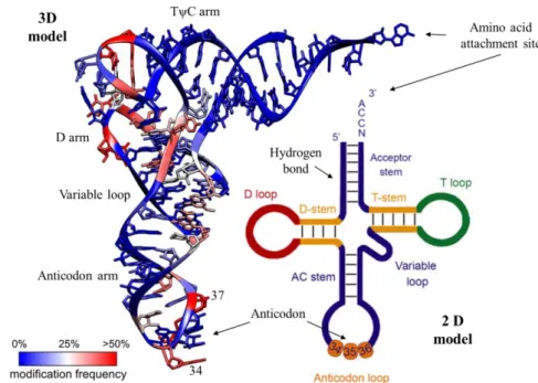
Figure 2: Crystal structure of the tRNAPhe from S. cerevisiae and its modification frequency (Adapted from (38)) with the correspondent 2D “clover leaf” model and the described identity elements of tRNA. Adapted from (31). ... 4
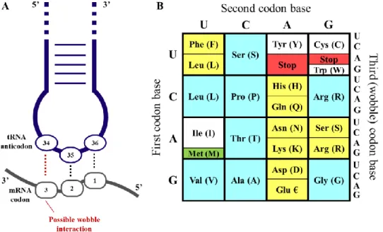
Figure 3: Representation of codon-anticodon interaction and Wobble Hypothesis. Adapted from (38). ... 5
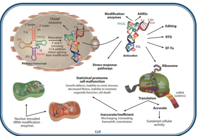
Figure 4: Essential roles of nucleosides modifications for tRNA function (25). ... 6
Figure 5: Chemical structures of tRNA modifications linked to human diseases. Adapted from (22). ... 7
Figure 6: Representation of the tRNA secondary structure with the respective tRNA modifications and associated tRNA modifying enzymes. Adapted from (61, 109). ... 12
Figure 7: Structure of the Elongator complex and its biological roles. Adapted from (63). ... 13
Figure 8: Representation of protein fates in the Proteostasis NetworkErro! Marcador não definido. Figure 9: The three divisions of the UPR (92). ... 17
Figure 10: Autophagy-lysosomal pathway (ALP) and ubiquitin-proteasome system (UPS) pathways under normal and pathological conditions (100) ... 19
Figure 11: Schematic representation of the functioning of the stable cell line expressing the HSP27:GFP reporter. ... 20
Figure 12: Schematic representation of a 24 well plate with the disposition of the SiRNAs. ... 244
Figure 13: Analysis of the insoluble fraction of transfected cells. ... 333
Figure 14: Analysis of protein ubiquitination ... 355
Figure 15: Analysis of total eIF2α and phosphorylated eIF2α expression ... 36
Figure 17: Analysis of protein synthesis of transfected cells with the respective bands
described ... 388
Figure 18: Relative proteasome activity in transfected cells. ... 399
Figure 19: Molecular interaction networks and biological pathways of the genes up-regulated (A) and down-up-regulated (B) after Elp3 knockdown on HeLa cells ... 40
III. List of Tables Table 1: Prominent modifications in the tRNA anticodon loop. Adapted from (39). ... 9
Table 2: Human disorders and associated tRNA modifications. Adapted from (22). ... 11
Table 3: ER transmembrane receptors. Adapted from (87). ... 16
Table 4: Reverse transfection solutions ... 25
Table 5: Human tRNA modifying enzymes tested ... 25
Table 6: Empigen lysis buffer (ELB) reagents and preparation. ... 26
Table 7: Reagents for 2 protein gels ... 27
Table 8: Antibodies used for Western Blot Analysis ... 28
Table 9: Recommended Samples for a gene engineering co-transfection experiment. ... 30
Table 10: Preparing transfection samples for gene editing experiment in a 24-well plate format ... 30
Table 11: Characterization of the two groups of enzymes studied. ... 32
Table 12: Summary of the achieved results of our tests with the correspondent tRNA modified residue. ... 41
CHAPTER
1
.
Introduction
1.1. Proteostasis in aging
Over the years, the advance of the scientific knowledge and the availability of drugs and therapies has allowed the population to live longer. Although this increase of life expectancy is a positive factor, age-related diseases such as neurological disorders and cancer are emerging with aging (1), which is described as the “persistent decline in the age-specific fitness components of an organism due to internal physiological degeneration” (2).
One of the hallmarks of aging and age-related diseases is the loss of proteostasis - protein homeostasis - characterized by a progressive decline in the ability of cells to maintain protein quality control, accompanied by the appearance of protein aggregates in several tissues. Proteostasis is maintained by processes such as protein synthesis, folding, aggregation, disaggregation and degradation, comprised in the proteostasis network (PN) (3,4). In short, the PN comprises mechanisms that are responsible for the stabilization of correctly folded proteins, particularly the heat-shock family of proteins, and mechanisms to restore the structure of misfolded proteins or to remove and degrade them by proteasomes or lysosomes in order to prevent them from accumulating in the cell (5). Moreover, when this unfolded, misfolded or aggregated proteins are chronically expressed, they contribute to the development of some age-related pathologies, such as Alzheimer’s and Parkinson’s disease, among others (1).
In Portugal, according to the Organization for Economic Co-operation and Development (OECD) statistics, between the years 2000 and 2016, people dying every year from Alzheimer’s disease (AD) had an increase of 43,8% and from Parkinson’s disease (PD) this value was about 62.9%. In the United States of America, in the same period, the data was very similar: from AD the increase of deaths per year was approximately 55.2% and from PD was 44.0%. This massive growth warns us of the urgency of understanding the underlying mechanisms of these diseases and finding new therapies not only to provide better quality of life for patients but also to prevent these diseases from developing and progressing rapidly.
1.2. Protein conformational diseases
In 1997, Carrel and Lomas came up with the concept of conformational disease to characterize the similar mechanism for the development of neurodegenerative disorders (Huntington’s, Alzheimer’s and Parkinson’s disease, Prion encephalopathies), cystic fibrosis, diabetes type II and systemic amyloidosis. This discovery was largely due to the fact that the onset and progression of the disease was caused by protein misfolding and conformational change. The conformational change can promote the disease in two ways, by causing a gain of a toxic activity or taunting the absence of biological function of the native folded protein (6,7). Each disease is associated to a specific protein or group of
proteins that could be unfolded, misfolded and/or aggregated and the accumulated protein deposits are pathological hallmarks for the corresponding disease (Figure 1), however the causative agents and toxicity mechanisms are still not very well understood (8).
There are two characteristics shared by the majority of neurodegenerative disorders, which are the presence of amyloid-like misfolded protein deposits and the impairment of neuronal function (9). Moreover, even though each disorder involves the amyloidogenic aggregation of distinct proteins, the biophysical and structural properties of amyloid fibrils are identical (10). Evidences suggest that the development of amyloid fibrils is related to the loss of protein function, a toxic gain of function or a functional reversible amyloid assembly (11,12). In the majority of these disorders, the formation of the amyloid structures is correlated with cell death but it is unclear if amyloid is cell pathogenic in a direct way or if it is the transition from a native folded protein to amyloid that triggers the toxic event (13).
The mechanisms that lead to the formation of particular protein aggregates are still unclear. It is known that post-translational modifications of proteins play a role in protein stability and structure, but loss of mRNA translation accuracy leads to amino acid misincorporations in nascent polypeptides that in turn may also lead to protein misfolding and accumulation of aggregated proteins (14,15). Among the various molecules involved in the translation process, transfer RNAs (tRNAs) stand out as one of the most relevant as they are the effector molecules of translation, whose function will be explored in more detail in the next topics.
Figure 1: Protein aggregation in neurodegenerative diseases. In several protein misfolding diseases, native
unfolded monomers form cross β-sheet assemblies, evolving into oligomers to form highly ordered fibrillary aggregates. This process produces insoluble protein deposits and it is associated with neurodegeneration (13).
1.3. tRNA – The “bridge” molecule between mRNA and proteins
tRNA is a key adaptor molecule, responsible for the conversion from RNA to protein, translating the genetic code by matching a codon in a messenger RNA (mRNA) with the amino acid (aa) it codes for during translation within the ribosome (16). Different types of tRNAs are floating around in a cell, each one with its own anticodon and corresponding amino acid; once inside the ribosomes, tRNAs bind to mRNA codons through complementarity with their anticodons, allowing the delivery of the amino acids to be added to the protein chain. Ribosomes possesses three slots for tRNAs: A, P and E site and tRNAs move through the A site to the P site and then to the E site as they deliver amino acids during translation (17).
Mature tRNAs are made from a single strand RNA of approximately 70-100 nucleotides long with a conserved three-dimensional shape that arises mostly from the interaction between bases in different regions of the RNA sequence, giving rise to the secondary “clover leaf” structure (Figure 2). This base pairing results in three double-stranded regions and stem-loops - the D loop, the Anticodon loop and the T loop – in an open-ended stem (amino acid attachment site) formed by the pairing of the 5’ and 3’ ends and it also has a vestigial projection called variable loop that it is between the Anticodon loop and the T-loop, like (Figure 2. The D-loop has this name because it has a D-hidro-uridine in position 16 and is responsible to bond to the aminoacyl tRNA synthetase. The T-loop or GTPCG (Thiamine-Pseudouridine(ψ)) T-loop is the ribosome recognition site and facilitates the connection of the amino acid to the A site. The Anticodon stem loop (ASL) is comprised between the positions 34, 35 and 36 of the tRNA and binds to complementary bases of the mRNA (18–20).
Figure 2: Crystal structure of the tRNAPhe from S. cerevisiae (3D model) and its modification frequency
(Adapted from (38)) with the correspondent 2D “clover leaf” model and the described identity elements of tRNA. The Acceptor stem (7bp), the D-stem (3-4bp) and the anticodon (AC) stem (5bp). The variable (V) region (4-23 nt) and the D-loop (4-12nt) confers some variety in the tRNA length, however, the anticodon in the anticodon loop is always numbered 34-36 and the CCA tail at the terminus is numbers 74-76. The 3’-CCA triplet is added post-transcriptionally by a 3’-CCA-adding enzyme. Adapted from (31).
The right amino acid binds to the right tRNA due to aminoacyl-tRNA synthetases; for each amino acid there is a different synthetase enzyme that recognizes only that amino acid and its correspondent tRNA. The cognate amino acid is only charged at the 3’-end after maturation of the tRNA. Through their anticodon loop, that contains the three nucleotide base sequence, tRNAs pair specifically with the codons in mRNAs during translation. Position 34 of tRNAs can wobble, meaning that it can pair with different nucleotides of the third position of mRNA triplets via non Watson-Crick interactions, giving some flexibility to the genetic code and allowing some tRNAs to decode different sets of codons for the same amino acid and even some codons to be recognized by more than one anticodon sequence (Figure 3A) (21,22).
To explain this possibility, Francis Crick proposed the Wobble Hypothesis, that elucidates how tRNAs recognize and read more than one codon, clarifying why cells have only 40 distinct tRNA species for 64 codons. In summary, the first two bases pair between tRNA nucleosides 36 and 35 and the coding triplet in mRNA are canonical (A-U; C-G or
vice versa) base-pairings. The third base pairing (position 34 of tRNA) can also be
canonical but to enlarge tRNA recognition of codons in protein synthesis, there are unconventional and non-canonical base pairings in the third base of the codon-anticodon triplet (18,23). The genetic code is illustrated in Figure 3B as well as its codon degeneracy.
The main function of tRNAs is to participate in protein synthesis as a key component of translation. However, this molecule participates in other processes such as
Figure 3: Representation of codon-anticodon interaction and Wobble Hypothesis. A) Interaction of the
anticodon bases (34–36) of a tRNA with the corresponding bases of the mRNA codons (3, 2, 1). A wobble interaction is possible between codon base 3 and anticodon base 34. The latter is frequently modified and directs the wobble interactions with the third codon base. B) The standard genetic code is illustrated as a simple decoding table; 2-fold degenerate codon boxes are colored yellow; 4-fold degenerate boxes are blue; special boxes are colored white. The start codon is colored green and stop codons are colored red. Adapted from (38).
inhibition of apoptosis via complexation of cytochrome c, amino acyl addition to membrane lipids, HIV-1 priming, antibiotics target and biosynthesis and can originate tRNA derived fragments (24).
1.4. tRNA Modifications
tRNAs are heavily chemically modified to maintain their proper structure, stability and function. In fact, tRNAs are the RNA molecules bearing, the largest number of post-transcriptional modifications. Their chemical diversity is, in this way, significantly augmented. Nucleoside modifications become an integral part of the process that ensures that tRNAs accomplish the deciphering of the genetic material. In fact, near 10% of the genes in the genome code for enzymes that are involved in tRNA modification (25), highlighting the relevance of such modifications for correct tRNA function. Figure 4 describes the essential roles of these modifications for tRNA function in the different compartments of the cell. Some modified nucleosides are present in a specific group of tRNA species while others are found in most tRNAs and for these nucleoside modifications to occur, one or more enzymes and enzymatic steps may be involved (26,27).
Figure 4: Essential roles of nucleosides modifications for tRNA function. Nucleoside modifications have
structural roles (e.g. correct formation of the tRNA L shape), are involved in tRNA interactions with translation machinery players (e.g. modification enzymes, ribosomes, mRNA codons, translation factor, aminoacyl-tRNA synthetases (AARSs), editing and RNA-degradation systems). Modifications can be introduced on tRNA substrates in the nucleus, cytoplasm or organelles (25).
Abbreviations: AARS, aminoacyl-tRNA synthetase; ASL, anticodon stem loop; DSL, D stem loop; EF-Tu, Elongator factor Tu; mRNA, messenger RNA; RTD, rapid tRNA decay pathway; TψCSL, TψC stem loop; TRAMP, Trf4/Air2/Mtr4p polyadenylation complex; tRNA, transfer RNA; VL, variable stem loop.
A total of 93 different tRNA modifications with their correspondent type and location have been identified and listed in the RNA Modification and MODOMICS databases, 51 of them belonging to Eukarya kingdom (http://mods.rna.albany.edu/) (28). The lack of a phenotype associated to tRNA modifications makes it difficult to classify their biological significance and function. However, the advance of technology allowed to describe numerous modification defects as well as their related phenotypes, which bring us insights of their biological roles (29). There are already tRNA modifications associated to human diseases whose chemical structures are illustrated in Figure 5. There has been an attempt to clarify some of the functions of these modifications and they are usually based on three principles: many modifications are in or around the anticodon loop, affecting translation and cellular growth; many modifications in the main body of tRNA molecules affect their folding and stability and some modifications at several positions affects specifically tRNA identity (30). Concrete examples of such functions are the stabilization of codon-anticodon interactions, increased tRNA capability to decode multiple synonymous mRNA codons, rapid tRNA response to environmental challenges like stress, increased protein synthesis fidelity, prevention of frame shift mutations, and codon reading tuning (24).
Figure 5: Chemical structures of tRNA modifications linked to human diseases. Unmodified nucleosides are
depicted in black. Atomic changes for each chemical structure upon modification are shown in red. Nucleobase (A-D) and ribose (E) modifications are shown. Adapted from (22).
The understanding of the diversity of the cellular functions of tRNAs has increased in the last years due to the development of new technologies, as well as due to the understanding of the mechanisms by which their expression is coordinated in specific tissues, making it a dynamic regulator of the stress response. tRNA mutated genes have been linked to several human pathologies, suggesting that its different abundance in certain tissues modulates the effect of such pathologies and their associated phenotypes, since tRNA diversity modulates the proteome depending on the tissue and the cell, even in the same genome (31).
1.4.1. Modifications at several positions of tRNAs
One of the simplest and most frequent modifications found in tRNAs is methylation that can occur in all positions of the target nucleotide; this type of modification destabilizes Watson-Crick interactions leading to massive structural changes in the global tRNA fold (32,33). For example, 1-methyladenosine at position 9 (m1A9) in human mitochondrial
tRNALys shifts the structural equilibrium from an alternative hairpin structure to the functional cloverleaf structure, while Pseudouridine (ψ) at positions 32 and 39 shape the anticodon stem loop (34). Other modifications like N4-acetyl-2'-O-methylcytidine (ac4Cm),
2'-O-methylguanosine (Gm) and N2,N2,2'-O-trimethylguanosine (m22Gm) are involved in
thermal stabilization of tRNA; this is relevant, for example, in case of hyperthermophiles that modify more extensively their tRNAs when growing at higher temperatures: survival at high temperature of Thermus thermophilus is dependent on the formation of 2-thioribothymidine (s2T) (35,36). Hypo modification of tRNAs usually causes their targeting for degradation, meaning that another role of tRNA modifications is to prevent tRNAs from entering into specific degradation pathways (22).
1.4.2. Modifications in or around the anticodon loop
Modifications of bases occurring at the wobble position in the anticodon or immediately next to the anticodon triplet frequently influence the decoding capacities of tRNAs by restricting and/or improving the codon-anticodon interactions, which can affect the behavior of tRNAs during gene translation and the maintenance of the reading frame (22,37). Modifications in the anticodon loop also have structural functions, since they reinforce a defined loop structure, necessary for a stable codon-anticodon interaction (38).
In addition to having a great diversity of hypermodified nucleotides at positions 34 and 37, modifications at position 34 are usually associated with decoding capacity because base modifications at this position are generally necessary for codon-anticodon wobbling to occur (39). However, these modifications may also prevent translational frameshifting and may even be required for amino acylation (40). Examples of wobble modifications include uridine (U) 34 modifications like the incorporation of methyl, hydroxyl and thiol groups, and adenosine (A) 34 modifications such as adenosine-to-inosine (A-to-I) editing,
as described in Table 1 (30,41). Modifications at position 37 help to stabilize codon-anticodon interactions by providing base-stacking interactions , preventing translational frameshifting (25).
Table 1: Prominent modifications in the tRNA anticodon loop. Adapted from (39).
Modification Characteristics
Queuosine (Q)
Occurs with GUN anticodons (N represents any nucleotide); mediated by the tRNA-guanine transglycosylase (TGT) complex; changes in Q abundance correlated with stress tolerance, cell proliferation and tumor growth (42,43).
Inosine
Post-transcriptional modification found in tRNAs residues 34, 37 and 57; results from a deamination reaction of adenines that is catalyzed by adenosine deaminases acting on tRNAs (ADATs); Adenine-to-inosine (A-to-I) editing allows the enlargement of the decoding capability of individual tRNAs and the limitation of the tRNA species number for codon-anticodon recognition; I34 hypomodification associated with myositis and missense mutation in ADAT3 gene associated with intellectual disability (44).
5-methoxycarbonyl methyl-2
thiouridine (mcm5)
Elongator (ELP) complex is needed; U34 base of cytoplasmic tRNA carries mcm5 or 5-carbamoylmethyl (ncm5) modifications; U34
modification associated with the enhancement of the translation efficiency and fidelity; cells lacking U34 modifications exhibit hallmarks of proteotoxic stress, like protein aggregation (45).
Wybutosine (yW)
G37 is methylated to form 1-methylguanosine (m1G); m1G is the first step for Wybutosine formation; Presence of yW provides base-stacking interactions of the tRNA anticodon with the A-site codon, playing a key function in reading frame maintenance since they prevent the propensity for ribosome sliding on phenylalanine codons (UUU and UUC) (46).
Threonyl- carbamoyl-adenosine
tRNA isopentenyl-transferases (IPTases) introduce an isopentenyl group onto N6 of adenine at position 37 (i6A37); i6A37 promotes translational efficiency and fidelity at cognate codons but decreases fidelity at non-cognate codons (47); mistranslation of several proteins due to the lack of such modifications associated to glucose intolerance and type 2 diabetes (48).
5-methylcytosine (m5C)
Positions 38, 48 and 49 are the most commonly modified; m5C protects tRNAs against endonucleolytic cleavage, conserving the stable levels of substrate tRNA and helping protein translation and differentiation; C38 tRNA methylation contributes to tRNA stability and translation accuracy (49,50).
1.4.3. Modifications in the main body of tRNAs
Some modifications outside the anticodon loop are important for the structure or stability of the tRNA and may also regulate the speed and fidelity of translation (51,52). There are cases suggesting that the loss of certain single modifications can be compensated by the presence of others, signifying the existence of some functional redundancy among certain tRNA modifications, contributing to the lack of phenotypes of single mutants, particularly in yeast (53).
Modifications taking place in the main body of tRNAs usually have a structural and stabilizing role in these molecules. For example, pseudouridine leads the sugar conformation of the nucleobase into the C3’-endo which will cause an increase in binding affinity and strengthens the tRNA structure while dihydrouridines are important to maintain a flexible tRNA structure and some others serve as identity elements for tRNAs (e.g., aminoacyl tRNA synthetase recognition) (20,25).
1.4.4. tRNA modifications in human diseases
The first tRNA mutation linked to a human disease was only discovered in 1990, although the participation of tRNA in the translation process has already been known since 1950s (54). Nowadays, disorders associated with defects in tRNA modifications can be divided, in a more extensive way, in X-linked intellectual disability, familial dysautonomia, type II Diabetes, mitochondrial disorders (MELAS - Mitochondrial encephalomyopathy, lactic acidosis and stroke-like episodes, MERRF - Myoclonic epilepsy with ragged-red fibers), Infantile hypertrophic cardiomyopathy, respiratory defects, myopathies, encephalopathies and MLASA (myoclonic epilepsy, myopathy, lactic acidosis and sideroblastic anaemia) (24). Examples of such modifications and associated diseases are described in Table 2.
In the early 1970s it was suggested that alterations in modification levels would be involved in modulating the expression of specific proteins, occasionally leading to observable phenotypes (e.g. MELAS is caused by the elimination of the naturally occurring taurine modification by a mutation in mitochondrial tRNALeuUUR)(55).
The existence of some cases in which mutants of tRNA modifying enzymes genes exhibit lethal or serious pleiotropic phenotypes raises questions such as: Does the absence of modifications in tRNA per se is causing these phenotypes due to a translation defect? If this is the case, it is crucial to identify the protein(s) affected by the translation defects. On the other hand, could the absence of the modification in a molecule other than tRNA (another RNA, protein) be causing the phenotypes? Did modifying enzymes own functions that are not associated with tRNA modifications?
Table 2: Human disorders and associated tRNA modifications. Adapted from (22).
Disease Category Disease Affecter tRNA
modification Gene involved
Neurological Intellectual disability 2’O-methylribose FTSJ1a m22G TRM1 m5C NSUN2 m7G WDR4b A-to-I editing ADAT3 Familial dysautonomia mcm5s2U IKBKAP (ELP1) Amyotrophic lateral sclerosis ELP3
Rolandic epilepsy ELP4 Cardiac Noonan-like
syndromec m
5C NSUN2
Respiratory Bronchial asthma mcm5s2U IKBKAP
Cancer Breast m5U TRMT2A Urothelial mcm5U HABH8 (HALKBH8) Epigenetic cancer treatment m 5C DNMT2
Metabolic Type 2 diabetes ms2t6A CDKAL1
Mitochondrial-linked
MELAS m5U mt tRNALeu(UAA) MERRF m5s2U mt tRNALys(UUU)
MLASA ψ PUS1
Infantile liver failure s2U MTU1 (TRMU) Abbreviations: mt, mitochondrial; m2
2G, N2,N2-dimethyl guanosine; m5C, 5-methylcytosine; m7G,
7-methylguanosine; mcm5s2U, 5-methoxycarbonylmethyl-2-thiouridine; m5U, 5-methyl uridine;
mcm5U, 5-methoxycarbonylmethyl uridine; ms2t6A, 2-methylthio-N6-threonyl
carbamoyladenosine; m5U, 5-taurinomethyluridine; m5s2U, 5-taurinomethyl-2-thiouridine; ψ,
pseudouridine; s2U, 2-thiouridine. aLinked to chromosome X
bMight be involved in Down’s syndrome (no direct link to the disease has been shown)
cHeart problems are one of the main features of the disease, but it is also characterized by specific
morphological phenotypes (widely set eyes, low set ears, webbed neck and chest deformity) and mental retardation in some cases.
For example, the urmylation pathway is a tRNA modification pathway with two functions with different cellular mechanisms: The C-terminal glycine thiocarboxylated ubiquitin-related modifier 1 (URM1) acts as a sulfur donor for thiolation of U34 and as a protein modifier when it is under oxidative stress (in yeast and humans), which means that defects in URM1 have pleiotropic phenotypes (56,57).
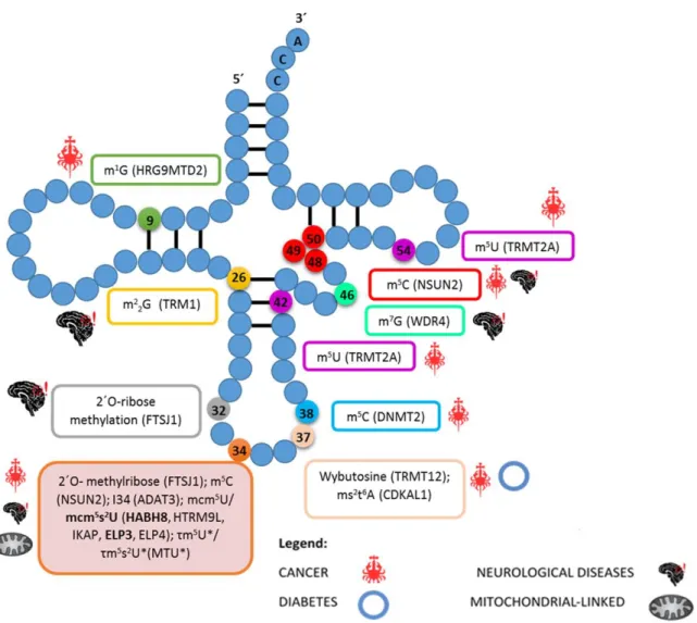
Another interesting cases are elp-defective mutants whose defects are redundant with those of URM1. The Elongator complex is composed by six different protein subunits – Elongator protein (ELP) 1 to 6- and has a role in transcription as an H3 and H4 histone acetylase. Beyond this, it also participates in diverse cellular processes such as transcriptional elongation, formation of modified wobble uridines in tRNAs, polarized exocytosis and telomeric gene silencing (58). In humans there are already several mutations in Elongator subunits genes linked to diverse pathologies concerning neurodegeneration, pinpointing a role of this complex in neurodevelopment and in protection against neurodegeneration (59,60). Figure 6 comprises data collected from several studies concerning defects in tRNA modifications associated with human diseases such as cancer, neurological disorders, Type II Diabetes and mitochondrial-linked disorders (61).
Figure 6: Representation of the tRNA secondary structure with the respective tRNA modifications and
associated tRNA modifying enzymes (in parenthesis) that have been linked to human pathologies – indicated near to each box. Connecting lines between RNA residues represents base pairing. Asterisks indicate modifications that are only found in mitochondrial tRNAs. Adapted from (61, 109).
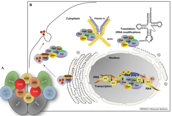
1.5. Elongator complex
Originally, the Elongator complex was described as a promoter of elongation of RNA polymerase II transcription (62) as well as other functions in different compartments of the cell (Figure 7B), with the acetyltransferase domain of ELP3 required in all of them (63). However, more recent studies have shown that its main cellular function is to promote the formation of 5-carbamoylmethyl (ncm5) and 5-methoxycarbonylmethyl (mcm5) side-chains on wobble uridines, once the conserved six-subunit elongator complex is crucial in the first step of the formation of ncm5 and mcm5 side chains, that frequently
contain uridines at the wobble position in eukaryotic cytoplasmic tRNAs (64,65).
As we can see in Figure7A, the Elongator complex is composed by six ELP proteins that form two distinct sub-complexes, a symmetric dimer composed by ELP1-ELP2-ELP3 and an heterohexameric ring composed by ELP4-ELP5-ELP6 that is bound to one of the two ELP1-ELP3 sub-complexes (66,67). All six ELP proteins are necessary for tRNA modifications, however, it is thought that the catalytic activity have its place in the ELP3 protein, since it contains both a lysine acetyltransferase (KAT) domain (earlier called a histone acetyltransferase (HAT)) and a radical S-adenosylmethionine (SAM)
Figure 7: Structure of the Elongator complex and its biological roles. A) Elongator complex structure;
B) Elongator complex functions. Is involved in protein translation fidelity by regulating tRNA modifications in the cytoplasm. In the nucleus, Elongator is associated with hyper phosphorylated RNA polymerase II and is required for histone H3 acetylation across the transcribed regions of multiple genes, including candidates coding for proteins involved in cell motility. It is also a filamin A-binding protein in the cytoplasm that regulates actin cytoskeleton organization. An interaction between ELP1 and Sec2p, a regulator of Rab activation, also negatively regulates exocytosis. Adapted from (63).
domain (64,68,69). This notion can be supported by the fact that only homologues of ELP3 and none of the other subunits are found in most archaea and some bacteria.
It was already possible to associate several mutations in genes for Elongator subunits to several human diseases, as is the case of Familial dysautonomia (FD), an autosomal recessive neurodegenerative disorder that is caused by a point mutation in IKBKAP (ELP1) gene which changes a splice site in one of the introns, leading to the skipping on an exon and the generation of frameshift and therefore a premature translation termination codon. FD patients show decreased levels of ELP1 protein, reduced levels of mcm5s2U in tRNA in brain tissue and fibroblast cell lines (70–72). Other mutations in genes for other Elongator subunits have been associated with other diseases. For example, allelic variants of ELP3 have shown strong association with axonal biology and neuronal degeneration in Amyotrophic lateral sclerosis (ALS), a progressive motor neuron disease that results in death from respiratory failure. Motor neurons are highly vulnerable to stress because they have an elevated threshold for activating heat-shock proteins (Hsps) and since ELP3 regulates Hsps expression (specifically HSP70) by histones (H3 and H4) acetylation, this ability of ELP3 to increase the Hsp70 transcription could explain the association between elevated levels of ELP3 expression and motor neuron protection from degeneration. In fact, ELP3 knockdown in zebrafish resulted in an anomalous splitting of neurons with no associated morphological abnormalities, two loss of function ELP3 mutations in Drosophila conferred irregular photoreceptor axonal targeting and synaptic development and, in humans, risk-associated ELP3 alleles were correlated with lower brain ELP3 expression (73–75). ALS pathogenesis is modified by ELP3 through the control and regulation of mcm5s2U wobble uridine tRNA modifications, affecting the aggregation of
vulnerable proteins, disturbing muscle enervation, disease onset and survival in the mutant SOD1 mouse model. Evidences of such correlation lies on the fact that lowering ELP3 levels reduces mcm5s2U levels and increasing of ELP3 expression restore the levels of this modification, attenuating mutant SOD1 aggregation (76).
Evidences correlated ELP4 to Rolandic epilepsy, the most common form of human epilepsy that affects children between 3 and 12 years old. One of the plausible explanations is the abolishment of the Elongator function in the central nervous system due to ELP4 mutations, since they affect cell motility and actin cytoskeleton genes and/or proteins during development. Although this association exists, no causative variant was already identified, but there are evidences suggesting that a non-coding mutation in ELP4 impairs brain-specific elongator-mediated interaction of genes that are implicated in brain development, which results in a predisposition to seizures (77). In the human genome, the gene locus of ELP4 is close to the brain-derived neurotrophic factor (BDNF) and recent reports demonstrate that ELP4 and BDNF genes act together, affecting cell motility, migration and adhesion of neurons, which may implicate them in the pathogenesis of benign epilepsy with centrotemporal spikes (BECTS) (78,79).
1.6. Mechanisms associated with proteostasis deregulation
Once tRNAs are a key element of the translation machinery, it is clear that their synthesis, posttranscriptional modifications and degradation is highly regulated and integrated into the cellular response circuit. In fact, the levels of many tRNA modifications are dependent of the rate of growth, oxygen levels and even the presence of vitamins and metals, reinforcing the potential role of tRNA modifications in the cellular response to environmental stimuli (80,81).
Although it is not yet fully understood how these modifications cause such diverse phenotypes and diseases, the deregulation of the proteostasis caused by changes in the tRNA decoding efficiency is a common feature of many of these diseases. Usually, the rate of protein synthesis decreases, cell growth and survival are affected and erroneous proteins aggregate, compromising the PN (5). This way, a great number of human disorders are considered proteostasis network disruptions, regardless of whether they arise from the chronic expression of instable mutant proteins or from mutations in PN genes (82).
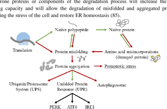
There are numerous signaling pathways controlling the PN, responsible for the improvement of the capability of the proteostasis network to enable and maintain cellular protein folding and function in spite of adverse challenges (83). When facing errors in protein folding, endoplasmic reticulum (ER) becomes stressed and this will activate a set of signaling pathways that will be described below (84). This PN regulation is represented in Figure 8. Activation of genes coding for folding enzymes, trafficking components, chaperone proteins or components of the degradation process will increase the cell’s folding capacity and will allow the degradation of misfolded and aggregated proteins, relieving the stress of the cell and restore ER homeostasis (85).
Figure 8: Representation of protein fates in the Proteostasis Network. Green lines represent the normal
course of a functional protein since their synthesis. Red arrows and lines represent mRNA translation accuracy lost, stress or mutations, triggering the production of non-functional proteins, causing proteotoxic stress to cells and the consequential degradation pathways.
1.6.1. Unfolded Protein Response (UPR)
Conditions like changes in intralumental calcium, nutrient deprivation, mutations, pathogen infection and aging disturb the protein folding in the ER lumen, threatening cell’s viability. To solve this problem, cells developed mechanisms to guarantee that misfolded proteins are discarded. The endoplasmic reticulum (ER) is the site where proteins that follow the secretory pathway enter and where the folding and assembling into subunit complex occur before they transit to the Golgi compartment. The accumulation of unfolded proteins in the ER leads to its stress, triggering the unfolded protein response (UPR), an adaptive signaling cascade activated by three ER-localized transmembrane signal transducers (86,87), described in Table 3.
Table 3: ER transmembrane receptors. Adapted from (87).
ER transmembrane receptors Description
PERK: The pancreatic endoplasmic reticulum-resident kinase pathway
Serine/threonine kinase that phosphorylates the translation initiation factor eIF2α (eIF2α-P), causing the reduction of the protein synthesis rate, releasing calcium from the ER and further activate pro-apoptotic pathways. Elevated levels of phosphorylated eIF2α indicate that the protein synthesis rate is reduced (88).
ATF6: The activating transcription factor 6
Basic Leucine zipper (bZIP) transcription factor that after being cleaved by local proteases will be responsible to activate the transcription of UPR-responsive genes (89).
IRE1: Inositol-requiring enzyme 1
Bi-functional protein required for UPR and also with a site-specific endoribonuclease (RNase) activity, important for RNA degradation to reduce protein synthesis (90).
Both PERK, ATF6 and IRE1 luminal domains are bound to the protein chaperone Binding immunoglobulin Protein (BiP), the main regulator of UPR, in non-stressed cells; when in stress, there is an accumulation of unfolded proteins that will bind to BiP, sequestering it and promoting its release from the UPR sensors (91,92). When it is localized in ER lumen but when it’s overexpressed it may become detectable on the cell surface (93). BiP expression is correlated with cancer, cell proliferation and histological grade (94). The three branches of the UPR, as well as their signaling cascades, are illustrated in Figure 9.
1.6.2. Ubiquitin/Proteasome System (UPS)
The discovery and characterization of the Ubiquitin/Proteasome pathway was made by Avram Hershko, Aaron Ciechanover and Irwin Rose and was worthy of a Novel Prize in 2004, demonstrating its importance (95). Ubiquitin molecule participates in the targeting of substrates in the most important protein degradation pathways – the proteasome, the lysosome and the autophagosome (95).
Ubiquitin is a member of a family of structural conserved proteins with 76 amino acids that can conjugate to other proteins/substrates by one process denominated ubiquitination that involves three steps carried out by three classes of enzymes: The Ubiquitin activating enzyme (E1) belongs to the initial activation step, forming a thio-ester bond with ubiquitin allowing the succeeding binding of ubiquitin to Ubiquitin conjugating enzyme (E2) – intermediate step - where the carboxy-terminus of ubiquitin forms a bond with the ε-amino group of a lysine residue on the substrate; The final step, facilitated by
Figure 9: The three divisions of the UPR (A to C). Three families of signal transducers (ATF6, PERK, and
IRE1) sense the protein-folding conditions in the ER lumen and transmit that information, resulting in production of bZIP transcription regulators that enter the nucleus to drive transcription of UPR target genes. Each pathway uses a different mechanism of signal transduction: ATF6 by regulated proteolysis, PERK by translational control, and IRE1 by nonconventional mRNA splicing. In addition to the transcriptional responses that largely serve to increase the protein-folding capacity in the ER, both PERK and IRE1 reduce the ER folding load by down-tuning translation and degrading ER bound mRNAs, respectively (92).
the Ubiquitin ligase enzyme (E3) is where ubiquitin reaches its last destination of the substrate amino group and it is the identity of E3 that provides substrate specificity (96).
After protein ubiquitination, they are targeted to the 26S proteasome for degradation or will suffer changes in their location or activity. The proteasome is an abundant complex found in the cytosol and nucleus of cells with an empty cylindrical structure composed of a central barrel-shaped core (20S) particle with many proteolytic sites and a regulatory (19S) particle at either or both of its ends that rules access to the core, selecting, preparing and translocating the substrates into the 20S core for degradation. The latter has three associated deubiquitinating enzymes (DUBs) – POH1/PSMD14, USP14 and UCH37 – with the major function of recover the ubiquitin from substrate protein in order to preserve the cellular ubiquitin pool (95). When the polypeptide substrate goes to the central chamber of the 20S particle it is cleaved by the six proteolytic sites resulting in small peptides, that are quickly digested by the cytosolic endopeptidases and aminopeptidases into constituent amino acids and then metabolized or reused to synthesize new proteins (97,98).
The UPS involves two stages, namely the conjugation of the multiple ubiquitin molecules with the substrate protein, marking them and the degradation of these proteins that are transported to the proteasome (99,100). Ubiquitin-mediated proteolysis also regulates other cellular processes, such as apoptosis, DNA transcription and repair, differentiation and development, immune response, ribosome biogenesis, viral infection and so on (101). Figure 10 demonstrates UPS pathway under normal and pathological conditions compared with autophagy-lysosomal pathway (ALP).
1.6.3. Autophagy-lysosomal pathway (ALP)
Autophagy means “self-eating” and it is a vital catabolic pathway responsible for cytoplasmic components degradation inside the lysosome that plays a key function in the quality control of the cell and also serves as an indispensable cytoprotective response to pathological stresses that occur during diseases (102). In eukaryotic cells, it comprises three main intracellular pathways - macroautophagy, microautophagy and chaperone-mediated autophagy (CMA) – that are mechanistically different but whose final destination is the lysosome (103).
The intracellular components degradation involves a multi-step process consisting of substrate recognition, its delivery to lysosomes, consequent degradation and following recycling of the breakdown products. Figure 10 demonstrate in a simple way this pathway under normal and pathological conditions, comparing to UPS pathway. During macroautophagy, the substrate is confiscated inside the autophagosomes, a double-membrane vesicle formed by intact organelles such as mitochondria and portions of the cytosol, for its delivery to lysosomes or endosomes through vesicular fusion, forming an autolysosome. Mammalian target of rapamycin (mTOR) complex is the central, but not exclusive, player of the machinery components involved in autophagosome formation and,
under normal nutrient conditions, active mTOR phosphorylates ULK1 and sequesters it in a complex, inhibiting autophagy (104).
In microautophagy, the cargo is internalized by single-membrane vesicles that are formed through invaginations in the surface of lysosomes (105). In the case of chaperone-mediated autophagy, there is no need for vesicles since unfolded, soluble proteins are identified by a cytosolic chaperone that delivers them to lysosomes for their internalization through a translocation complex formed by the multimerization of the CMA receptor protein LAMP-2A (106).
AMP-activated protein kinase (AMPK) is the main positive regulator of autophagy that is activated by a high ratio of AMP to ATP (107). mTOR and AMPK also control cell growth and metabolism, coupling these processes to autophagy and malfunctions of this catabolic pathway have been associated with a wide range of human diseases, arising mostly from its role in quality control of the proteome and the maintenance of the proteostasis. For example, when autophagy is disrupted in post-mitotic tissues, such as neurons, it leads to altered proteins accumulation and its activation is part of the cellular response to stress (102).
Figure 10: Autophagy-lysosomal pathway (ALP) and ubiquitin-proteasome system (UPS) pathways under
normal and pathological conditions. Proteins are tagged with ubiquitin conjugates through a sequential enzymatic mechanism involving three classes of enzymes (E1, E2, E3). Under normal conditions, ubiquitylated substrates are recognized by ubiquitin receptors present in ALP and UPS pathways and efficiently eliminated. In the UPS, substrates are subsequently deubiquitylated, a key step for substrate degradation and amino acid recycling. Free-Ub chains formed promote ALP function. Ubiquitin receptors in the ALP form oligomers to facilitate substrate recognition and autophagosomal recruitment. Under aging and Alzheimer’s disease conditions there is a decrease in the function of the ALP and the UPS that reduces substrate degradation and amino acid recycling (100)
1.7. Hela cells and the Aggregation Reporter System
Hela cells are human cervical cancer cells that became very important and helpful to medical research because they grow rapidly when in the right medium (with the appropriate nutrients and conditions) and with proper space. Compared to normal cells, they multiply and grow quickly and are extremely resilient and, under the right conditions, they form an immortal cell line, dividing indefinitely (108).
To monitor protein aggregation in human cells, our team has previously developed a fluorescence-based sensor assay that consists in HeLa stable cell line expressing a HSP27:GFP chimeric reporter protein characterized by the fusion of the heat-shock protein 27 (HSP27) with a fluorescent protein, in this case, the Green Fluorescent Protein (GFP), as schematized in Figure 11. HSP27 (HSPB1) is a human small heat shock protein and represent the first line of defense in proteostasis, being activated by diverse triggers such as temperature, pH or post-translational modifications. HSP27 is recruited by misfolded proteins and binds to them in an ATP-independent manner, forming an HSP27-substrate complex which allows the refolding by the larger ATP-dependent chaperones (HSP70 and HSP90) or leads the proteins to degradation (109). If there are misfolding proteins, the GFP fluorescence is re-localized to foci. This cell line was used to perform fluorescence-based genetic screenings. Based on the small interfering RNA (siRNA) technology, our group performed experiments where the expression of the human tRNA modifying enzymes was knocked-down and observed the consequences of their absence for protein misfolding. The stable HeLa HSP27:GFP cell line can provide valuable information to identify modulators of proteostasis, identify compounds that lead to proteostasis deregulation and find novel targets that modulate protein aggregation (110).
Figure 11: Schematic representation of the functioning of the stable cell line expressing the HSP27:GFP
1.8. Motivations and Aim of the study
Evidences show that a growing number of tRNA modifying enzymes are implicated in diseases where proteostasis is affected, specifically neurological disorders, cancer and mitochondrial-linked diseases. However, the implication of deregulation of tRNA modifying enzymes to human diseases is not very clear and the role of these enzymes in proteostasis deregulation, proteotoxic stress and protein aggregation as the causal mechanism of disease has not been fully experimentally demonstrated in mammalian cells.
Our hypothesis is that the deregulation of tRNA modifying enzymes results in protein aggregation and consequent activation of UPR, characteristic of protein conformational diseases.
To test this hypothesis and making use of the fluorescence-image based siRNA screen developed by our team in the past, the main goal is to identify human tRNA modifying enzymes involved in proteostasis, pinpoint the most relevant for protein aggregation and elucidate which protein quality control pathways are affected.
ELP3 was identified in this study as the most relevant tRNA modifying enzyme for proteostasis in human cells, highly involved in protein aggregation, with alterations at the level of protein synthesis and increased ubiquitination.
This data reinforces the viability of our initial hypothesis and additional tests are now undergoing to further disclose the molecular consequences of ELP3 deregulation and validate it as a therapeutic target for conformational disorders.
CHAPTER
2
.
Metodology
2.1. Cell Culture
A stable HeLa cell line expressing HSP27-GFP was cultured in Dulbecco’s Modified Eagle Medium (DMEM) complemented with 10% of Fetal Bovine Serum (FBS) and 1% of Pen-Strep-Glut (a combination of the antibiotics penicillin and streptomycin and the amino acid glutamine). These cells were maintained in culture in a culture chamber at 37ºC with 5% of CO2 and 95% of humidity. Except for fluorescence and proteostat assays,
cells were detached from plates using TrypLe Express (ThermoFisher Scientific) and incubated 5 minutes at 37ºC. The subsequent cell suspension was centrifuged for 3 minutes at 3000 rpm at Room Temperature (RT), the supernatant was discarded and the pellet was ressuspended in fresh medium. To perform reverse transfection with siRNAs, cells were counted in an optical microscope using a Neubauer chamber, diluting 2μL of suspended cells with 18μL of trypan blue.
2.2. Reverse transfection with siRNAs
SiGenome SMARTpool human siRNA targeting different RNA modifying enzymes (Table 5) were obtained from Dharmacon (Thermo scientific) and reverse transfected in triplicate into the stable HSP27-GFP HeLa cell line in 24 well plates, as exemplified in figure 12.
Figure 12: Schematic representation of a 24 well plate with the
In each well 12μL of 500nM siRNA duplex and 88μL of Opti-MEM were added, followed by the addition and mixing of 1μL/well of the mix of Lipofectamine RNAimax and an incubation period of 30 minutes. After this time, 500μL of 2x10⁴ cells solution was added to each well and incubated for 72 hours at 37ºC in a CO2 incubator (Solutions
preparation described in Table 4).
Table 4: Reverse transfection solutions
siRNA aliquots Lipofectamine mix Cells solution
CiVi = CfVf (=) 5.4x10⁴ x Vi = 500x103 x 100μL (=) Vi = 2.5μL of SiRNA stock + 97.5μL of TE (0.5μL lipofectamine + 0.5μL Opti-MEM) x Number of wells CiVi = CfVf (=) 5.4x10⁴ x Vi = 2x104 x 2.4mL (=) Vi = 889μL of cells + 2.399mL of DMEM without antibiotics
For experiments with the positive control condition MG132 (carbobenzoxy-Leu-Leu-leucinal), 5μM of MG132 was added to cells 16h before collecting the pellets. For protein synthesis study by SUnSET, 15 min before collecting the pellets, cells were incubated with puromycin (10μg/mL) and after that time the collection and protein extraction processes were the same.
Table 5: Human tRNA modifying enzymes tested
Human tRNA modifying enzyme Modification
IKBKAP (Elp1) mcm5U 34, mcm5s2U34, ncm5U34, ncm5Um34 Elp2 mcm5U 34, mcm5s2U34, ncm5U34, ncm5Um34 Elp3 mcm5U 34, mcm5s2U34, ncm5U34, ncm5Um34 Elp4 mcm5U 34, mcm5s2U34, ncm5U34, ncm5Um34 Elp5 (Orf81) mcm5U 34, mcm5s2U34, ncm5U34, ncm5Um34 Elp6 (Orf75) mcm5U 34, mcm5s2U34, ncm5U34, ncm5Um34 TRMT1 m2,2G 26 URM1 mcm5s2U 34 TRMT61A m1A TRMT2A m5U 54 TRMT5 m1G 37, m1I37, yW37 ALKBH8 (TRM9) mcm5U 34, mcm5s2U34 TRDMT1 m5C 34
2.3. Total protein extraction and quantification
During all the procedures, the cells remained on ice to avoid the proteases activity. Extracts were prepared by sonication of the pellets in 100μL of Empigen lysis buffer (ELB) (Table 6) for 2 cycles, at a 60% frequency during 15 seconds each and centrifuged at 200G during 20 minutes at 4ºC. After centrifugation, supernatants were kept for the following stage of total protein quantification.
Total protein quantification was performed using Pierce™ Bovine Serum Albumin (BCA) Protein Assay Kit (Thermo Scientific) following the manufacturer’s instructions, using a 20x dilution for our samples. The absorvance was read at 575nm using Microplate Manager 6 Bio-Rad Software. The concentration values provided by the software were in μg/mL, so the conversion to μg/μL was made by multiplying each value by 20 (20x dilution) and division by 1000 (mL to μL).
Table 6: Empigen lysis buffer (ELB) reagents and preparation.
For SDS-PAGE of total protein extracts, the volume equivalent to 10μg of total protein was diluted in MilliQ Water to a final volume of 10μL, 3μL of Loading Buffer (LB) 6x were added and samples were denaturated at 95ºC during 5 minutes. For SDS-PAGE of total protein extracts for western blot analysis, the volume equivalent to 30μg of total protein was diluted in MilliQ Water to a final volume of 25μL, 5μL of LB 6x were added and samples were denaturated at 95ºC during 5 minutes.
For proteomic labeling and analysis, 100µg of total protein extract of SiCtrl and SiElp3 transfected cells were send to I3S. Dr. Hugo Osório perform a quantitative mass spectrometry using iTRAQ and used the software Proteome Discoverer to analyse the data. The data show the up- and down-regulated genes when compared to control.
ELB (10mL) - Triton X-100: 50μL - Hepes (1M, pH 7): 500μL - NaCl (5M): 500μL - H2O: 8.95mL Complete ELB (10mL) - ELB: 9.29mL - DTT (1M): 10μL - Naf (1M): 10μL - EDTA (0.5M): 40μL - EGTA (100mM, pH 8): 100μL - Na3VO4 (100mM): 100μL - *Roche 50x - *PMSF (40nM)
Lysis buffer was prepared without Roche 50x and PMSF (*) and aliquots of 955μL were made. When needed, it was added to each aliquot 20μL Roche 50x and 25μL PMSF.