Marker Annexin A4 in Cancer Cell Proliferation Using
Exon Arrays
Li-Ling Lin1, Hsuan-Cheng Huang2*, Hsueh-Fen Juan1*
1Department of Life Science, Institute of Molecular and Cellular Biology, National Taiwan University, Taipei, Taiwan,2Institute of Biomedical Informatics, Center for Systems and Synthetic Biology, National Yang-Ming University, Taipei, Taiwan
Abstract
Gastric cancer is a malignant disease that arises from the gastric epithelium. A potential biomarker for gastric cancer is the protein annexin A4 (ANXA4), an intracellular Ca2+sensor. ANXA4 is primarily found in epithelial cells, and is known to be involved in various biological processes, including apoptosis, cell cycling and anticoagulation. In respect to cancer, ANXA4-overexpression has been observed in cancers of various origins, including gastric tumors associated withHelicobacter pylori infection. H. pylori induces ANXA4 expression and intracellular [Ca2+]i elevation, and is an important risk factor for carcinogenesis that results in gastric cancer. Despite this correlation, the role of ANXA4 in the progression of gastric tumors remains unclear. In this study, we have investigated whether ANXA4 can mediate the rate of cell growth and whether ANXA4 downstream signals are involved in tumorigenesis. After observing the rate of cell growth in real-time, we determined that ANXA4 promotes cell proliferation. The transcription gene profile of ANXA4-overexpressing cells was measured and analyzed by human exon arrays. From this transcriptional gene data, we show that overexpression of ANXA4 regulates genes that are known to be related to cancer, for example the activation of hyaluronan mediated motility receptor (RHAMM), AKT, and cyclin-dependent kinase 1 (CDK1) as well as the suppression of p21. The regulation of these genes further induces cancer cell proliferation. We also found Ca2+could regulate the transmission of downstream signals by ANXA4. We suggest that ANXA4 triggers a signaling cascade, leading to increased epithelial cell proliferation, ultimately promoting carcinogenesis. These results might therefore provide a new insight for gastric cancer therapy, specifically through the modification of ANXA4 activity.
Citation:Lin L-L, Huang H-C, Juan H-F (2012) Revealing the Molecular Mechanism of Gastric Cancer Marker Annexin A4 in Cancer Cell Proliferation Using Exon Arrays. PLoS ONE 7(9): e44615. doi:10.1371/journal.pone.0044615
Editor:Eric Y. Chuang, National Taiwan University, Taiwan
ReceivedJune 6, 2012;AcceptedAugust 6, 2012;PublishedSeptember 7, 2012
Copyright:ß2012 Lin et al. This is an open-access article distributed under the terms of the Creative Commons Attribution License, which permits unrestricted use, distribution, and reproduction in any medium, provided the original author and source are credited.
Funding:This work was supported by the National Science Council of Taiwan (NSC 99-2621-B-002-005-MY3, NSC 99-2621-B-010-001-MY3), the National Taiwan University Cutting-Edge Steering Research Project (10R70602C3) and the National Health Research Institute, Taiwan (NHRIEX100-9819PI). The funders had no role in study design, data collection and analysis, decision to publish, or preparation of the manuscript.
Competing Interests:The authors have declared that no competing interests exist. * E-mail: [email protected] (HCH); [email protected] (HFJ)
Introduction
Gastric cancer is the second leading cause of cancer deaths worldwide and shows high prevalence in Asian populations. Although the incidence of gastric cancer is declining, the overall 5-year survival rate remains low [1]. Determining the most efficient gastric cancer therapies and developing early-stage diagnostic tools are important strategies in affecting clinical outcomes. The comprehensive investigation of the molecular mechanisms that underlie gastric carcinogenesis could provide assistance in de-veloping useful therapeutic strategies for this disease.
Helicobacter pyloriis a gastric pathogen and is the predominant etiological factor for gastric carcinogenesis. Approximately half of the world’s population is infected withH. pylori, and more than 60% of gastric cancer patients have a history ofH. pylori-positivity [2,3,4]. Recent studies have showed thatH. pylorican induce both the proliferation of gastric cancer cells and mucosal inflammatory responses [5,6]. Thus, in order to investigate the molecular mechanisms underlying gastric carcinogenesis, it is necessary to investigate the role and mechanisms of H. pylori in gastric carcinogenesis.
Annexins are ubiquitously expressed in most organisms, in-cluding animals, plants, fungi and protists. It is associated with a variety of physiological functions [7]. Based on the structure of their conserved core domain, annexins are considered to be intracellular Ca2+ sensors and phospholipid binding proteins. They have been observed to stimulate membrane trafficking and vesicle aggregation in response to increased intracellular [Ca2+
]i [8,9]. In humans, annexins have been observed to have a range of cellular functions that have been implied in cytoskeletal organi-zation, exocytosis, endocytosis, ion channel regulation, inflamma-tion, apoptosis, fibrinolysis and coagulation [8]. Annexins are also considered to be involved in cancer, diabetes and inflammation [10]. Recently, more and more studies have emerged that implicate the involvement of annexins in carcinogenesis, as well as promoting proliferation [11,12], invasion [13] and metastasis [14,15]. However, the relationship between all members of the annexins family with cancer has not been characterized.
[25,26]. These events indicate that ANXA4 has a tumorigenic function by regulating cell growth rate.
In this study, we intended to elucidate the molecular mechanism by which ANXA4 induces carcinogenesis. To accomplish this, we monitored the growth rate of gastric cancer cells with different expression levels of ANXA4. We also evaluated the transcriptional expression profile of ANXA4-overexpressing cells and used exon arrays to analyze the downstream signaling of ANXA4. From these studies, we identified 9 cancer-related genes from ANXA4 downstream signals by using the Ingenuity Pathway Analysis (IPA) database. Furthermore, we demonstrated that ANXA4 regulates the activation of RHAMM, AKT, CDK1 and the suppression of p21, and therefore proposed an ANXA4-regulated cellular proliferation pathway.
Results
The Activation of ANXA4 Promotes Cell Proliferation We have previously reported that ANXA4 is overexpressed in
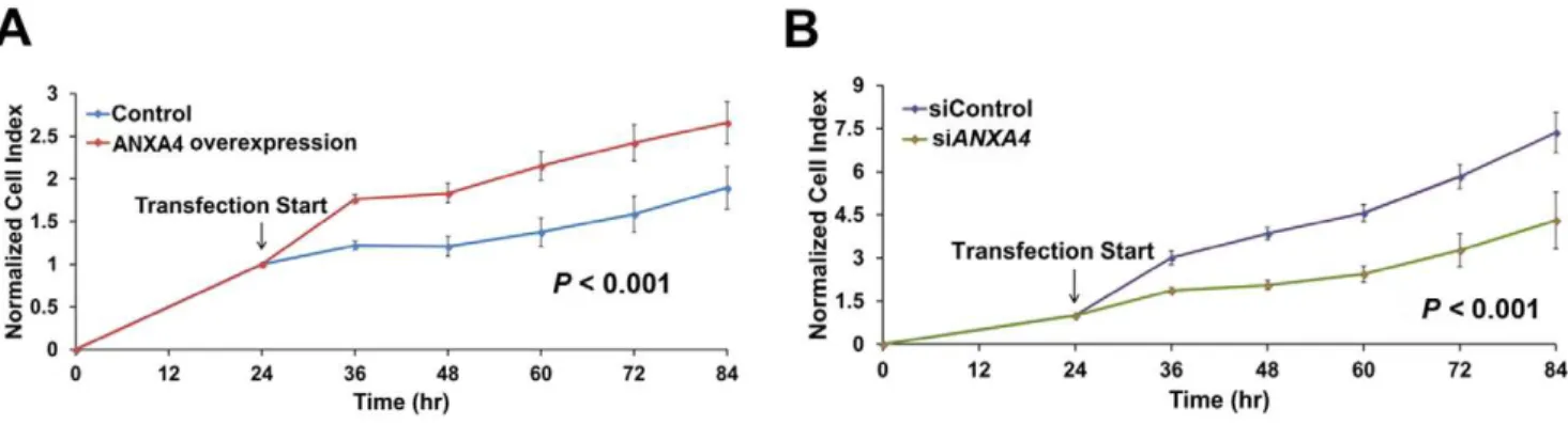
H. pyloriinfected gastric tumor tissue [17]. To further investigate the relationship between ANXA4 and carcinogenesis, we aimed to determine whether ANXA4 promotes cell proliferation. Cells were transfected with either full-length ANXA4 cDNA to increase ANXA4 expression or specific siRNA intended to silence ANXA4 expression. After transfection, we monitored the growth rate of AGS gastric cancer cells using a real-time cell analysis (RTCA) system. Cell numbers were recorded as a cell index (CI) value. The growth curves of AGS cells were modified following transfection. The overexpression of ANXA4 significantly increased the cell growth rate in AGS cells compared with control cells (P,0.001; Figure 1A), whereas ANXA4 knockdown significantly decreased the growth rate (P,0.001; Figure 1B). These results suggest that ANXA4 has the potential to promote cell proliferation.
ANXA4 Increases Expression of the Membrane Proteins, RHAMM and LAMP2
We also examined the difference in gene expression between ANXA4-overexpressing cells and control cells expressing an empty vector. We examined this using exon array analysis, which offers a more accurate view of gene-level expression by using four probes per exon, compared to the conventional 39arrays [27]. In Figure S1, theX-axis of the scatter plot displays the intensities of probe expression measured in one experiment and theY-axis displays the intensities of probe expression measured in the other experiment. These results indicate a correlation between the results of both experiments and therefore indicate a consistency between our duplicate microarrays. The gene expression levels were calculated from the intensities measured via probe sets. Overall, the expression of 1,052 genes were found to be significantly different (P,0.05) between ANXA4-overexpressing cells and control cells.
(Figure S2 and File S1). Here, transcriptional expression of the
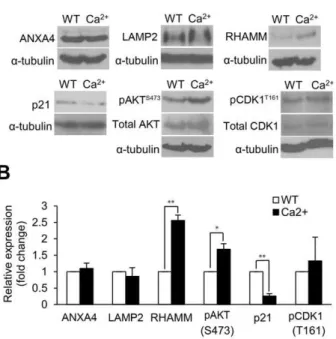
LAMP2 also showed an increase in expression (1.8-fold; Table S1) when measured by exon array analysis. In this study, ANXA4 was overexpressed or silenced (Figure 2A) and then analyzed by immunoblotting to check the protein expression of RHAMM and LAMP2. ANXA4 overexpression increased the expression of RHAMM (Figure 2B) and LAMP2 (Figure 2C) and, consistent with this, the knockdown of ANXA4 expression decreased their levels of expression (Figure 2B and 2C).
ANXA4 Overexpression Regulates Cancer-related Gene Expression
We used the Ingenuity Pathway Analysis (IPA) database to perform a gene function analysis of the exon array data and found that 25 of the 42 genes were eligible for network function analysis ($2-fold differential expression,ttest,P,0.05) (Table 1). Three functional networks were significantly associated with ANXA4-regulated genes. The most strongly associated network was cancer, cell cycle, and reproductive system disease (P,0.05; Figure 3A), and the top-ranked of diseases or disorders was
cancer (P,0.05; Figure 3B). There were 9 genes classified as cancer-related genes in ANXA4-overexpressing cells including 7 genes that were up-regulated in our experiments. These genes are eukaryotic translation initiation factor 4E (EIF4E), succinate dehydrogenase complex, subunit C, integral membrane protein, 15 kDa (SDHC), cyclin-dependent kinase 1 (CDK1), deleted in lymphocytic leukemia 2 (DLEU2), chromatin modifying protein 5 (CHMP5), TIMELESS interacting protein (TIPIN), PDZ-binding kinase (PBK). Chorionic somatomammotropin hormone 1 (placental lactogen) (CSH1) and interferon, alpha 2 (IFNA2) were down-regulated by ANXA4. Based on our results, we propose that ANXA4 has a function in inducing cell pro-liferation. Moreover, CDK1 and PBK (Figure 3B and Table 1) were considered to be involved in the ANXA4 proliferation-inducing model. Since CDK1 activation is associated with the cell proliferation in developing gastric MALT lymphoma [28]. The mutual activation of CDK1 and PBK has been previously reported [29]. In this study, the up-regulation ofCDK1andPBK
observed in ANXA4-overexpressing cells was validated by quantitative real-time polymerase chain reaction (qRT-PCR) analysis (Figure S3).
ANXA4 Regulates the Activation of AKT, CDK1 and the Suppression of p21
phosphor-ylation of serine 473 on AKT and threonine 161 on CDK1 and decreased the expression of p21 (Figure 4A). In addition to this, the knockdown of ANXA4 expression decreased phospho-AKT and phospho-CDK1 and increased the expression of p21 (Figure 4B). This data suggests that AKT, phospho-CDK1 and p21 are regulated by ANXA4 and are downstream signals of ANXA4.
Ca2+Mediates ANXA4 Downstream Signaling Transduction
In carcinogenesis, Ca2+ is involved in causing signal trans-duction to mediate a wide variety of biological processes including invasion, proliferation, angiogenesis and metastasis [32]. It has been reported that annexins can mediate some physiological mechanisms in a Ca2+-dependent manner [9]. In previous studies, intracellular [Ca2+]
i elevation was induced by
H. pylori infection, which, in turn, up-regulates ANXA4 expression [17,33]. To determine whether an increase in intracellular [Ca2+]
i mediates the transmission of downstream signals from ANXA4, AGS cells were treated with ionomycin. Ionomycin is a Ca2+ ionophore and was added to the culture media in our AGS cell model to induce a sustained elevation of intracellular [Ca2+
]i [34]. The protein expressions of ANXA4, LAMP2, RHAMM, p21, phospho-AKT and phospho-CDK1 were measured by immunoblot analysis (Figure 5A). Similar to our observations with ANXA4 overexpression, increased [Ca2+
]i levels significantly up-regulated the expression of RHAMM (P,0.01) and phospho-AKT (Ser 473) (P,0.05) and significant-ly down-regulated the expression of p21 (P,0.01) (Figure 5B). CDK1 activation was slightly increased by [Ca2+]
i elevation. However, elevated [Ca2+]
ilevels had no effect on the expression of ANXA4 and LAMP2. In our previous study, ANXA4 and LAMP2 can localize to plasma membrane after H. pylori
Figure 1. ANXA4 induces cell proliferation.To measure cell proliferation, AGS cells were cultured in a 16-well microtiter E-plate. After incubation for 24 h, the cell growth rate of (A) cells overexpressing ANXA4, and (B) cells containingANXA4-specific siRNA were measured. It was observed that ANXA4 regulated the cell index in a time-dependent manner. (A and B) Data were normalized from measurements taken at 24 h, which was when transfection was initiated. The detection time from three independent experiments is represented as mean6SD, n = 3.Pvalues were calculated using the two-sample Kolmogorov-Smirnov test.
doi:10.1371/journal.pone.0044615.g001
Figure 2. ANXA4 up-regulates RHAMM and LAMP2 expression.(A) AGS cells were transfected with either an empty vector (pcDNA3.1), full-lengthANXA4(pcDNA3.1/ANXA4), control siRNA (siControl), orANXA4siRNA (siANXA4). (B–C) RHAMM and LAMP2 expressions were measured in ANXA4-overexpressing cells or cells containingANXA4-specific siRNA by immunoblotting analysis. (B) The expression of RHAMM was significantly up-regulated (P,0.05) after overexpressing ANXA4 and significantly down-regulated (P,0.01) after silencing ANXA4 expression. (C) The expression of LAMP2 was up-regulated after overexpressing ANXA4 and significantly down-regulated (P,0.05) after silencing ANXA4 expression. Data are represented as mean6SD, n = 3. *P,0.05 vs. control treatment values.
infection with intracellular [Ca2+]
ielevation (Figure S4 and File S1). These results suggest that Ca2+ just changes intracellular location of ANXA4 and LAMP2, but not regulates their expression.
Discussion
In cancer research, the identification of biomarkers and the subsequent clarification of their mechanistic relationships with tumorigenesis can contribute to the development of useful
GPN3* GPN-loop GTPase 3 2.5 0.025
HIGD2A HIG1 hypoxia inducible domain family, member 2A 2.4 0.017
RPS27L* ribosomal protein S27-like 2.4 0.002
CDK1*{ cyclin-dependent kinase 1 2.4 0.041
DLEU2*{ deleted in lymphocytic leukemia 2 (non-protein coding) 2.4 0.041
CHMP5*{ charged multivesicular body protein 5 2.3 0.019
MRPL39 mitochondrial ribosomal protein L39 2.3 0.030
FCF1 FCF1 small subunit (SSU) processome component homolog (S. cerevisiae) 2.3 0.010 NDUFA4* NADH dehydrogenase (ubiquinone) 1 alpha subcomplex, 4, 9 kDa 2.2 0.013
FBXO16* F-box protein 16 2.2 0.036
LYPLA1* lysophospholipase I 2.2 0.004
FSD1L fibronectin type III and SPRY domain containing 1-like 2.2 0.042
TIPIN*{ TIMELESS interacting protein 2.1 0.019
hypothetical LOC727817 2.1 0.018
ZDHHC20 zinc finger, DHHC-type containing 20 2.1 0.045
EXTL2* exostoses (multiple)-like 2 2.1 0.044
COX16 COX16 cytochrome c oxidase assembly homolog (S. cerevisiae) 2.1 0.045
NMI* N-myc (and STAT) interactor 2.1 0.040
PBK*{ PDZ-binding kinase 2.0 0.030
ANKRA2* ankyrin repeat, family A (RFXANK-like), 2 2.0 0.036
PHOSPHO2 phosphatase, orphan 2 2.0 0.017
WBP5* WW domain-binding protein 5 2.0 0.016
PSMA3* proteasome (prosome, macropain) subunit, alpha type, 3 2.0 0.012
C17orf78 chromosome 17 open reading frame 78 0.5 0.030
C15orf2 chromosome 15 open reading frame 2 0.5 0.048
CSH1*{ chorionic somatomammotropin hormone 1 (placental lactogen) 0.5 0.037 C1QTNF9 C1q and tumor necrosis factor-related protein 9 0.5 0.008
IFNA2*{ interferon, alpha 2 0.5 0.048
KRTAP5-5 keratin-associated protein 5–5 0.5 0.024
TSPYL6 TSPY-like 6 0.4 0.037
SRP14* signal recognition particle 14 kDa (homologous Alu RNA-binding protein) 0.4 0.020 OR13A1 olfactory receptor, family 13, subfamily A, member 1 0.4 0.030 OR10G7 olfactory receptor, family 10, subfamily G, member 7 0.4 0.048
PRH1* proline-rich protein HaeIII subfamily 1 0.3 0.028
KRTAP4-12* keratin-associated protein 4–12 0.3 0.036
*Gene eligible for network function analysis.
{
diagnostic tools and result in optimal therapeutic strategies. Recently, more and more studies have shown that the biomarker candidate proteins in the annexins family are potentially in-strumental in the progression of various cancers; e.g., ANXA1 for clear cell renal cancer, ANXA2 for gastric cancer and colorectal cancer, ANXA4 for colorectal cancer, ANXA8 for breast cancer, ANXA10 for hepatocellular cancer, ANXA11 for ovarian cancer and colorectal cancer [35]. ANXA4, a member of the annexins family, has been observed to be overexpressed in gastric tumor tissues and is also associated with the gastric cancer-related H. pyloriinfection [17]. In addition, another annexins family member, ANXA2, has also been observed to be overexpressed in gastric cancer and is related to poor clinical outcome, making it a potential prognostic factor [36]. Taken together, these findings suggest the involvement of ANXA4 in tumorigenesis; however, its exact mechanism in the process remains unclear. In order to further evaluate the cellular function of ANXA4 in the progression of gastric cancer, we explored the link between the two and subsequently demonstrated that ANXA4 may regulate cancer-related genes and propagate the path to cell proliferation.
In the present study, we found that carcinogenesis-associated proteins such as RHAMM, AKT, p21, PBK, and CDK1 are regulated by the overexpression of ANXA4. A schematic representation of ANXA4-induced downstream signals related to
cell proliferation is shown in Figure 6.HMMR(RHAMM) is an oncogene that is overexpressed in several cancers, including gastric cancer, and has been implicated in many cellular processes, such as cell signaling, cell proliferation, and tumorigenesis [37,38].
It has been reported that RHAMM induces the RAS signaling cascade and activates AKT [39]. The RAS signaling cascade transduces downstream signals by activating phospho-AKT through phosphoinositide 3-kinases. This activation of AKT has also been reported as a marker for gastric cancer progression [40]. We have previously reported thatH. pyloriinfection is associated with ANXA4 overexpression [17]. H. pylori can deliver the cytotoxin associated gene A (CagA) into host cells, resulting in AKT activation, and thus promoting cell proliferation [41]. In terms of cell survival, AKT suppresses apoptosis by stimulating NF-kB [42]. Recent studies have also shown that ANXA4
interacts with p105 (the NF-kB p50 precursor protein) and
suppresses the transcriptional activity of NF-kB to induce an
anti-apoptotic effect [25]. Taken together, these observations indicate that ANXA4 plays an important role in cell survival and cell growth.
CDK1-cyclin B1 complexes regulate the cell cycle G2/M phase and have also been implicated in promoting tumorigenesis [43]. A recent study revealed that increased expression of CDK1 is associated with the progression fromH. pylori-associated gastritis to
Figure 3. The significance of ANXA4-induced gene expression was analyzed using the Ingenuity Pathway Analysis (IPA) database. (A) 25 of the 42 candidate genes showed a significant difference in expression (a fold increase of$2 or a fold decrease of#0.5,P,0.05) and were categorized among the three top-ranked networks. (B) Genes were classified according to their documented/established roles in various disease states and disorders.
mucosa-associated lymphoid tissue lymphoma [28]. In this study, CDK1 activation was up-regulated by ANXA4. These events suggest that ANXA4 could mediate the downstream signal pathway, leading to tumorigenesis in gastric cancer patients with aH. pyloriinfection.
In addition to its involvement in the progression of cancer, ANXA4 has also been linked to acquired chemoresistance to anticancer drugs [44,45,46]. In a paclitaxel-resistant cell line (H460/T800), ANXA4 expression is increased and localized in the nucleus [44]. In clear cell carcinoma of the ovary and mesothelioma cells, ANXA4 expression is elevated and associated with resistance to treatment with carboplatin, and is considered to be a biomarker for susceptibility to cisplatin [45,46]. Moreover, ANXA4 is an intracellular Ca2+ sensor, and Ca2+ plays an important role in neurotoxicity and heart failure [47,48]. Recent studies have shown that the increased amount of ANXA4 is not only associated with cancer but also Alzheimer’s disease, heart failure and cell lesion caused by ethanol [49,50,51].
Ca2+
messenger system is also required for cell proliferation process and can regulate cell cycle [52,53]. It has been reported that Ca2+ can activate AKT pathway to promote cell survival [54]. In this study, we found that the expression of RHAMM, phospho-AKT and the suppression of p21 were significantly increased by [Ca2+
]i elevation (Figure 5). H. pylori infection is associated with ANXA4 overexpression and intracellular [Ca2+] i elevation (Figure S4 and File S1) [17,33]. These results indicate that H. pyloriinfection might induce some downstream signaling
of ANXA4 by stimulating intracellular [Ca2+
]i elevation. Nevertheless the detail mechanism in the process might be complex and remains unclear. These could provide elucidation of gastric tumorigenesis process underlying H. pylori stimulation. Taken together, this evidence suggests that Ca2+
might assist ANXA4 to transduce signaling and promote tumorigenesis in gastric patients withH. pylori infection.
In conclusion, our results show that the silencing of ANXA4 decreases epithelial cell proliferation, while its overexpression increases proliferation. Furthermore, ANXA4 induces down-stream signals that promote cell growth. We hypothesize that these downstream signals and activation of host cell division may be pathogenic events in H. pylori-induced carcinogenesis. In current clinical therapy, AKT, p21 and CDK1 have been used as an anticancer drug target. AKT inhibitor, Perifosine, is an oral anti-cancer agent and has anti-proliferation activity in several tumor models [55,56,57]. Paclitaxel (Taxol) and vincris-tine can induce and increase p21 expression to decrease G2/M arrest and block cell proliferation [58,59,60,61]. Flavopiridol is an inhibitor of several CDKs including CDK1 to induce apoptosis and anti-angiogenesis [62]. These studies indicate that the elevation of ANXA4 in patients could be considered as a drug target for gastric cancer therapy. Using multiple drugs in combination might provide more effective treatment for blocking signals in the pathway. Taken together, this study could provide new insights into the development of therapeutic strategies for gastric cancer.
Figure 4. ANXA4 induces downstream signal transduction.(A–B) Protein levels of p21, phospho-AKT (Ser473) and phospho-CDK1 (Thr161) in AGS cells, as determined by immunoblotting analysis. (A) Cells were transfected with empty vector or full-lengthANXA4. (B) Cells were transfected with siControl or siANXA4. Representative data from three independent experiments are presented as mean6SD.a-tubulin was used as an internal
Materials and Methods
Cell Lines and Culture Conditions
Human stomach adenocarcinoma AGS cells (CRL-1739, ATCC) were grown in 90% RPMI 1640 medium (Biological Industries, Beth-Haemek, Israel) that was supplemented with 1% penicillin/streptomycin and 10% fetal bovine serum (Biological Industries, Beth-Haemek, Israel). Cells were cultured at 37uC in a controlled humidified atmosphere in an incubator containing 5% CO2.
Plasmids and Transfections
Full-lengthANXA4was amplified by PCR using the primer pair
ANXA4-F (59atataagcttgccaccatggccatggcaaccaaa 39) andANXA4 -R (59 gcgcgggaattcttaatcatctcctccaca 39), and the amplification product was inserted into the HindIII/EcoRI sites of pcDNA 3.1(+) (Invitrogen, Carlsbad, CA). ANXA4-specific siRNA and negative control Stealth siRNA (Stealth RNAiTM) were purchased from Invitrogen (Carlsbad, CA, USA). Cells were cultured in six-well plates or on coated cover slips for 24 h. Cells were then transiently transfected with pcDNA 3.1(+)/ANXA4(8mg for a six-well plate; 0.4mg/mL for a 96-well E-plate) or ANXA4 siRNA (100 pmol for a six-well plate; 10 pmol for a 96-well E-plate) using Lipofectamine 2000 (Invitrogen) according to the manufacturer’s instructions. The efficiency of expression vector and siRNA transfection was analyzed by immunoblot. After transfection for 48 h, the differential expression of proteins and genes was detected.
Antibodies
The mouse monoclonal antibodies used in this study were as follows: CDK1 p34 (sc-51578) from Santa Cruz Biotechnology (Santa Cruz, CA, USA); LAMP2 (ab25631) and RHAMM
Figure 5. Ca2+mediates the expression of RHAMM, phospho-AKT and p21. (A) Cells were treated with ionomycin to increase intracellular Ca2+levels, and the expression levels of ANXA4, LAMP2, RHAMM, phospho-AKT (Ser473), p21, and phospho-CDK1 (Thr161) were showed by immunoblotting. (B) The histogram shows the related levels of (A). The relative expressions of RHAMM (P,0.01), phospho-AKT (Ser473) (P,0.05) and p21 (P,0.01) were significantly different. Data are taken from three independent experiments (mean6SD). *P,0.05, **P,0.01 vs. control treatment values.
doi:10.1371/journal.pone.0044615.g005
Figure 6. Schematic representation of the molecular mechanism is induced by ANXA4.ANXA4 binds to the plasma membrane in a Ca2+ -dependent manner and induces downstream signaling transduction. ANXA4 up-regulates LAMP2, a lysosomal marker involved in exocytosis, and RHAMM. Previous reports have showed that RHAMM activates RAS and PI3K, which subsequently leads to the induction of AKT. ANXA4 up-regulates AKT and CDK1 activation, PBK gene expression and down-regulates p21. Ca2+
also up-regulates RHAMM and phospho-AKT, and down-regulates p21. This signal cascade might eventually lead to cell hyperproliferation. Solid lines with arrows and blue circles indicate confirmed regulation; dashed lines with arrows and purple circles indicate references or unconfirmed interactions.
[Ca ]i, ionomycin (5mM) was added to the cells for 1 h. To study the effects of inhibition of Akt phosphorylation, the cells were starved for 1.5 h following a 48-h transfection and were treated with 5mM of the AKT inhibitor VIII (Merck KGaA, Darmstadt, Germany) for 1 h. Samples were separated by 10% SDS-PAGE and then transferred onto polyvinylidene difluoride (PVDF) membranes (Millipore, Billerica, MA, USA). After blocking in 5% nonfat milk and tris-buffered saline (TBS) containing 0.1% Tween 20 (JT Baker, Phillipsburg, NJ, USA) for 1 h at RT with gentle rocking, the following primary antibodies were applied: anti-ANXA4 (1:1000), anti-p21 (1:500), anti-AKT (1:500), anti-pAKT (Ser473; 1:500), anti-CDK1 (1:1000), anti-pCDK1 (Thr 161; 1:500), and anti-RHAMM antibody (1:400). Membranes were incubated with secondary goat anti-mouse conjugated IgG antibodies (Sigma) or goat anti-rabbit conjugated IgG (Rockland, Gilbertsville, PA), respectively. a
-tubulin antibody (1:4000) was used as an internal control. Immunoblots were developed using enhanced chemiluminiscence (ECL) detection kit (Millipore) and visualized on X-ray films. The intensity of the observed bands was normalized to the intensity of thea-tubulin band. Densitometric analysis was performed using
Kodak 1-D Image Analysis software version 3.6 (Eastman Kodak, London, UK).
Exon Array Hybridization and Analysis
To study the downstream genes of ANXA4, we compared the gene expression profiles between the cells transfected with pcDNA3.1 (+)/ANXA4and control cells transfected with an empty vector using an exon array. Total cellular RNA was extracted using TRIzolH Reagent (Invitrogen), and RNA purity was confirmed by spectrophotometry (A260/A280 ratio) as well as by capillary electrophoresis (Agilent 2100 Bioanalyzer, Agilent Technologies, Palo Alto, CA, USA). RNA processing and hybridization were performed using the Affymetrix Human Exon 1.0 ST arrays (Affymetrix, Santa Clara, CA, USA) according to the manufacturer’s protocol. Each array has 28,869 well-annotated genes with 764,885 different probes. The array contains approximately 26 probes for each gene. Affymetrix probe sets information are displayed on NetAffx Web site (http://www. affymetrix.com) [63]. Microarray analysis (n = 2 per group) was performed using the Partek Genomics Suite version 6.5 (Partek Inc., St Louis, MO, USA). The raw data (CEL files) were normalized using the robust multichip averaging (RMA) algorithm and analyzed usingttests. Analysis of the function and biological mechanism of the differentially expressed genes was performed using the Ingenuity Pathway Analysis (IPA) software version 7.5; a score of 3 or above was considered statistically significant (P,0.01) to annotate the information. We have submitted the array data to the GEO database, and the series record number is GSE33620.
Cell Proliferation Assay
AGS cells were loaded in each well of a 16-well microtiter E-plate. Each well contained microelectronic sensor arrays at the base to detect the cell index (CI). For transfection experiments, after incubation for 24 h, AGS cells were transfected with expression vectors or siRNAs for 6 h and monitored for a total of 84 h. The E-plate was placed in the Real-Time Cell Analyzer (RTCA) system and incubated in an incubator containing 5% CO2at 37uC. The level of cell proliferation was represented as CI, which was based on the electrical impedance measured using the xCELLigence system (Roche, Mannheim, Germany).
Statistical Analysis
Data were expressed as mean 6 standard deviation (SD). Difference between independent groups was analyzed using a two-tailed Student’s t test. Data obtained from the cell proliferation assay were analyzed using the two-sample Kolmogorov-Smirnov test. APvalue of less than 0.05 indicated statistical significance. Supporting Information
Figure S1 Scatter plot of the probe intensities in the
repeated exon array experiments.The probe intensities of two repeated experiments were presented separately on anX-axis and Y-axis. Each probe was represented by a single dot in the scatter plot. These results showed the consistency in our duplicate exon array experiments.
(TIF)
Figure S2 ANXA4 participates in plasma membrane
repair by recruiting exocytotic membrane.Representative flow cytometric analyses demonstrated the presence of LAMP2 in
H. pylori-infected cells. (A) ANXA4-overexpressing cells were compared with (B)ANXA4-silenced cells. The results indicate that ANXA4 promotes LAMP2 expression on the surface ofH. pylori -infected cells. ANXA4 overexpression, Over-ANXA4; Control siRNA, siControl;ANXA4siRNA, siANXA4.
(TIF)
Figure S3 ANXA4 induces downstream signal
trans-duction.A qRT-PCR assay of ANXA4-overexpressing AGS cells (black boxes) was performed to confirm the data obtained from exon arrays (gray boxes). The relative mRNA levels ofCDK1and
PBKwere measured and normalized toGAPDHmRNA levels. (TIF)
Figure S4 Intracellular Ca2+
elevation, ANXA4 and LAMP2 localization upon H. pylori infection. (A) H. pylori-infected AGS cells were loaded with Fluo-3/AM to monitor intracellular Ca2+
AGS and SC-M1 cells (yellow arrow) stained with Hoechst 33258. (C) LAMP2 fluorescence on the surface ofH. pylori-infected AGS cells was more enhanced than on the surface of non-infected cells. (TIF)
Table S1 ANXA4-upregulated genes (fold-change $1.5)
in AGS cells based on an exon array classified as plasma membrane proteins with the IPA database.
(PDF)
Table S2 List of primer sequences used for qRT-PCR.
(PDF)
File S1 Supplementary Materials and Methods.
(PDF)
Acknowledgments
We would like to thank Cho-Yi Chen for assistance with statistical analyses, and the staff of the National Taiwan University Hospital for technical support.
Author Contributions
Conceived and designed the experiments: HCH HFJ. Performed the experiments: LLL. Analyzed the data: LLL HCH HFJ. Contributed reagents/materials/analysis tools: LLL HCH HFJ. Wrote the paper: LLL HCH HFJ.
References
1. Jemal A, Bray F, Center MM, Ferlay J, Ward E, et al. (2011) Global cancer statistics. CACancer J Clin 61: 69–90.
2. Herrera V, Parsonnet J (2009) Helicobacter pylori and gastric adenocarcinoma. Clin Microbiol Infect 15: 971–976.
3. Kim SS, Ruiz VE, Carroll JD, Moss SF (2011) Helicobacter pylori in the pathogenesis of gastric cancer and gastric lymphoma. Cancer Lett 305: 228–238. 4. Kusters JG, van Vliet AH, Kuipers EJ (2006) Pathogenesis of Helicobacter pylori
infection. Clin Microbiol Rev 19: 449–490.
5. Fan XG, Kelleher D, Fan XJ, Xia HX, Keeling PW (1996) Helicobacter pylori increases proliferation of gastric epithelial cells. Gut 38: 19–22.
6. Polk DB, Peek RM Jr (2010) Helicobacter pylori: gastric cancer and beyond. Nat Rev Cancer 10: 403–414.
7. Gerke V, Moss SE (2002) Annexins: from structure to function. Physiol Rev 82: 331–371.
8. Gerke V, Creutz CE, Moss SE (2005) Annexins: linking Ca2+signalling to membrane dynamics. Nat Rev Mol Cell Biol 6: 449–461.
9. Monastyrskaya K, Babiychuk EB, Hostettler A, Rescher U, Draeger A (2007) Annexins as intracellular calcium sensors. Cell Calcium 41: 207–219. 10. Fatimathas L, Moss SE (2010) Annexins as disease modifiers. Histol Histopathol
25: 527–532.
11. Ortiz-Zapater E, Peiro S, Roda O, Corominas JM, Aguilar S, et al. (2007) Tissue plasminogen activator induces pancreatic cancer cell proliferation by a non-catalytic mechanism that requires extracellular signal-regulated kinase 1/2 activation through epidermal growth factor receptor and annexin A2. Am J Pathol 170: 1573–1584.
12. Khau T, Langenbach SY, Schuliga M, Harris T, Johnstone CN, et al. (2011) Annexin-1 signals mitogen-stimulated breast tumor cell proliferation by activation of the formyl peptide receptors (FPRs) 1 and 2. FASEB J 25: 483–496. 13. Zhai H, Acharya S, Gravanis I, Mehmood S, Seidman RJ, et al. (2011) Annexin A2 promotes glioma cell invasion and tumor progression. J Neurosci 31: 14346– 14360.
14. de Graauw M, van Miltenburg MH, Schmidt MK, Pont C, Lalai R, et al. (2010) Annexin A1 regulates TGF-beta signaling and promotes metastasis formation of basal-like breast cancer cells. Proc Natl Acad Sci U S A 107: 6340–6345. 15. Tanaka T, Akatsuka S, Ozeki M, Shirase T, Hiai H, et al. (2004) Redox
regulation of annexin 2 and its implications for oxidative stress-induced renal carcinogenesis and metastasis. Oncogene 23: 3980–3989.
16. Dreier R, Schmid KW, Gerke V, Riehemann K (1998) Differential expression of annexins I, II and IV in human tissues: an immunohistochemical study. Histochem Cell Biol 110: 137–148.
17. Lin LL, Chen CN, Lin WC, Lee PH, Chang KJ, et al. (2008) Annexin A4: A novel molecular marker for gastric cancer with Helicobacter pylori infection using proteomics approach. Proteomics Clin Appl 2: 619–634.
18. Lin LL, Huang HC, Juan HF (2012) Discovery of biomarkers for gastric cancer: A proteomics approach. J Proteomics 75: 3081–3097.
19. Shen J, Person MD, Zhu J, Abbruzzese JL, Li D (2004) Protein expression profiles in pancreatic adenocarcinoma compared with normal pancreatic tissue and tissue affected by pancreatitis as detected by two-dimensional gel electrophoresis and mass spectrometry. Cancer Res 64: 9018–9026. 20. Miao Y, Cai B, Liu L, Yang Y, Wan X (2009) Annexin IV is differentially
expressed in clear cell carcinoma of the ovary. Int J Gynecol Cancer 19: 1545– 1549.
21. Toyama A, Suzuki A, Shimada T, Aoki C, Aoki Y, et al. (2012) Proteomic characterization of ovarian cancers identifying annexin-A4, phosphoserine aminotransferase, cellular retinoic acid-binding protein 2, and serpin B5 as histology-specific biomarkers. Cancer Sci 103: 747–755.
22. Zimmermann U, Balabanov S, Giebel J, Teller S, Junker H, et al. (2004) Increased expression and altered location of annexin IV in renal clear cell carcinoma: a possible role in tumour dissemination. Cancer Lett 209: 111–118. 23. Duncan R, Carpenter B, Main LC, Telfer C, Murray GI (2008) Characterisa-tion and protein expression profiling of annexins in colorectal cancer. Br J Cancer 98: 426–433.
24. Xin W, Rhodes DR, Ingold C, Chinnaiyan AM, Rubin MA (2003) Dysregulation of the annexin family protein family is associated with prostate cancer progression. Am J Pathol 162: 255–261.
25. Jeon YJ, Kim DH, Jung H, Chung SJ, Chi SW, et al. (2010) Annexin A4 interacts with the NF-kappaB p50 subunit and modulates NF-kappaB transcriptional activity in a Ca2+-dependent manner. Cell Mol Life Sci 67: 2271–2281.
26. Masuda J, Takayama E, Satoh A, Ida M, Shinohara T, et al. (2004) Levels of annexin IV and V in the plasma of pregnant and postpartum women. Thromb Haemost 91: 1129–1136.
27. Kapur K, Xing Y, Ouyang Z, Wong WH (2007) Exon arrays provide accurate assessments of gene expression. Genome Biol 8: R82.
28. Banerjee SK, Weston AP, Zoubine MN, Campbell DR, Cherian R (2000) Expression of cdc2 and cyclin B1 in Helicobacter pylori-associated gastric MALT and MALT lymphoma : relationship to cell death, proliferation, and transformation. Am J Pathol 156: 217–225.
29. Gaudet S, Branton D, Lue RA (2000) Characterization of PDZ-binding kinase, a mitotic kinase. Proc Natl Acad Sci U S A 97: 5167–5172.
30. Smits VA, Klompmaker R, Vallenius T, Rijksen G, Makela TP, et al. (2000) p21 inhibits Thr161 phosphorylation of Cdc2 to enforce the G2 DNA damage checkpoint. J Biol Chem 275: 30638–30643.
31. Liang J, Slingerland JM (2003) Multiple roles of the PI3K/PKB (Akt) pathway in cell cycle progression. Cell Cycle 2: 339–345.
32. Monteith GR, McAndrew D, Faddy HM, Roberts-Thomson SJ (2007) Calcium and cancer: targeting Ca2+transport. Nat Rev Cancer 7: 519–530. 33. Marlink KL, Bacon KD, Sheppard BC, Ashktorab H, Smoot DT, et al. (2003)
Effects of Helicobacter pylori on intracellular Ca2+signaling in normal human gastric mucous epithelial cells. Am J Physiol Gastrointest Liver Physiol 285: G163–176.
34. Liu C, Hermann TE (1978) Characterization of ionomycin as a calcium ionophore. J Biol Chem 253: 5892–5894.
35. Mussunoor S, Murray GI (2008) The role of annexins in tumour development and progression. J Pathol 216: 131–140.
36. Emoto K, Sawada H, Yamada Y, Fujimoto H, Takahama Y, et al. (2001) Annexin II overexpression is correlated with poor prognosis in human gastric carcinoma. Anticancer Res 21: 1339–1345.
37. Li H, Guo L, Li JW, Liu N, Qi R, et al. (2000) Expression of hyaluronan receptors CD44 and RHAMM in stomach cancers: relevance with tumor progression. Int J Oncol 17: 927–932.
38. Maxwell CA, McCarthy J, Turley E (2008) Cell-surface and mitotic-spindle RHAMM: moonlighting or dual oncogenic functions? J Cell Sci 121: 925–932. 39. Hall CL, Yang B, Yang X, Zhang S, Turley M, et al. (1995) Overexpression of the hyaluronan receptor RHAMM is transforming and is also required for H-ras transformation. Cell 82: 19–26.
40. Cinti C, Vindigni C, Zamparelli A, La Sala D, Epistolato MC, et al. (2008) Activated Akt as an indicator of prognosis in gastric cancer. Virchows Arch 453: 449–455.
41. Chen YR, Juan HF, Huang HC, Huang HH, Lee YJ, et al. (2006) Quantitative proteomic and genomic profiling reveals metastasis-related protein expression patterns in gastric cancer cells. J Proteome Res 5: 2727–2742.
42. Madrid LV, Wang CY, Guttridge DC, Schottelius AJ, Baldwin AS Jr, et al. (2000) Akt suppresses apoptosis by stimulating the transactivation potential of the RelA/p65 subunit of NF-kappaB. Mol Cell Biol 20: 1626–1638.
43. Hunter T, Pines J (1991) Cyclins and cancer. Cell 66: 1071–1074.
44. Han EK, Tahir SK, Cherian SP, Collins N, Ng SC (2000) Modulation of paclitaxel resistance by annexin IV in human cancer cell lines. Br J Cancer 83: 83–88.
45. Kim A, Enomoto T, Serada S, Ueda Y, Takahashi T, et al. (2009) Enhanced expression of Annexin A4 in clear cell carcinoma of the ovary and its association with chemoresistance to carboplatin. Int J Cancer 125: 2316–2322. 46. Yamashita T, Nagano K, Kanasaki SI, Maeda Y, Furuya T, et al. (2012)
53. Berridge MJ, Lipp P, Bootman MD (2000) The versatility and universality of calcium signalling. Nat Rev Mol Cell Biol 1: 11–21.
54. Yano S, Tokumitsu H, Soderling TR (1998) Calcium promotes cell survival through CaM-K kinase activation of the protein-kinase-B pathway. Nature 396: 584–587.
55. Kondapaka SB, Singh SS, Dasmahapatra GP, Sausville EA, Roy KK (2003) Perifosine, a novel alkylphospholipid, inhibits protein kinase B activation. Mol Cancer Ther 2: 1093–1103.
56. Sampson HA, Leung DY, Burks AW, Lack G, Bahna SL, et al. (2011) A phase II, randomized, doubleblind, parallelgroup, placebocontrolled oral food
1093.
61. Shinwari Z, Manogaran PS, Alrokayan SA, Al-Hussein KA, Aboussekhra A (2008) Vincristine and lomustine induce apoptosis and p21(WAF1) up-regulation in medulloblastoma and normal human epithelial and fibroblast cells. J Neurooncol 87: 123–132.
62. Zhai S, Senderowicz AM, Sausville EA, Figg WD (2002) Flavopiridol, a novel cyclin-dependent kinase inhibitor, in clinical development. Ann Pharmacother 36: 905–911.