Universidade de Aveiro 2009
Departamento de Biologia
Maria João Teixeira
Ribeiro de Magalhães
Projecções encefálicas do núcleo reticular ventral do
bolbo raquidiano
Brain projections from the medullary ventral reticular
nucleus
Universidade de Aveiro 2009
Departamento de Biologia
Maria João Teixeira
Ribeiro de Magalhães
Projecções encefálicas do núcleo reticular ventral do
bolbo raquidiano
Brain projections from the medullary ventral reticular
nucleus
Dissertação apresentada à Universidade de Aveiro para cumprimento dos requisitos necessários à obtenção do grau de Mestre em Biologia Molecular e Celular, realizada sob a orientação científica do Professor Doutor Armando Alberto Nova Pinto Almeida, Professor Associado, Escola de Ciências da Saúde da Universidade do Minho, e co-orientação do Professor Doutor Mário Guilherme Garcês Pacheco, Professor Auxiliar, Departamento de Biologia da Universidade de Aveiro.
o júri
presidente Profª. Doutora Maria Adelaide de Pinho Almeida
Professora Auxiliar do Departamento de Biologia da Universidade de Aveiro
Prof. Doutor Armando Alberto da Nova Pinto de Almeida Professor Associado da Escola de Ciências da Saúde da Universidade do Minho
Prof. Doutor Mário Guilherme Garcês Pacheco
Professor Auxiliar do Departamento de Biologia da Universidade de Aveiro
Profª Doutora Isaura Ferreira Tavares
Professora Associada com Agregação da Faculdade de Medicina da Universidade do Porto
Agradecimentos Ao Professor Doutor Armando Almeida pela oportunidade profissional de trabalhar no seu grupo, orientação e conhecimentos partilhados.
Ao Professor Doutor Mário Pacheco pela presença, disponibilidade e por ter aceite ser co-orientador nesta importante etapa académica.
Ao Hugo pela convivência diária, amizade, motivação, paciência, por tudo o que me ensinou e pelo apoio prestado neste trabalho.
À Vera Cardoso e à Paula, pela disponibilidade e amizade.
À Margarida Dourado e à Filipa Ribeiro, pela amizade, motivação, ajuda… A todos os NeRD, pela ajuda, amizade e excelente ambiente de trabalho.
A todo o ICVS.
À minha família, principalmente aos meus pais e à minha irmã, que acreditam em mim, me compreendem e me apoiam incondicionalmente.
Palavras-chave Sistema nervoso; formação reticular; vias da dor; subunidade B da toxina da cólera; dextrano-amina biotinilado; projecções eferentes encefálicas.
Resumo O Sistema Nervoso (SN) dos mamíferos é uma rede complexa de células especializadas na recepção, transmissão e integração de informação. O desempenho de cada um dos subsistemas depende da forma como a comunicação entre as diferentes áreas se organiza. Várias ferramentas têm vindo a ser desenvolvidas para o efeito, sendo actualmente os traçadores neuroanatómicos aquelas de uso mais abrangente. Após injecção do traçador seleccionado na área pretendida do SN, este é incorporado e subsequentemente transportado de forma retrógrada ou anterógrada de acordo com as suas propriedades. O estudo dos sistemas supraespinais de controlo da dor tem em muito beneficiado do uso desta tecnologia. Várias áreas do encéfalo e, em particular, da formação reticular do tronco cerebral, participam na modulação supraspinal da dor. O núcleo reticular ventral (VRt) do bolbo raquidiano continua a ser uma área pouco explorada do encéfalo, contrariamente ao seu homólogo dorsal (DRt), cujo envolvimento na modulação da dor se encontra bem estabelecido. No presente trabalho, as projecções encefálicas (eferentes e aferentes) do VRt são analisadas no rato, recorrendo-se para tal a injecções intracerebrais de traçadores neuronais anterógrados e retrógrados, respectivamente o dextrano-amina biotinilado (BDA) e a subunidade B da toxina da cólera (CTb). Verificou-se que os neurónios do VRt recebem projecções e projectam para áreas do encéfalo implicadas no processamento somatosensitivo, emocional e cognitivo da dor. Estes resultados corroboram com o papel do VRt na modulação da dor. As projecções encefálicas do VRt e DRt para o tronco cerebral são em si muito semelhantes, com o VRt a projectar para áreas mais restritas do diencéfalo. O papel de cada um dos núcleos na modulação da dor poderá estar relacionado com as diferenças observadas nas projecções dos núcleos.
Keywords Nervous system; medullary reticular formation; pain pathways; cholera toxin subunit B; biotinylated dextran; brain efferents.
Abstract The nervous system (SN) of a mammal is a complex network of cells
specialized for the reception, transmission and integration of information. The performance of each subsystem depends on how the communication between different areas is organized. Several tools have been developed for this purpose, being currently the neuroanatomical tracers those of wider use. After selected tracer injection in the desired area of the SN, this one is incorporated and subsequently transported anterogradely and retrogradely according to their properties. The study of the supraspinal pain control systems has greatly benefited from the use of this technology. Several areas of the brain and, in particular, the reticular formation of the brainstem, are involved in supraspinal pain modulation. The ventral portion of the caudal reticular formation (VRt) remains a relatively unexplored area of the brain contrary to its dorsal counterpart (DRt), whose involvement in pain modulation is well established. In the present work, the VRt brain connections (efferent and afferent projections) are investigated in the rat, using iontophoretic injections of the anterograde tracer biotinylated-dextran amine (BDA) and the retrograde tracer cholera toxin-subunit (CTb). It was found that neurons from the VRt receive and project to areas of the brain involved in somatosensitive, emotional and cognitive pain processing.
The set of brain projections observed in VRt is compatible with a role in pain modulation. VRt and DRt brain projections to the brainstem are similar; however, concerning to the diencephalon, VRt has a narrower set of targets. It remains unclear how these differences relate to differential roles in pain modulation.
TABLE OF CONTENTS
Index of Abbreviations --- 5
Chapter 1: Introduction --- 8
1.1 – Overview of the Nervous System --- 9
1.1.1 Cellular Elements of Nervous System --- 9
1.1.2 How Neurons Communicate --- 12
1.1.3 Basic Mechanisms of Axonal transport--- 14
1.3.4 A Brief Approach to Tract-tracing Neuronal Circuits --- 16
1.2 – Pain Control --- 18
1.2.1 Pain Definition --- 18
1.2.2 Peripheral Afferent Fibers --- 18
1.2.3 Nociceptors --- 20
1.2.4 From Periphery to Thalamus --- 21
1.2.5 Brainstem Control of Spinal Nociceptive Processing --- 22
1.3 – Brainstem Reticular Formation and Pain --- 24
1.3.1 The Brainstem Reticular Formation --- 24
1.3.2 Neurotransmitters in the Reticular Formation --- 26
1.3.4 The Ascending Reticular Activating Systems Mediates Consciousness and Arousal --- 27
1.3.5 The Reticular Formation and Nociception --- 28
1.3.6 The ventral reticular nucleus --- 29
Chapter 2: Experimental Procedures --- 31
2.1 – Ethical guidelines --- 32
2.2 – Anterograde Tracing Experiments --- 32
2.3 – Retrograde Tracing Experiments--- 33
2.4 – Image analysis and illustrations --- 33
Chapter 3: Results --- 34
3.2 – Anterograde Tracing Experiments --- 36
3.3 – Retrograde Tracing Experiments--- 37
Chapter 4: Discussion and Conclusion --- 41
4.1 – Specificity of the Tract-tracing Methodology --- 42
4.2 – Specificity of the VRt Brain Projection Patterns --- 45
4.3 - The VRt integrated in the medullary reticular formation – a comparative study with the DRt --- 47
4.4 – Functional Considerations --- 48
Chapter 5: References --- 50
LIST OF FIGURES
Figure 1 – Glia-neuron interactions --- 11
Figure 2 - Structure of myelinated axons --- 11
Figure 3 – The synapse --- 11
Figure 4 – Chemical transmission of a nerve impulse at the synapse --- 11
Figure 5 – Axonal transport on microtubules --- 11
Figure 6 – Different nociceptors detect different types of pain --- 11
Figure 7 – The nociceptor --- 11
Figure 8 – The neural pathway of nociception from primary afferent neurons (PANs) to the superficial lamina in the dorsal horn of the spinal cord --- 11
Figure 9 – The brainstem reticular formation --- 11
Figure 10 – Photomicrographs of a representative iontophoretic BDA injections in the VRt --- 35
Figure 11 - Photomicrographs of a representative iontophoretic CTb injections in the VRt --- 36
Figure 12 – Camera lucida-like drawings of a representative BDA injection along three successive rostro-caudal (A-C) levels of the VRt --- 36
Figure 13 – Camera-lucida-like drawings (A–D) (and two photomicrographs) of four coronal brain sections presenting significant amount of BDA labelled fibers originated from the VRt. --- 37
Figure 14 – Photomicrographs depicting retrogradely labeled cells in areas along the medulla oblongata, diencephalon and telencephalon, following CTb injections in the VRt --- 40
LIST OF TABLES
Table 1 - The differnt types of neurons found in the nervous system --- 10 Table 2 - CTb troubleshooting table --- 46
Index of Abbreviations
The abbreviations are listed in alphabetical order. Each abbreviation is followed by the structure name. The nomenclature and abbreviations used to designate brain nuclei and fiber tracts are in accordance with those used by Paxinos and Watson (1998) or result from a simplification of it, except for a few exceptions assigned with (*).
3 layer 3 of cortex 3V 3rd ventricle 7 facial nucleus
10N dorsal motor nucleus of vagus 12N hypoglossal nucleus
A
A5/A7 noradrenaline cells ABC avidin–biotin complex Amb ambiguous nucleus Amy amygdaloid nucleus AP area postrema
APT anterior pretectal nucleus Aq aqueduct
Arc arcuate hypothalamic nucleus
B
BDA biotinylated dextran
BST bed nucleus of the stria terminalis BSTLV bed nucleus of the stria terminalis,
lateral division, ventral part
BSTMA bed nucleus of the stria terminalis, medial division, anterior part
C
CC central canal
CeC central amygdaloid nucleus, capsular part
CeCv central cervical nucleus of the spinal cord
CeL central amygdaloid nucleus, lateral division
CeM central amygdaloid nucleus, medial division
CG central gray
CL centrolateral thalamic nucleus CM central medial thalamic nucleus CnF cuneiform nucleus
CTb cholera toxin subunit B Cu cuneate nucleus
D
DCDp dorsal cochlear nucleus, deep core DK nucleus of Darkschewitsch
DLPAG dorsolateral periaqueductal gray DPGi dorsal paragigantocellular nucleus DpMe deep mesencephalic nucleus DR dorsal raphe nucleus
DRt* dorsal reticular nucleus
E
ECu external cuneate nucleus Eth ethmoid thalamic nucleus
F
G
Gi gigantocellular reticular nucleus Giα gigantocellular reticular nucleus alpha
part
GiV gigantocellular reticular nucleus ventral part
GP globus pallidus Gr gracile nucleus
H
HDB nucleus of the horizontal limb of the diagonal band
I
IG indusium griseum
IMLF interstitial nucleus of the medial longitudinal fasciculus
IO/IOM inferior olive/medial nucleus IP interpeduncular nucleus IRt intermediate reticular nucleus
K
KF Kölliker-Fuse nucleus
L
LC locus coeruleus LH lateral hypothalamus
LL nuclei of the lateral lemniscus LM lateral mammillary nucleus LPGi lateral paragigantocellular nucleus LPO lateral preoptic nucleus
LRt lateral reticular nucleus
LS/LSI lateral septal nuclei/intermediate part ltg lateral tegmental tract
M
Me5 mesencephalic trigeminal nucleus mlf medial longitudinal fasciculus
MnA median accessory nucleus of the medulla
Mo5 motor trigeminal nucleus MPO medial preoptic nucleus MS medial septal nucleus
N
NTS* nucleus tractus solitarius
P
PAG periaqueductal gray PB parabrachial nuclei PBS saline phosphate buffer
PBS-T 0.1 M saline phosphate buffer containing 0.3% Triton X-100
PC paracentral thalamic nucleus PCRt parvicellular reticular nucleus Pe periventricular hypothalamic nucleus PF parafascicular thalamic nucleus PH posterior hypothalamic area PMn paramedian reticular nucleus Pn pontine nuclei
PnC pontine reticular nucleus, caudal part PnO pontine reticular nucleus, oral part PnV pontine reticular nucleus, ventral part Po posterior thalamic nuclear group
Pr prepositus nucleus
PV paraventricular thalamic nucleus PVN* paraventricular hypothalamic nucleus py pyramidal tract
R
R red nucleus
Re reunions thalamic nucleus Rh rhomboid thalamic nucleus RIP raphe interpositus nucleus RMg raphe magnus nucleus RPa raphe pallidus nucleus ROb raphe obscurus nucleus Rt reticular thalamic nucleus RVM rostral ventromedial medulla
S
SGe supragenual nucleus SHi septohippocampal nucleus SNC substantia nigra, compact part SNR substantia nigra, reticular part
SolC nucleus of the solitary tract, commissural part
SolM nucleus of the solitary tract, medial part
SolV nucleus of the solitary tract, ventral part
SolVL nucleus of the solitary tract, ventrolateral part
sp5 spinal trigeminal nucleus
Sp5C spinal trigeminal nucleus, caudal part SPF subparafascicular thalamic nucleus
SpVe spinal vestibular nucleus SubC subcoeruleus nucleus
V
VA ventral anterior thalamic nucleus VDB nucleus of the vertical limb of the diagonal band
VL ventrolateral thalamic nucleus VLH ventrolateral hypothalamic nucleus VLMlat* lateral portion of the caudal
ventrolateral medulla
VM ventromedial thalamic nucleus VMH ventromedial hypothalamic nucleus VP ventral pallidum
VPL ventral posterolateral thalamic nucleus
VPM ventral posteromedial thalamic nucleus
VRt* ventral reticular nucleus VTA ventral tegmental area
X
Chapter 1
1.1 – Overview of the Nervous System
What distinguishes the mammals from other animals is the possession of a more or less elaborate system for rapidly receiving, integrating and transferring information through the body in the form of electrical signals, or nervous impulses. Anatomically, the nervous system is divided in the central nervous system (CNS) that comprises the brain and spinal cord, and the peripheral nervous system (PNS), where afferent sensory nerves transmit information to the CNS, while efferent motor nerves convey instructions from it. The fundamental units of the nervous system are the neurons, which working together form complex and organized networks for communication and information processing. In addition to neurons, there are glial cells that play a supporting role (Kandel et al., 1991) and can even modify communication between neurons (Auld and Robitaille, 2003).
1.1.1 Cellular Elements of Nervous System
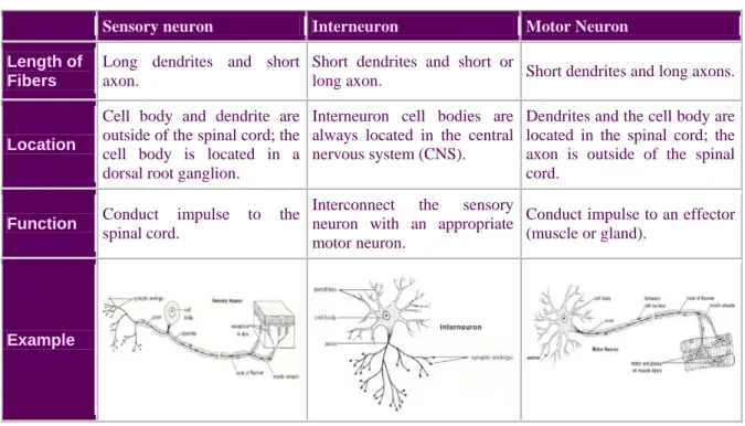
The nervous system is made up of more than 100 billion nerve cells. These cells are classified as either neurons or glial cells, each of which has several sizes and shapes.
Neurons are specialized secretory cells composed of a cell body (soma, perikaryon), dendrites and an axon (Zigmond et al., 1999). Neurons usually receive messages from other neurons through the dendrites that pick up messages and carry them to the neuron's cell body. The axon carries outgoing messages from the cell. Some axons are covered with a myelin sheath, made up of glial cells, the Schwann cells at the periphery and oligodendrocytes centrally. This asymmetric organization allows the neurons to send and receive electrochemical signals and release of signaling molecules. As described in Table 1, according to their function within the nervous system, neurons can be divided into: a) sensory (afferent) neurons - cells that carry messages from the sense organs to the brain or spinal cord, b) motor (efferent) neurons - cells that carry messages from the brain or spinal cord to the muscles and glands, and c) interneurons - association neurons carrying messages from one neuron to another.
Table 1 – The different types of neurons found in the nervous system.
Sensory neuron Interneuron Motor Neuron Length of
Fibers
Long dendrites and short axon.
Short dendrites and short or
long axon. Short dendrites and long axons.
Location
Cell body and dendrite are outside of the spinal cord; the cell body is located in a dorsal root ganglion.
Interneuron cell bodies are always located in the central nervous system (CNS).
Dendrites and the cell body are located in the spinal cord; the axon is outside of the spinal cord.
Function Conduct impulse to the
spinal cord.
Interconnect the sensory neuron with an appropriate motor neuron.
Conduct impulse to an effector (muscle or gland).
Example
The three types of neurons are arranged in circuits and networks, the simplest of which is the reflex arc. In a simple reflex arc, such as the knee jerk, a stimulus is detected by a receptor cell, which is located in the peripheral branch of a sensory neuron. The sensory neuron carries the impulse from site of the stimulus to the CNS, where it synapses with an interneuron. The interneuron synapses with a motor neuron, which carries the nerve impulse out to an effector, such as a muscle, which responds by contracting.
Glial cells far outnumber neurons, comprises the other major cellular constituent of the nervous system. During many years it was thought that functionally, these cells were only involved in support, protection and nutrition of neurons, and also to have a facilitatory role in conduction for the neurons they surround (Auld and Robitaille, 2003; Ndubaku and Bellard, 2008). Lately, it has been shown that these cells have other functions: they participate in synaptic transmission and modulation, as key regulators of neurotransmitter release, and also as instructors for the development, maintenance, and recovery of synapses (Fields and Stevens-Graham, 2002; Auld and Robitaille, 2003; Allen and Barres, 2009). Glial cells have also been implicated in other neuron-glial interactions that contribute to glial proliferation, differentiation, myelination, among others (Auld and Robitaille, 2003; for reviews, see Barres and Raff, 1999; Fields and Stevens-Graham, 2002).
There are three types of glial cells in the CNS – astrocytes, oligodendrocytes and microglia – and one type in the PNS – Schwann cells – that fill up the spaces between neurons with layers of myelin membrane around axons to insulate them for impulse conduction (Figure 1) (Kandel et al., 1991; Fields and Stevens-Graham, 2002).
Astrocytes are known by their star-like shape and by the extensive end-feet on their processes. They perform many functions, including biochemical support of endothelial
cells which form the blood-brain barrier, the provision of nutrients to the nervous tissue, maintenance of extracellular ion balance, and a principal role in the repair and scarring process of the brain and spinal cord following traumatic injuries. Microglia are nonneuronal cells found in the brain that respond to injury or disease by surrounding cellular “trash” and activating inflammatory responses. Recent studies have shown that microglia can respond to neural impulse activity, thus mediating neuroimmune interactions, i.e. in chronic pain conditions (Watkinis et al., 2001; Brodal, 2004). Oligodendrocytes can be distinguished from astrocytes by having less and thinner processes. They form myelin sheaths around axons in the CNS, by enveloping them with concentric layers of plasma membrane. In PNS, these functions are performed by Schwann cells, forming myelin around PNS axons, ensheathing synaptic junctions, and bundling small-diameter axons together (Fields and Stevens-Graham, 2002). Myelin forms an insulating sheath around an axon, leaving small areas of axonal membrane exposed between successive myelin segments called nodes of Ranvier (Figure 2) (Watkinis et al., 2001; Poliak and Peles, 2003).
Figure 1 – Glia-neuron interactions. Different types of glia interact with neurons and the surrounding blood vessels. Oligodendrocytes wrap myelin around axons to speed up neuronal transmission. Astrocytes extend processes that ensheath blood vessels and synapses. Microglia keeps the brain under surveillance for damage or infection (from Allen and Barres, 2009).
1.1.2 How Neurons Communicate
Communication between neurons is dependent on the properties of neuronal membranes. In a normal situation, substances will move from areas of high concentration to areas of low concentration (osmosis law), until they reach an equilibrium. However, in neuronal membranes, differences between intra and extracellular environments, prevents molecules to “walk” freely from one side to the other (Anthea et al., 1997). The diffusion of these molecules is due to their attachment to proteins that form ions channels through which some ions, such as sodium (Na+), chloride (Cl-), potassium (K+) and calcium (Ca2+), can diffuse. These transmembrane proteins or pumps transport ions bidirectionnaly. For example, the sodium-potassium pump uses transporter molecule that forces the Na+ to leave the cell and K+ to entry the cell. Due to these pumps, it is possible to say that neurons have at least two moments: a moment where the neuron is “resting”, where there is a greater concentration of K+ inside the cell than outside, and a greater concentration of Na+, Cl- and Ca2+ outside the cell than inside, and a second one, where any changing in the permeability of the membrane will cause an influx or an efflux of these Figure 2 - Structure of myelinated axons. Myelinating glial cells, oligodendrocytes in the central nervous system (CNS) or Schwann cells in the peripheral nervous system (PNS), form the myelin sheath by enwrapping their membrane several times around the axon. Myelin covers the axon at intervals (internodes), leaving bare gaps — the nodes of Ranvier. Oligodendrocytes can myelinate different axons and several internodes per axon, whereas Schwann cells myelinate a single internode in a single axon (adapted from Poliak and Peles, 2003).
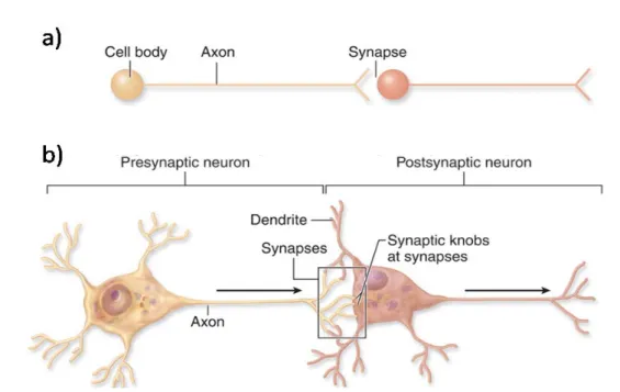
ions, until the system establish a balance between the inside and the outside of the cell. Due to the electrical charge of the ions, this concentration gradient will create an electrical potential (about -70 millivolt) between the two sides of the cell. The movement of ions across the cell membrane is controlled by both chemical and electrical gradients. To carry out any cognitive or motor task, whether memory formation or the execution of a movement, neurons will evaluate the inputs arriving in the form of ever-changing combinations of synaptic potentials, to determine if and when an action potential should be initiated. Neurons are polarized cells characterized by their membrane domains, including a single extensive axon and multiple dendritic processes which contain thousands of individual synapses (Figure 3a). Synapses are specialized junctional structures through which neurons communicate. According to the direction of synaptic transmission, neurons can be classified as “pre-” or “post-” synaptic neurons (Figure 3b). Synapses are composed of a pre-synaptic terminal, a synaptic cleft and a postsynaptic specialization (Kandel et al., 1991).
The process of synaptic transmission involves neurotransmitter releasing from the presynaptic nerve terminal, into the synaptic cleft that interact with postsynaptic membrane that gate ion channels (Figure 4). For example, the release of glutamate will open postsynaptic Na+ channels, and thus the influx of Na+ will decrease the electrical potential at the channels location. This local depolarization is referred to as an excitatory
postsynaptic potential (EPSP). On the other hand, neurotransmitters as GABA (gamma aminobutyric acid) exhibit inhibitory effects, as they interact with receptors to open Cl- and K+ channels. The influx of Cl- or exflux of K+ results in an increase in the resting potential at the channels location. This local hyperpolarization is referred to as an inhibitory postsynaptic potential (IPSP) (Katz, 1969). As illustrated in figure 4, the release of the neurotransmitter from the presynaptic terminal involves (1) depolarization of the terminal and (2) the presence of ions in the extracellular fluid.
1.1.3 Basic Mechanisms of Axonal Transport
The mammalian nervous system includes billions of neurons, which are interconnected in several neural circuits (Horowitz el al., 1999). Neuronal function and survival involves cytoskeletal elements and constant transport of proteins and organelles to and from the cell body. The proteins used include kinesin, which is specific for anterograde transport, and dynein for retrograde transport (Ström et al., 2008).
Neuronal cytoskeleton comprises microtubules, actin and intermediate filaments (Figure 5). Microtubules and actin filaments provide the neuron with structural support, but also give conduits for intracellular transport (Ström et al., 2008; Chevalier-Larsen and Holzbaur, 2006). Microtubules provide long-range pathways for fast-anterograde movement (from the cell body to the axon terminals) of kinesin motor proteins and the Figure 4 – Chemical transmission of a nerve impulse at the synapse (from 2002 Encyclopædia Britannica, Inc).
retrograde movement (toward cell body) of the dynein motor complex, whereas actin filaments are used by myosin motor proteins for short-range, dispersive distribution of vesicles, and\or organelles to the cell periphery. Roughly speaking, cargos are transported along microtubules and then transferred to the actin cytoskeleton for the final part of their journey (Akhmanova and Hoogenraad, 2005; Lansbergen and Akhmanova, 2006).
As discussed above, anterograde transport is mediated by kinesin-family proteins and is used in the translocation of membranous organelles (e.g., mitochondria) and vesicles as well as of macromolecules, such as actin, myosin, and clatrin, and some of the enzymes necessary for neurotransmitter
synthesis at the axon terminals. In turn, retrograde transport is mediated by cytoplasmic dyneins and include transport of protein building blocks of neurofilaments, subunits of microtubules, soluble enzymes and materials taken up by endocytosis (e.g., viruses and toxins) (Oztas, 2003).
The rate of transport is specific for each class of substances and also independent of electrical activity within an axon (Ochs, 1972). Fast axonal transport occurs in both anterograde and retrograde ways at a rate of 0.5–10 μm/sec and includes the transport of membrane-bound
organelles, mitochondria,
neurotransmitters, channel proteins, multivesicular bodies and endosomes (Shah et al., 2002). Slow axonal transport occurs only in the anterograde direction at a rate of 0.01– 0.001 μm/sec, and conveys cytoskeletal components, such as neurofilaments, tubulin, and actin, as well as proteins such as clathrin and cytosolic enzymes (Heidemann et al., 1981; Shah et al., 2002).
Figure 5 – Axonal transport on microtubules. The motors for anterograde and retrograde fast axonal transport are the kinesins and dynactin complex proteins, respectively; microtubules provide the tracks for these motors. Vesicles for transport are sorted and loaded onto transport motors both in the cell body and the distal nerve terminal. The former are transported not only into the axon but also into dendrites. Those in the distal nerve terminal permit uptake and axosomatic movement of substances such as trophic proteins. Mutations in dynactin (humans), dynein (mice) and three different forms of kinesin all provoke motor neuron degeneration (from Pasinelli and Brown, 2006).
1.3.4 A Brief Approach to Tract-tracing Neuronal Circuits
The discovery of the axonal transport triggered years of study into the structural basis behind this mechanism. Neuroanatomical tract-tracing methods are considered one of the best approaches to study connections between neurons located in different areas of the nervous system and to obtain data on the processing of information within a particular area (Merighi and Carmignoto, 2002). These anatomical connections can be determined by using axonal tracers that rely on intracytoplasmic movement along an axon, by retrograde transport towards the neuronal cell body and dendritic tree, or by anterograde transport towards a synapse (Köbbert et al., 2000; Lanciego and Wouterlood, 2000; Reiner et al., 2000). Thus, retrograde axonal transport allows identification of the cells of origin of afferent nerve fibers to a specific target zone, whereas anterograde axonal transport show us the projection targets of groups of cells to be charted within the CNS (Köbbert et al., 2000). There is a multitude of tracers available for the study of neuronal connections: a) horseradish peroxidase (HRP) or cholera toxin subunit B (CTb) (retrograde tracers) and low or high molecular weight dextran amines (anterograde tracers); (b) micelles and/or membrane vesicles containing lipophilic dyes; and (c) fluorescein-labeled microparticles (for review see for review see Lanciego et al., 1999; Vercelli et al., 1999; Köbbert et al., 2000).
Comparing to HRP that is passively taken up by neurons, CTb binds specifically to surface receptors of neurons (GM1-ganglioside receptor) and is actively taken up and transported by the axons, which may explain the high sensitivity of CTb as a tracer (Luppi et al., 1987; George-Chandy et al., 2001). However, CTb receptors are found to be roughly distributed either in neurons, or across cell types and also in species that may influence the labelling of the neurons according to the species on study or even in the different pathways in the same animal (Sabin, 1938). Since its discovery, in 1977, CTb has been strongly chosen for retrograde studies. As a tracer, CTb produces intense retrograde labelling from small injection sites (Dederen et al., 1994; Datiche et al., 1995; Angelucci et al., 1996; Cobos et al., 2003). CTb can also be transported anterogradely, and thus, with a single injection it is possible to study the efferent and afferent inputs of a certain area in the CNS (Chen and Aston-Jones, 1995).
Biotinylated dextran amines (BDAs) are one of the tracers mostly used in anterograde tract-tracing studies of the nervous system. BDAs are known for their
versatility and sensitivity, and depending on their molecular weight, they can be used for both anterograde and retrograde studies (Fritzsch, 1993; Kaneko et al., 1996; Medina et al., 1997a). High-molecular-weight biotinylated dextran amines (BDAs; 10 kDa) provides high-quality of labeling of axons and terminals, whereas low-molecular-weight BDAs (3 kDa) provides a detailed Golgi-like retrograde labeling of neurons. The stability of the molecular structure of BDA makes them ideals for long-term storage and examination, and their visualizations can be done by simple histochemical methods. Moreover, due to its flexibility with fixatives, BDA can be visualized at light or electronic microscopic level (for review see Reiner et al., 2000).
As CTB, BDA can be delivered into the nervous system iontophoretically or pressure-injected (Reiner et al., 2000). After injection into the CNS, BDA yields extensive and detailed anterograde labeling of axons and terminals (Veenman et al., 1992, 1995; Brandt and Apkarian, 1992; Rajakumar et al., 1993). These tracers can CTb toxin B fragment, fluorescents dextran amines or intracellular labeling (review see Reiner et al., 2000).
For anatomical tracing, cells have to be “alive”, i.e. they cannot be used in fixed tissues and for long-distance tracing in juveniles and adults, or require the presence of active transport mechanisms instead of simple passive diffusion (Lanciego and Wouterlood, 2000; Reiner et al., 2000). Using different techniques of visualization, neuronal tracing not only provide information on the morphology or afferent and efferent connectivity of neurons, but may also show the synaptic contact of neurons. However, there are some problems associated to the conventional tracing methods: 1) because the tracers must be delivered to neurons by microinjection or local application, it is difficult or impossible to selectively label small populations of neurons of a given phenotype, and 2) axons passing through the application region can be damage and become labeled, leading to a misinterpretation of results (for review see Lanciego et al., 1999; Vercelli et al., 1999; Köbbert et al., 2000). Recently, it was demonstrated that it is possible to use proteins as transneuronal tracers when its expression is genetically targeted to a subset of neurons, and thus avoid these problems (Horowitz et al., 1999).
1.2 – Pain Control
1.2.1 Pain DefinitionFor better or worse, we all feel pain (except in some pathologies). The International Association for the Study of Pain (IASP) define pain as a sensory and emotional experience associated with real or potential injuries, or described in terms of such injuries [International Association for the Study of Pain (IASP) - 1994 definition, reviewed in 2008]. Painful experience exists so the body can recognize that something is threatening it and leads to behavior that will remove the organism from the source of potential injury (Landrieu et al., 1990). Pain is a key process for our nervous system to learn from and react to the environment (King et al., 1997). Certain tissues have specialized sensory receptors, called nociceptors that are activated by noxious stimulus to peripheral nerves. Upon activation, they transmit the message, by action potentials and neurotransmitters release, to the spinal cord dorsal horn for processing and transmission to the brain (Costigan et al., 2009). It is known that a noxious stimulus can result in a real or potential injury, without causing pain. In same cases, noxious stimulus can lead to pain sensation, characterized as nociceptive pain. However, painful experience can be spontaneous, such as the nonnociceptive pain characterized by the reduction of the receptor thresholds as a result of alterations of the central nervous system (CNS) (Casey, 2000). According to this, nociception and pain have different means; nociception refers to the neurophysiologic manifestations produced by noxious stimulus, while pain involves the perception of an aversive stimulus, which requires the capacity of abstraction and the elaboration of sensory impulses (Millan, 1999).According to the IASP definition, the relation between pain and degree of injury is not obligatory. Alert function is applied only to an acute manifestation, i.e., the one that follows damage to the tissue. Acute pain is delimited in time and disappears with the settle of the pathological process. On the other hand, chronic pain is characterized as persistent, and is associated with chronic pathological processes (Almeida et al., 2004).
1.2.2 Peripheral Afferent Fibers
The most important fibers for pain perception are the axons of afferent nociceptors. The afferent nociceptors (Figure 6a) consist of thermal nociceptors that are activated at
temperatures above 45ºC (C-fibers) or lower than 5ºC (Aδ -fibers), high-threshold mechanical nociceptors that transmit information indicating injurious force on the skin (Aδ- and some Aβ-fibers) and polymodal nociceptors that are activated by thermal, mechanical and chemical stimuli (C-fibers). According to their diameter, structure and conduction velocity, C-fibers are characterized as thin (0.4-1.2 µm in diameter), unmyelinated and slowly-conducting (0.5-2.0 m sec-1) fibers; Aδ-fibers as medium (2-6 µm), myelinated and of intermediate velocity (12-30 m sec-1) fibers; and Aβ-fibers as large (>10 µm), myelinated and fast (30-100 m sec-1) fibers (Millan, 1999). Each one of these classes encodes sensory information, however they respond differentially to noxious and innocuous stimuli, in the sense that the three fibre types transmit non-nociceptive information, but only C and Aδ fibers transmit nociceptive information in the normal tissue (nociceptors; Giordano, 2005).
Following a noxious stimulus, primary nociceptive afferents respond with differentiated patterns of propagation (Figure 6b). The myelinated Aδ-fibers transmit
Figure 6 – Different nociceptors detect different types of pain. a) Peripheral nerves include small-diameter (A) and medium- to large-diameter (A,) myelinated afferent fibres, as well as small-diameter unmyelinated afferent fibres (C); b) The fact that conduction velocity is directly related to fibre diameter is highlighted in the compound action potential recording from a peripheral nerve. Most nociceptors are either A or C fibres, and their different conduction velocities (6–25 and 1.0 m s-1, respectively) account for the first (fast) and second (slow) pain responses to injury (from Julius and Basbaum, 2001).
impulses much faster than do the unmyelinated C-fibers. The Aδ-fibers transmit what is called the first pain, i.e., sharp and highly localizable. Impulses on C-fibers are responsible for what is called second pain. Second pain is slower in arriving, duller and endures after the stimulus end (Casey, 2000).
1.2.3 Nociceptors
To guard against tissue damage, it is important that the body is alerted of potentially damaging stimuli. This awareness is attained by a noxious stimulus-detecting sensory system (Costigan et al., 2009). Nociceptors are physiological receptors located all over the body - skin, internal organs, joints, muscles and tendons. When activated, either by noxious stimuli, tissue injury or acute inflammation, the propagation of nociception is initiated and afferent information is send to the dorsal horn of the spinal cord where synaptic transmission to ascending pathways is subject to modulation by descending pathways, local neuronal circuits and different kinds of neurochemicals (Figure 7) (Almeida et al., 2004). Some nociceptors are thinly myelinated (Aδ-fibers) but most are unmyelinated (C fibers), and these slowly conducting afferents represent the majority of sensory neurons in the PNS. Like all primary sensory neurons in the somatosensory system, nociceptors have their cell bodies located in the dorsal root ganglia (DRG) or trigeminal ganglia, give rise to a single axon that bifurcates into a peripheral branch that innervates peripheral target tissue, and a central axon that enters the CNS to synapse on nociceptive second order neurons. Morphologically, nociceptors are similar to other neurons; they have a peripheral terminal that transduce external stimuli and initiates action potentials, a axon that conducts action potentials, a cell body that controls the identity and integrity of the neuron and a central terminal that forms the presynaptic element of the first synapse in the sensory pathway in the CNS (Figure 7) (Woolf and Ma, 2007).
There are three major classes of nociceptors – thermal, mechanical, and polymodal – as well as a class classified as silent nociceptors. Aδ mechanical nociceptors, respond to noxious mechanical stimuli that damage or threaten to damage tissue. C-polymodal nociceptors, react to noxious mechanical, noxious thermal (>44°C) and noxious chemical stimuli. Silent (or sleeping) nociceptors, which do not respond to acute noxious stimulation of uninjured tissue, become active after tissue injury. The information from nociceptors is
cord where they synapse onto second-order spinal cord neurons, which transmit the information to supraspinal sites (e.g., the thalamus in the brain) (Willis and Westlund, 1997).
1.2.4 From Periphery to Thalamus
Pain can be understood as a complex entity that does much more than simply activate a “pain center”, resulting from complex and interactive series of mechanisms integrated at all levels of the neuroaxis, from the periphery, via the spinal dorsal horn to higher cerebral structures (Talbot et al., 1991; Casey et al., 1994; Derbyshire et al., 1997; Millan, 1999).
The dorsal horn of the spinal cord is the location of the first synapse in pain pathways, and as such, offers a powerful target for regulation of nociceptive transmission (Heinricher et al., 2008). Primary afferent fibers form synapses with dorsal horn sensory neurons, which send ascending projecting fibers and make synapses with neurons located at supraspinal sites, such as the thalamic nuclei (Zhuo, 2007). Nociceptive information ascends from the spinal cord to the thalamus in the contralateral spinothalamic tract (STT), to the medulla and brainstem via the spinoreticular and spinomesencephalic tracts, to the hypothalamus via the spinohypothalamic tract, to the supraspinal autonomic control centers via the spinohypothalamic tract and to the nuclei in the midbrain, ventroposterior lateral and posteriomedial nuclei of the thalamus in the cervicothalamic tract (Millan, 1999; Willis and Westlund, 1997; Craig, 2003; Pralong et al., 2004).
Figure 7 – The nociceptor. The operational components of the nociceptor include a peripheral terminal that innervates target tissue and transduces noxious stimuli, an axon that conducts action potentials from the periphery to the central nervous system, a cell body in the dorsal root ganglion, and a central terminal where information is transferred to second order neurons at central synapses (from Woolf, and Ma, 2007).
1.2.5 Brainstem Control of Spinal Nociceptive Processing
The descending pain modulatory system, also known as endogenous pain control system, is a well-characterized anatomical network that enables us to regulate nociceptive processing in differents situations to produce either facilitation (pro-nociception) or inhibition (antinociception) (Hagbarth and Kerr, 1954; Fields and Basbaum, 1999; Treed et al., 1999; Heinricher et al., 2009). This modulatory system may facilitate or inhibit nociceptive input by three major mechanisms: 1) the modification of synaptic strength in the spinal dorsal horn may increase or decrease transmission of nociceptive signals to the brain; 2) local dorsal horn interneurons provide both feed-forward and feed-back modulation to spinothalamic and spinobulbar projection neurons; and 3) descending systems initiating in the brainstem exert top-down modulation of nociceptive input at the spinal level (Seifert et al., 2009).
In 1906 it was demonstrated, for the first time, that brain can modulate in a ‘‘top-down’’ way spinal cord excitability via a tonically active influence, most of the time inhibitory in function. This idea came from Sherrington work that showed that nociceptive reflexes were improved after transaction of the spinal cord (Sherrington, 1906). Later on, data from 1969 came to emphasized the relevance of this experience by showing that focal electrical stimulation in the rat midbrain periaqueductal gray (PAG) produced analgesia strong enough to allow surgery without anesthetics or analgesics (Reynolds, 1969). Electrophysiological, anatomical, and pharmacological studies have shown that these descending influences on spinal nociceptive processing were modulated at the rostral ventromedial medulla (RVM), which includes the medial nucleus raphe magnus (Porreca et al, 2002; Gebhart, 2004). The RVM receives inputs from the PAG and, in turns, projects to the dorsal horn, primarily to the superficial layers, where it can influence spinal nociceptive transmission. RVM cells have two major types of neurons that may explain the role of the RVM in pain modulation: ON-cells, which facilitate nociception via descending axons projecting to the spinal cord (pro-nociception), and OFF-cells, which inhibit nociceptive information directly at the level of the spinal cord (antinociception) (Fields et al., 1983; Fields and Heinricher, 1985).
The antinociceptive nature of brainstem areas, such as the PAG (Bodnar, 2000), the RVM (Mason, 2001), the locus coeruleus (LC; Jones, 1991), the lateral portion of the caudal ventrolateral medulla (VLMlat; Tavares and Lima, 2002), the dorsal reticular nucleus (DRt; Bouhassira et al., 1992; Almeida et al., 1996; 1999), and the nucleus tractus solitarius (NTS; Randich et al., 1988) is well established in the literature. The inhibitory antinociceptive nature of the system was latter questioned by the observation of pronociceptive effects from areas classically considered as antinociceptive, as the RVM (Porreca et al., 2002), the NTS (Wiertelak et al., 1997) and the DRt (Almeida et al., 1996; Almeida et al., 1999; Dugast et al., 2003), which play an additional profound nociceptive facilitating effect upon acute, inflammatory and chronic pain (Sotgiu et al., 2008). The idea of a primary pronociceptive centre in the endogenous pain control system leaded to a new concept of pain modulation as a dynamic and flexible process, resulting from balance between excitatory and inhibitory actions as the way of adapting to the various unsteady pain determinants (Lima and Almeida, 2002). In conclusion, pain control results from the balance between inhibiting and facilitating outflows from the brainstem upon spinal nociceptive transmissions and a disruption of this balance may constitute the basis for
Figure 8 – The neural pathway of nociception from primary afferent neurons (PANs) to the superficial lamina in the dorsal horn of the spinal cord. Second-order neurons in the dorsal horn convey the noxious signal to the brainstem, midbrain, and thalamus. Finally, third-order neurons relay the electrical signal to the somatosensory/cingulate cortex and limbic system. Descending modulatory influences arrive in the spinal cord dorsal horn (dashed lines) and are derived from the midbrain periaqueductal gray (PAG), the locus ceruleus, and the rostral ventromedial medulla (RVM) (from White et al., 2007).
chronification of pain (Urban and Gebhart, 1999; Porreca et al., 2002; Vanegas and Schaible, 2004; Heinricher et al., 2009).
1.3 – The Brainstem Reticular Formation and Pain
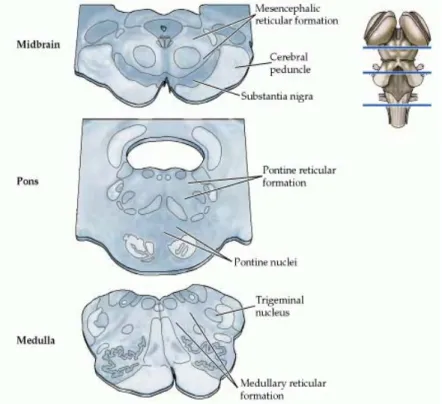
1.3.1 The Brainstem Reticular FormationThe reticular formation (RF) is the name given to the collection of small nuclei and fiber tracts that run through the core of the brainstem. The RF extends from the caudal medulla, where it is continuous with the spinal cord reticular formation, to the mesencephalon (Figure 9). The different RF areas receive afferents from most of the sensory systems and projects for almost all parts of the nervous system (Hendelman, 2000). Several studies demonstrated that some subgroups of reticular neurons that receive inputs from peripheral receptors, including skin, muscle, bone and joint receptors, integrate them and link to the vestibular and cerebellar circuits that will determine posture and movement. This information is also integrated by higher brain structures in the visual, somatosensory, and motor systems to develop the complex motor patterns of adaptive behaviour (Squire, 2003).
The heterogenic morphology of the RF may explain the multifaceted role in the CNS; it exerts important functions on the sleep/wake cycle, regulation of visceral activity, control of movement, behaviour, alertness and modulation of pain (Rhoades and Bell, 2008). According to the function performed, it is possible to delimit different subgroups within the RF:
• Cardiac and respiratory “centers”: subsets of neurons within the medullary reticular formation responsible to control the vital functions of heart rate and respiration;
• Motor areas: the motricity is controlled by both pontine and medullary nuclei of the RF via the cortico-reticulo-spinal system;
• Ascending projecting system: fibers from the RF ascend to the thalamus and project to differents nonspecific thalamic nuclei. Here, fibers ascend diffusely to the cerebral cortex. This system is related with consciousness and has been termed reticular activating system (ARAS);
• Pre-cerebellar nuclei: several nuclei in the brainstem placed within the boundaries of the RF that project to the cerebellum.
A different way to describe this area is according to the spatial positioning of the neurons. Topographically, neurons can be arranged in three longitudinal sets, each of them with different cytoarchitecture, connections and physiological functions. The lateral group is formed by small neurons that receives inputs to the RF, including those from the
anterolateral (pain and temperature) and trigeminal systems, and the auditory and visual input (Kiernan J., 2008). The central group consists of big neurons, and gives rise to long ascending and descending fibers pathways, some of them projecting both rostrally into the thalamus and caudally to the sacral levels of the spinal cord, thus influencing the axial and proximal limb muscles. Within this group are the nucleus gigantocellularis of the medulla and the pontine reticular nuclei, positioned at caudal (lower) and oral (upper) parts, forming the two reticulo-spinal tracts. Several staining studies have shown that the medial RF contains medium-to-giant-bodied neurons characterized by far-reaching bifurcating axons running rostro-caudally, with long branches contacting either the forebrain or spinal Figure 9 – The brainstem reticular formation. The location of the reticular formation in relation to some other major landmarks at different levels of the brainstem. Neurons in the reticular formation are scattered among the axon bundles that course through the medial portion of the midbrain, pons, and medulla (from Purves et al., 2007).
cord (Scheibel and Scheibel, 1967; Newman, 1965). These cells are referred as projection neurons based on the idea that all of them project outside the medial RF. Electrophysiological studies have demonstrated that synaptic connections between these neurons, formed by terminals of the axons collaterals, are enough to induce excitatory post-synaptic potentials (Ito and McCarley, 1987), suggesting a anatomically and functionally connection between these projection neurons (Humphries et al., 2006). Some evidences suggests a second cell-type in the medial RF, referred as interneurons, however not conclusive (Ito and McCarley, 1987). Finally, the midline region is occupied by a group of neurons, known as raphe nuclei. The nucleus raphe magnus is one of the nucleus of this group, which plays an important role in the descending pain modulatory system (Hendelman, 2000).
1.3.2 Neurotransmitters in the Reticular Formation
Chemical neurotransmitters have been identified and localized in groups of cells within the RF, acting in cortical activation and behavioural arousal. Norepinephrine (NE) is contained in neurons of the pons and medulla (A1-A7 cells groups). The A5 and A7 cells groups (in rostral part) projects caudally to the brainstem and spinal cord, whereas the A1-A3 cell groups (in caudal part) projects rostrally to the brainstem, hypothalamus and basal forebrain. Together, they form the lateral tegmental area, apparently involved in hypothalamic regulation and motor control (Moore and Card, 1984). Serotonin or 5-HT is contained in neurons located in the midbrain nuclei, dorsal raphe nuclei, and the median raphe nuclei (B8 and B9). These neurons project rostrally, innervating nearly the entire forebrain, thus suggesting a role in regulation of behaviour state. The acetylcholine (ACh) is known as the neurotransmitter of motor neurons, and apparently is associated to nonmotor brain areas. Cholinergic neurons may be found either in the pontine nuclei (the laterodorsal tegmental nucleus and the pedunculopontine nucleus), projecting to the brainstem RF, hypothalamus, thalamus and basal forebrain, or in the medial septum (nucleus of the diagonal band and the substantia innominata-nucleus basalis complex), which projects to the limbic forebrain, including the hippocampus, and to the neocortex (Squire, 2003). In summary, these neurotransmitters are produced by modulatory neurons, whereas neurons involved in sensorimotor integration produce either the excitatory
1.3.3 The Ascending Reticular Activating Systems Mediates Consciousness and Arousal
The RF receives sensory information from many systems of the body and it is broadly interconnected with the cerebellum and the limbic system (Gilman et al., 2003). The limbic system consists of a group of deep brain structures, including the hippocampus, amygdala, gyrus fornicatus and connecting structures, which support a variety of functions such as emotion, behaviour, long term memory, and olfaction. Nearly all the neural fibers carrying information into and out of the RF have a crucial role in initiation and control of sleep-wake cycle (Siegel, 2002).
Individual reticular neurons communicate with higher brain areas such as hypothalamus, thalamus, cerebellum and spinal cord, making reticular neurons ideal for leading the arousal of the brain as a whole. For example, some reticular neurons, if not inhibited by other brain areas, are sending continuously impulses (via thalamic relays) to the cerebral cortex, keeping it alert and conscious and improving its excitability. This function of the RF is called the reticular activating system (RAS) also known as “activating system” and its activity is crucial for maintaining the state of consciousness and sleepiness (Marieb and Hoehn, 2007). The interest in the sleep-wakefulness cycle begins in 1949, when Giuseppe Moruzzi and Horace Magoun discovered that by stimulating the reticular formation, they could awaken animals from normal sleep (Butkov and Lee-Chiong, 2007). On the same year, Donald Lindsley authored a paper showing that the loss of arousal was related to the rostral section of the RF in the mesencephalon (Siegel, 2002). However, in 1950, a new study on cats, has shown that these animals were capable to be awakened from coma with auditory and tactile stimuli, despite the destruction of large neuronal fiber tracts that where projecting from the RF to thalamus and then to the cortex (Siegel, 2002).
The RAS consist of specific thalamic nuclei, parts of the hypothalamus, the ventral tegmental area, the parabrachial nuclei, the periaqueductal gray, the nucleus locus coeruleus, the raphe nuclei, and the RF itself, most of them involved in arousal and cortical tone modulation (Solms and Turnbull, 2002). Impulses coming from ascending sensory tracts synapse with RAS neurons, maintaining them active and improving their arousing effect on the cerebrum. These pathways involve three to four synapses: a peripheral receptor responds to a specific sensory stimulus, as touch, hearting or vision, and transmits the information to cells of the RF that projects to the intralaminar nuclei of thalamus,
which innervate large areas of the cerebral cortex and limbic system (Rhoades and Bell, 2008). This system is inhibited by sleep centers located in the hypothalamus and other neural regions, and is depressed by alcohol and drugs, including tranquilizers. RAS stimulation makes the cortex more alert and aware. However, any injury to the brain, mainly in the brainstem, like head injuries, oxygen deprivation of the brain, drugs, and electrolyte changes can influence these centers, resulting in permanent unconsciousness or even coma (Marieb and Hoehn, 2007).
RAS can be divided into two components: the first one, ventral, lower and arousal circuit, running parallel to the reticular-thalamic-cortical circuit, and the second one, more rostral, is in the basal forebrain, making part of the ventral arousal circuit (for more detail see book Marieb and Hoehn, 2007). Besides RAS, there is also a descending element of arousal, which function as a physiological activator of the body, and supporting increase in activity that normally occurs with waking (Siegel, 2002). These two systems functioning together form a positive loop that, if uncontrolled, can result in a severe arousal state (Butkov and Lee-Chiong, 2007). In summary, the reticular activating system is responsible to transmit sensory input. Nerve impulses, as light and noise, project from the cerebral cortex and course via thalamus, stimulating the reticular activating system, resulting in arousal and awaken. The sleep-wakefulness cycle results from a feedback system of communication between the reticular activating system and the cerebral cortex (Butkov and Lee-Chiong, 2007).
Additionally, some nuclei of the RF are motor nuclei, which project to motor neurons in the spinal cord through the reticulospinal tracts. Some nuclei assist in the control of skeletal muscles during coarse limb movement, and other, as vasomotor, cardiac, and respiratory centers of the medulla, function as autonomic centers regulating visceral motor functions (Marieb and Hoehn, 2007).
1.3.4 The Reticular Formation and Nociception
It is know that nuclei from the reticular formation play an important role in the processing of nociceptive information (Bowsher, 1976). This idea is based on anatomic studies performed in various species (Villanueva et al., 1988), including man (Bowsher, 1957; Bowsher, 1962), where most of spinal afferents that travel through the anterolateral
quadrant terminate within the brainstem reticular formation. In addition, several areas throughout the brainstem reticular formation contain neurons responsive to noxious stimuli (Villanueva et al., 1988). For example, neurons recorded within the nucleus gigantocellularis are activated by noxious mechanical, electrical, or chemical stimulation of their peripheral receptive fields (Casey, 1969; Goldman et al., 1972; Guilbaud et al., 1973; Gokin et al., 1977; Leblanc and Gatipon, 1974; Pearl and Anderson 1978). On the other hand, focal electrical stimulation of this area has been shown to elicit escape behaviour (Casey, 1971). Other authors have also reported the existence of neurons responding exclusively to noxious mechanical, thermal, or electrical stimulation in more caudal areas, such as the caudal bulbar reticular formation (Benton, 1968; Benjamin, 1970; Rose, 1975; Mayer and Hill, 1978; Blair, 1985); others have shown that neurons in this region can also respond to high threshold visceral stimulation (Gokin et al., 1977) and noxious cardiac stimulation (Blair, 1985).
RF structures have a well-characterized role in descending modulation of pain. For instance, the RVM is involved in the development and maintenance of central sensitisation and secondary hyperalgesia in animals (Urban and Gebhart, 1999). The PAG and the nucleus cuneiformis (NCF) are the main sources of input to the RVM (Basbaum and Fields, 1984; Behbehani and Zemlan, 1986), and are in an ideal position to modulate its output, i.e. modulate spinal nociception. These nuclei have a physiological substrate for bidirectional modulation of pain processing, due to different functionally classes of cells, which either facilitate (ON-cells) or inhibit (OFF-cells) nociception (Fields et al., 1983; Haws et al., 1989; Heinricher et al., 1987). Later on, other nuclei were shown to have anatomical inter-bulbar and bulbo-spinal connections (Lee et al., 1991; Mtui et al., 1993, Esteves et al., 1993, Tavares and Lima, 1994, reviewed by Tavares and Lima, 2002, Lima and Almeida, 2002; Almeida et al, 2006). Areas like the DRt are involved in modulatory actions that are based on a reciprocal circuitry with the spinal cord that allows a rapid adaptation to any change occurring in the system (reviewed by Tavares and Lima, 2002, Lima and Almeida, 2002, Almeida et al., 2006).
1.3.5 The ventral reticular nucleus
involvement in pain modulation is well established. The VRt is the caudal continuation of the nucleus reticularis gigantocellularis and continues caudally through the deep lamina of the spinal dorsal horn. It is located ventrally to the trigeminal subnucleus caudalis and the DRt. The potential involvement of VRt in nociceptive processing was initially proposed based on the fact that VRt neurons could be activated bilaterally by noxious stimulation of the face (Yokota et al., 1991). VRt neurons project mainly to the ventral horn (laminae VII-X), via the ventral funiculi and to laminae IV-V (Tavares and Lima, 1994; Villanueva et al., 1995) and receives projections from laminae V-VII (Raboisson et al., 1996).
VRt is also connected with other reticular areas, including the VLM (Cobos et al., 2003) and the DRT (Almeida et al., 2002). The participation of VRt in pain modulation has been demonstrated by electrophysiological studies. The first one reported two different groups of VRt neurons: 1) neurons exhibiting spontaneous activity which was unaffected by innocuous mechanical stimulation and either unaffected or inhibited by noxious peripheral mechanical stimulation and 2) neurons displaying regular, rhythmic activity which was synchronous with the rate of ventilation (Villanueva et al., 1988). A second study shows that electrical stimulation of the VRt induced analgesia and attenuation of the responses of spinal neurons (Aicher and Randich, 1990). A third study reported that animals with lesions in VRt seem to feel more pain in behavioral tests, which is the opposite of results from lesioning the DRt, thus excluding VRt involvement in the facilitation of nociception and suggesting its involvement in antinociception (Almeida et al., 1999).
This thesis aims to analyze the overall pattern of brain connections of the VRt, using local iontophoretic injections of the anterograde tracer biotinylated-dextran amine (BDA) or the retrograde tracer cholera toxin subunit B (CTb).
Chapter 2
2.1 – Ethical guidelines
Surgical procedures were performed under pentobarbital anesthesia (50 mg/kg, i.p.) on Wistar male rats (Charles River Laboratories, Barcelona, Spain) weighing 280–320 g. Animals were placed in a stereotaxic device (Stoelting, Wood Dale, IL, USA) and a craniotomy was performed. Coordinates for brain injections followed the stereotaxic parameters of (Paxinos and Watson, 1998). The experiments were in accordance with the regulation of local authorities for handling laboratory animals and the European Community Council Directive 86/609/EEC. The number of animals used and their suffering were minimized.
2.2 – Anterograde tracing experiments
Twenty one rats received iontophoretic injections (positive direct current of 3.0 µA; 5 s on/5 s off, lasting for 10 min) of 10% BDA (10,000 MW; Vector Laboratories, Burlingame, USA) in the left VRt through glass micropipettes with 15–20m diameter tips. After completion of the injection period the micropipettes were left in situ for 10–15 min before being slowly retracted to avoid tracer reflux along the pipette tract. Two to three weeks later, animals were reanesthetized with eutasil (1 mL/kg body weight) and perfused through the ascending aorta, first with 100 mL of saline phosphate buffer (PBS) 0,1 M, pH 7.2 and then with 1000 mL of 4% paraformaldehyde in PBS. The entire brain was removed, immersed in the same fixative for 4 h (RT) and then in 8% sucrose in PBS at 4 °C for 1–2 days. Coronal sections of the entire brain were serially cut on a vibratome at 50 µm and incubated with 3.3% H2O2 in order to inhibit endogenous peroxidase. Two in every three successive brain sections were immunoreacted with avidin–biotin complex (ABC, 1:200; Vector Laboratories) for 1 h and then BDA was revealed with 0.0125% diaminobenzidine tetrahydrochloride (DAB; Sigma Immunochemicals, St. Louis, USA) and 0.02% H2O2 in Tris–HCl buffer 0.05 M, pH 7.6. Half of these sections were counterstained using the formol-thionin technique (Donovick, 1974) and the remaining was left without any counterstaining. Sections with and without counterstaining were then serially placed in SuperFrost Plus slides (Menzel-Gläser, Braunschweig, Germany), dehydrated and mounted in Entellan (Merck, Darmstadt, Germany).
2.3 – Retrograde Tracing Experiments
Twenty Wistar male rats were iontophoretically injected with 1% CTb (List Biological Products, Campbell, CA, USA) using the same procedures described above for BDA. One week after the injection they were reanesthetized and perfused as above. After the inhibition of endogenous peroxidase, serial brain sections were left overnight at 4 ºC in a goat antibody against CTb (List Biological Products) at 1:40,000 in 0.1 M PBS containing 0.3% Triton X-100 (PBST). After several washes in PBST sections were incubated for 1 h in PBST containing a biotinylated anti-goat antibody raised in horse (1:200; Vector Laboratories). Sections were washed again in PBST and then incubated in PBST containing ABC (1:200). This and subsequent steps were similar to those described above for BDA experiments.
2.4 – Image analysis and illustrations
All the photographic material presented in this study was obtained using a digital camera (AxioCam HRc) connected to a microscope (Axioskop 2 Plus), both from Carl Zeiss (Göttingen, Germany). Images were captured in a computer using AxioVision 3.1.2.1 software and the brightness/contrast of each image was improved using Adobe Photoshop 7.0.1. software. For illustrative proposes, the brain areas receiving efferent projections from the VRt were drawn using a sequence of selected formol–Thionin-stained coronal sections of one illustrative animal injected with BDA. A motorized microscope (Axioplan2, Carl Zeiss) connected to a digital camera (Sony 3CCd DSP, Japan) was used to capture the image of the selected brain sections. For each section, the limits of the brain nuclei and the labeled fibers were drawn under a 1.25x or 40x objective lens, respectively, using Stereo Investigator 4.34 software (MicroBrightField, Inc, Willinston, VT, USA).
The nomenclature/abbreviations used to designate brain nuclei and fiber tracts are in accordance with those used by Paxinos and Watson (1998, 2005) or result from a simplification of it.
Chapter 3
3.1 – Injection sites
Of the 21 animals that received iontophoretic BDA injections, 5 had injection sites that were located in the VRt. All of the BDA injection sites presented a central core of labelled neuronal somata and fibers surrounded by a peripheral region containing scattered neuronal perikarya with dendrites that extended into the core, resulting from retrograde transport. Only those injections whose central dark core and surrounding halo were located inside the VRt were considered valid for the present study. Two representative injection sites are illustrated in figure 1: animal UMHugo23 (Figure 10A), located medially to Sp5C, ventrally to IRt and dorsal medially to LRt, and UMHugo24 (Figure 10B), more centred within the nucleus. The injection centers are small and as shown in Figures 1 and 3, they are found to be located within the boundaries of the VRt as defined by Paxinos.
As to the CTb injections, from the 20 animals that were injected only 2 had injection sites located in the VRt. CTb injection sites appeared as a compact dark zone, bounded by a halo in which dark areas intermingled with lighter zones. Nearby the peripheral halo were observed a small number of retrogradely labelled cells, probably due to uptake from the more central areas. According to previous studies (Ericson and Blomqvist 1988; Lima et al., 1991), the injection sites will be delimited only by the central core and the peripheral halo. Selection of CTb injection (Figure 11) followed the same principle.
B
A
A
Figure 10 - Photomicrographs of representative iontophoretic BDA injections in the VRt. Scale bar = 200 μM
3.2 – Anterograde Tracing Experiments
BDA administration to the VRt. Following BDA injections restricted to the VRt, anterogradely labelled fibers and terminal boutons appeared along the medulla oblongata, pons, mesencephalon, and in some restricted areas of the diencephalon (Figure 12). BDA labelled fibers were found mainly ipsilaterally to the injection, with a contralateral predominance to diencephalic areas (Figure 13). A detailed analysis of the brain areas receiving projections from the VRt will be further presented.
Figure 12 - Camera lucida-like drawings of a representative BDA injection along three successive rostro-caudal (A-C) levels of the VRt. Red areas represent the core of the injection. Figure 11 - Photomicrographs of a representative iontophoretic CTb injections in the VRt. Scale bar = 500 µM.
3.2 – Retrograde Tracing Experiments
CTb-labelled neurons projecting to the VRt were found to be distributed throughout all the rostrocaudal extension of the VRt, mainly in the ipsilateral hemisection. A first analysis to the brain sections shows that at most caudal part, the higher number of neurons was
Figure 13 - Camera-lucida-like drawings (A–D) (and two photomicrographs) of four coronal brain sections presenting significant amount of BDA labelled fibers originated from the VRt. Note that in the black/gray area (scheme D) fibers have not been represented individually due to their high density resulting from the proximity to the injection site. RF, reticular formation; PB, parabrachial nucleus; RVM, rostroventral medulla; PAG, periaqueductal gray; PF, parafascicular nucleus.
presented at areas of the brainstem, as PAG and DRt, which are known to be implicated in pain modulation. As to higher areas, such as PVN (Figures 14E, F) and MGP (Figures 14G, H) CTB-labelled neurons were mainly found ipsilateral to the injection site, despite a high labelling in the contralateral part. In subcortical telencephalic areas, a small number of labelled neurons was located in the ipsilateral bed nucleus of the stria terminalis (BST; Figure 14C), contrary to its contralateral part, where rare or none neurons appeared (Figure 14D). In the amygdala, a very strong projection was shown to occur, mainly along the entire rostrocaudal extent of the central amygdaloid nucleus (CeC), ipsilaterally (Figure 14I). Contralaterally, none CTb-labelled neurons were found (Figure 14J). In the motor cortex, a higher number of neurons were present in the DLO and in secondary (M2) motor cortice, mainly on the ipsilateral hemisphere (Figure 14A, B).
A
B
G
H
I
J
O
P
Figure 14 - Photomicrographs depicting retrogradely labeled cells in areas along the medulla oblongata, diencephalon and telencephalon, following CTb injections in the VRt. Panels at the left side are ipsilateral to the injection site, and contralateral at the right side. Scale bars = 200μM. All the panels are at the same magnification.