EVALUATION OF HEADACHE INTENSITY IN
MIGRAINOUS PATIENTS WITH VISUAL HANDICAP
THROUGH THE TACTILE ANALOGICAL SCALE (TAS)
Elcio Juliato Piovesan, Marcos Christiano Lange, Pedro André Kowacs,
Carlos Pacheco, Lineu Cesar Werneck
ABSTRACT - The tactile analogue scale (TAS) was elaborated to be used in blind subjects or those who can not use the vision during their crises. The objective of this study was to characterize, from TAS, the architecture of migraine attacks in subjects with visual disability. For that, 11 migrainous with visual disturb (MVD) subjects were studied and 22 migrainous subjects with no visual disability as a control group. All patients fulfilled the criteria for migraine and the patients of the group studied showed visual acuteness less than 20/200. To evaluate the results, the patients of the group MVD were subdivide within two groups, according to their visual acuteness: subgroup A subjects with subnormal vision and subgroup B amaurotic ones. In subgroup A measurement 46 attacks with average of the migraine attacks of the 56.50 mm, in the subgroup B 45 attacks with average of the 59.58mm and in the control group 92 attacks with average of the 49.88mm. When subgroup B and control group were compared there was a significant statistic difference (p=0.022). Through these outcomes we can observe that the migrainous subjects with no visual afference show a higher pain intensity during the migraine crises comparing to those subjects with no visual handicap. The study suggests that, as in other forms of sensibility, the total visual loss can also interfere in the nociceptive control of the pain during the migraine attacks.
KEY WORDS: analogue scale, intensity, migraine, pain, visual acuteness.
Comportamento das crises de migrânea em pacientes com deficiência visual, utilizando a escala analógica táctil para a dor
RESUMO - A escala analógica táctil para dor (TAS) foi elaborada para ser utilizada em cegos ou em indivíduos que não podem utilizar a visão durante suas crises. O objetivo deste estudo foi caracterizar, a partir da TAS, a arquitetura das crises de migrânea em indivíduos portadores de deficiência visual. Foram estudados 11 pacientes com deficiência visual e migranosos (DVM) e, como controle, 22 pacientes migranosos sem deficiência visual. Todos os pacientes preenchiam os critérios para migrânea e os pacientes do grupo DVM apresentavam acuidade visual menor que 20/200. Para avaliação dos resultados, os pacientes do grupo DVM foram separados em dois subgrupos: subgrupo A, pacientes com visão subnormal e o subgrupo B, pacientes amauróticos. No subgrupo A avaliamos 46 ataques com intensidade média das crises de cefaléia de 56,50 mm; no subgrupo B foram avaliados 45 ataques com média de 59,58 mm; no grupo controle 93 ataques com média de 49,88 mm segundo a TAS. Quando comparados o subgrupo B e o grupo controle, ocorreu diferença estatísticamente significativa (p=0,022). Através destes resultados, podemos observar que os pacientes migranosos sem aferências visuais apresentam maior intensidade de dor durante as crises de migrânea em relação a pacientes sem deficiência visual. O estudo sugere que, como em outras formas de sensibilidade, a perda visual total pode interferir nos controles nociceptivos da dor.
PALAVRAS-CHAVE: dor, escalas analógicas, intensidade, migrânea, perda visual.
Unidade de Cefaléia, Especialidade de Neurologia do Departamento de Clínica Médica do Hospital de Clínicas da Universidade Federal do Paraná (UFPR), Curitiba PR, Brasil.
Received 12 February 2001, received in final form 6 June 2001. Accepted 12 June 2001.
Dr. Elcio Juliato Piovesan - Serviço de Neurologia, Hospital de Clínicas da UFPR - Rua General Carneiro 181 / 12 andar - 80060-900 Curitiba PR - Brasil. FAX 55 41 264 3606. E-mail: [email protected]
The evaluation of pain in subjects with headache, during scientific studies or clinical practice, can be carried out by different methods. The analogue scales constitute one of the principal ones, when the stud-ies require sharp measurements of the pain and its
influence on the functional capacity1. Piovesan et al.
have structured and tested in subjects with normal vision a new scale named Tactile Analogue Scale for pain (TAS)2. Although it is addressed for patients with
Eleven visual handicapped patients (median age 27.9 years old; 10 female and 1 male) with visual handicap that filled the International Headache Societys diagnostic cri-teria for migraine were evaluated3. The patients were di-vided in two groups according to their deficiency degree4: the subgroup A (five subnormal vision) and subgroup B (six amaurotic patients). The patients of the subgroup B were dexterous and have used in average the Brailles read-ing for four hours daily in the last two years. The visual deficiency origin was neoplasic in three patients; cataract, pigment retinose in one; association of congenial ract and rubella in one; association of glaucoma and cata-ract in one; and association of catacata-ract and retina mispla-cing in one patient. Three patients ignored the cause of their visual handicap. No one had ophtalmologics disturbs that could provocate pain or influence intensity of the mi-graine crises. The subgroup A showed an average of 25.8 + 11.7 years old, the visual disability stopped increasing at 14.4 years and have remained stable for the last 11.4 + 10.4 years. Subgroup B showed an average age 29.6 + 4.9 years old, amaurosis started at 9.8 + 11.3 years old (two of the patients were born amaurotic) and the aver-age time duration of the amaurotic was 19.8 + 10.2 years.
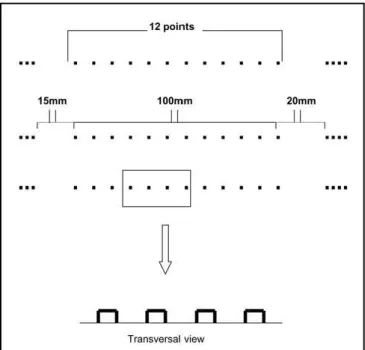
TAS was formed by 5 pages in Braille, containing the first one instruction to fulfil the scale. The other four pages showed 31 divisions composed by lines in high relief measu-ring 100 mm. These lines were constituted by points in high relief, in a total 12 points intercalated by normal spaces (low relief) (Fig 1). Before the line there was, in Braille, the expression No Pain followed by 15 mm space and at the end a 20 mm space with the expression Worst Pain in Braille. To fulfil, the patient received all orientation in the first interview. During the migraine crises the patients were orientated to put their distal phalanx of the left little finger on the first point on the scale (no pain reference point) and fore-finger on the last point (worst pain reference point) and with distal phalanx of the forefinger of the right hand the patients marked with the nail on the line, the imaginable place where the pain was. After that, the patients marked the TAS they higlighted with the last point on the scale. Such procedure was repeated during all migraine attacks. The days that there were not crises they did not fulfil the form, but they highlighted with the nail the first point on the scale. The absence of a point, in the beginning or at the end, orientates the patient to fill the next line and so there was no risk of the same line be fulfilled twice.
Statistical analysis
The statistic analysis was applied through Mann-Whit-ney test for the visual handicaped group and the control group. It was evaluated the confidence rate (95%) for sub-groups A and B.
The study was approved by the Ethic Committee in Human Beings of the Hospital de Clínicas da Universidade Federal do Paraná.
RESULTS
The individual analysis of the attacks in each pa-tients of the subgroup A was characterised by at-tacks of medium intensity 58.17 + 26.24 mm (Gra-phic 1). Observing collectively subgroup A, 46 at-tacks were evaluated with an average intensity of 56.49 mm + 31.19 mm with an average duration of 1.3 + 0.67 days (Graphic 2). Subgroup B showed an average intensity of 55.79 + 18.32 mm (Graphic 1) their attacks. Observing collectively the attacks in this group, there were 45 migraine attacks with average
intensity of 59.58 + 24.11 mm. The average dura-tion was 1.77 + 1.38 days. In the control group, the migraine attacks average intensity individually was 47.88 + 17.06 mm. The global analysis of all pa-tient was consituted by 93 attacks with average in-tensity 49.88 + 27.36 mm. The attacks average du-ration was 1.6 + 0,8 days.
Analyzing the data with a confidence rate of 95% for the subgroup A headache average intensity, we got the minimum value of 25.6 mm and the maxi-mum value 90.8 mm, and all patients showed at-tacks average intensity in this range. When we analysed the same characteristics in subgroup B, we can realize a minimum value 36.6 mm and a maxi-mum value 75 mm. In this group, a patient showed his average attacks below the confidence rate (28.5 mm). However, this value could occurred by the little sample that was used for the study, not invalidating the statistics analyses.
Graphic 2. Comparison between average of the duration and mediana duration (days) of the migraine attacks in the subgroup A and B and control. Average.
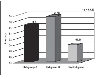
Graphic 3. Measurement of the intensity of the migraine attacks between subgroup A, B and control.
Graphic 1a. Measurement of the average intensity of the migraine attacks in the patients of the subroup A (n=-5).
When comparing the migraine crises average in-tensity between subgroups A and B, there was not statistic difference (p=0.787) for crises average in-tensity (56.5 mm versus 59.58 mm). When sub-groups A and control group were compared, there was not a significant statistic difference (p=0.238). However, analysing subgroups B and the control group we got a significant statistic value (p=0.022) (Graphic 3).
DISCUSSION
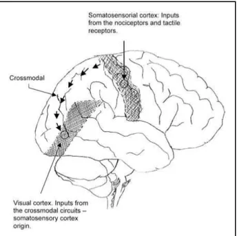
The outer layer of the cerebral cortex is divided into several areas specialised in detecting and pro-cessing sensory signals from the eyes and ears and from receptors for touch and nociceptors. Evereday experience illustrates that, despite their differences, the sensory regions of the cortex must be co-oper-ating with each other by integrco-oper-ating the sensory sti-muli they receive from the outside world5. The
inte-gration among different sensory forms (visual, tac-tile and nociceptive) is processed through multimo-dal neurones and through cortical association cross modal.
The multimodal integration in the neurone cells is a physiologic characteristic that occurs in some cortical structures. Areas containing bimodal neuro-nes are located in the ventral premotor area, puta-men, poscentral gyro, parietal area 7b and ventral intraparietal6. These neurons can respond to tactile
and visual stimuli simultaneously. This way the vi-sual stimulus reaches the vivi-sual receptive field and extend secondarily to the tactile receptive field at the same time and vice-versa7,8. The bimodal cells
show visual and tactile receptive fields distributed predominantly on the face, arm and parts of the su-perior trunk. With the integration between tactile and visual perception the human brain structures visual-spatial maps for the hand, arm and face9-11.
The visual spatial perceptive integration for the face involves the optic nerve and trigeminal nerve. The total visual loss produces an adaptation in this via, promoting exclusivity in the tactile afferences for the
tex anatomy remained unchangeable13. So cerebral
function, when it is analysed through photon sion computed tomography (SPECT), positron emis-sion tomography (PET) and functional resonance nu-clear magnetic (fRNM), demonstrated a recruiting of the occipital cortex for other neurones commands, including other sensitive forms as tactile and audi-tory14-18. The metabolic activity of the visual cortex
in blind people does not show any difference in the cerebral blood flow, glucose metabolism and oxy-gen consumption in relation to the control group with no visual deficiency, demonstrating electrical activity in the area19-21.
This cortical plasticity does not occur just in the occipital cortex but also in other areas analysed (mo-tor, sensory, tactile), being dependent of frequency and using time of the analysed area22-24. This
plastic-ity cross modal probably helps to improve the tac-tile perception abilities in blind people16,25,26. The
characteristic of the plasticity is represented by an alteration in the cortical topography and an increase of the cortical representation in the analysed area, due to an increase in the peripheral afference asso-ciated to an adaptive phenomenon of the cortical representation of the organisation24,27.
As a visual experience is important for a preco-cious organization of the visual cortex, a possible re-organization will be associated directly with the visual loss time28,29. It is known that the
re-organiza-tion of the occipital cortex is interrupted at about 14 years old30. The age group coincides with the
pu-berty and is associated with a reduction of supernu-merary synapses inside the visual cortex31,32. Another
associative via that occurs between the somatosen-sory cortex (touch) and visual cortex is through cros-smodal mechanism. This crossmodal effect arises even if the tactile cues are tasked irrelevant and do not predict the location of the visual targets, sug-gesting an exogenous (stimulus driven) attentional mechanism32. Recent studies that analysed the
cor-tex via back projection through association areas in the parietal lobe33. Such back-projections according
to recent theorical proposals may play a crucial role in crossmodal links in spatial attention34. The
exis-tent relation between primary and secondary visual cortex and the tactile sensory cortex is justified by the bimodal neurones and crossmodal connections. The lost of the visual inputs to the occiptal cortex also influences the trigeminal nociceptive discrimi-nation12. It permits to draw a conclusion that the
visual cortex is involved in other tasks that are not just visual, as reading in Braille, tactile and trigemi-nal nociceptive discrimination12,14,35.
These studies show that subjects totally blind, with visual loss before 14 years, show, in their mi-graine attacks, an average intensity superior to migraneous subjects without visual loss. We suggest that the increase of the cortical activity for a tactile sensibility could interfere on the cortical nociceptive modulation during the migraine attacks.
Recent studies show that the secondary soma-tosensory cortex is located on the superior edge of the Sylvian fissure as part of the front-parietal oper-culum (SII) (Figs 2 and 3)36. Studies using PET and
fMRI after tactile stimuli in human beings, reveal an activation of multiple somatosensory areas that ex-tend on the superior edge of the Sylvian fissure in-side the retroinsular cortex and anterior insula37-39.
Both of them, the parietal operculum and insula (pla-ced on the opposite side of front-parietal opercu-lum of the insulas sulcus) contains areas of soma-tosensory association (nociceptive and tactile)36.
The potential register evoked from stimuli utilising the laser suggest that the nociceptive area inside the parasylvian cortex is located deeply in the parietal
operculum surface. This area is next to the interme-diate part between SII and insula36-40. One of the first
electro-physiological studies that try to establish connections between the tactile and nociceptive sen-sibilities inside the cerebral cortex and posterior re-gion of SII includes nociceptive and polisensory neurones. However SII anterior portion is only asso-ciated to tactile neurons41. An extensive study trying
to codify the nociceptive neurones properties in mon-keys has identified a greater number of cells inside the posterior parietal region 7b than in the region SII42. Studies utilizing high-resolution fMRI after
vibratactile and painful thermos stimuli showed only an activation in common (painful nociceptive sensi-bility and tactile sensisensi-bility) of 30% of the neurones located inside the parietal operculum43.
This study reveals that migraneous blind subjects before 14 years old showed an average intensity of their crises higher than the migraneous subjects with partial visual loss and normal migraneous ones. The duration and frequency of their attacks did not suf-fer any intersuf-ference by the visual loss. Probably the patients with partial loss, keeping their visual affe-rences, incapacitate the neuroplasticity on the tac-tile and nociceptive sensibility. We concluded that the visual loss can interfere directly or indirectly on the nociceptive mechanisms of the migraneous pa-tients, and so determining a greater intensity in their attacks. The study can not establish the real nature of these findings. However, we speculate that the nociceptive cerebral neuroplasticity was interfered after losing the visual afferences. The relation
be-Fig 2. Confluences between tactile sensitiveway and nocicep-tiveway.
logue scale (TAS), a new scale. Cephalalgia 2000;20:323.
3. Headache Classification Committee of the International Headache So-ciety. Classification and diagnostic criteria for headache disorders, cra-nial neuralgia, and facial pain. Cephalalgia 1988;8(Suppl7):1-96. 4. OMS. Classificação estatística internacional de doenças e problemas
relacionados a saúde CID 10. 4.Ed. São Paulo: EDUSP, 1997;1:442-443. 5. Gelder B. More to seeing than meets the eye. Science
2000;289:1148-1149.
6. Rizzolatti G, Luppino G, Matelli M. The organization of the cortical motor system: new concepts. Eletroencephalogr Clin Neurophysiol 1998;106:283-296.
7. Iriki A, Tanaka M, Iwamura Y. Coding of modified body schema during tool use by macaque postcentral neurones. Neuroreport 1996; 7:2325-2330.
8. Rizzolatti G, Luppino G, Matelli M, Gentilucci M. Afferent properties of periarcuate neurons in macaque monkeys: II. Visual responses. Behav Brain Res 1981;2:146-163.
9. Di Pellegrino G, Làdavas E, Famè A. Seeing where your hands are. Nature 1997;388:370.
10. Làdavas E, Zeloni G, Farnè A. Visual peripersonal space centred on the face in humans. Brain 1998;121:2317-2326.
11. Farnè A, Pavani F, Meneghello F, Làdavas E. Left tactile extinction fol-lowing visual stimulation of a rubber hand. Brain 2000;123:2350-2360. 12. Piovesan EJ, Lange M C, Pacheco CG, Fameli H, Kowas PA, Werneck LC. Influence of visual loss on the threshold of pain perception. Neu-rology 2001;56(Suppl 3):A65.
13. Breitenseher M,Uhl F, Wimberger DP, Deecke L, Trattnig S, Kramer J. Morphological dissociation between visual pathways and cortex: MRI of visually-deprived patients with congenital peripheral blindness. Neuroradiology 1998;40:424-427.
14. Sadato N, Pascual-Leone A, Grafman J, Deiber MP, Ibañez V, Hallet M. Neural networks for Braille reading by the blind. Brain 1998;121: 1213-1229.
15. Sadato N, Hallet M. fMRI occipital activation by tactile stimulation in a blind man. Neurology 1999;52:423.
16. Cohen LG, Celnik P, Pascual-Leone A, et al. Functional relevance of cross-modal plasticity in blind humans. Nature 1997;389:180-183. 17. Kujala T, Huotilainen M, Sinkkonen J, et al. Visual cortex activation in blind
humans during sound discrimination. Neurosci Lett 1995;183:142-146. 18. Piovesan EJ, Lange MC, Minguetti G, et al. Análise estrutural e funcional do lobo occipital em pacientes cegos totais: perda antes dos 14 anos. Arq Neuropsiquiatr 2000;58 (Supl-2):43.
19. Ishikawa N, Nishua K, Satou M, Takeda T, Itai Y. Study on the pri-mary cortex of visually impaired subjects by means of 123-I-IMP SPECT and MRI. Ann Nucl Med 1995;9:105-108.
20. De Volder AG, Bol A, Blin R, et al. Brain energy metabolism in early blind subjects: neural activity in the visual cortex. Brain Res 1997; 750:235-244.
recognition. Neurology 1999;52:1413-1417.
27. Werring DJ, Bullmore ET, Toosy A, et al. Recovery from optic neuritis is associated with a change in the distribution of cerebral response to visual stimulation: a functional magnetic resonance imaging study. J Neurol Neurosurg Psychiatry 2000;68:441-449.
28. Hubel DH, Wiesel TN, Functional architecture of macaque monkey visual cortex. Proc R Soc Lond Biol Sci 1977;198:1-59.
29. Price DJ, Ferrer JM, Blakemore C, Kato N. Postnatal development and plasticity of corticocortical projections from area 17 to area 18 in the cat’s visual cortex. J Neurosci 1994;14:2747-2762.
30. Cohen LG, Weeks RA, Sadato N, Celnik P, Ishi K, Hallet M. Period of susceptibility for cross-modal plasticity in the blind. Ann Neurol 1999;45:451-460.
31. Bourgeois JP, Rakic P. Changes of synaptic density in the primary vi-sual cortex of the macaque monkey from fetal to adult stage. J Neurosci 1993;13:2801-2820.
32. Veraart C, De Volder AG, Wanet-Defalque MC, Bol A, Michel C, Gof-finet AM. Glucose utilization in human visual cortex is abnormally elevated in blindness of early onset but decreased in blindness of late onset. Brain Res 1990;510:115-121.
33. Macaluso E, Frith CD, Driver J. Modulation of human visual cortex by crossmodal spatial attention. Science 2000;289:1206-1208.
34. Driver J, Spence C. Crossmodal attention. Curr Opin Neurobiol 1998; 2:245-253.
35. Sathian K, Zangaladze A, Hoffman JM, Grafton ST. Feeling with the mind’s eye. Neuroreport 1997;8:3877-3881.
36. Treede RD, Apkarian AV, Bromm B, Greenspan JD, Lenz FA. Cortical representation of pain: functional characterization of nociceptive areas near the lateral sulcus. Pain 2000;87:113-119.
37. Burton H, Videen TO, Raichle ME. Tactile-vibration-activate foci in insular and parietal-opercular cortex studied with positron emission tomography: mapping the 2nd somatosensory area in humans. Somatosens Motor Res 1993;10:297-308.
38. Hodge CJ, Huckins SC, Szeveryl NM, Fonte MM, Dubroff JG, Davuluri K. Patterns of lateral sensory cortical activation determined using func-tional magnetic resonance imaging. J Neurosurg 1998;89:769-779. 39. Disbrow E, Roberts T, Krubitzer L. Somatotopic organization of
corti-cal fields in the lateral sulcus of homosapiens: evidence for SII and PV. J Comp Neurol 2000;418:1-21.
40. Greenspam JD, Lee RR, Lenz FA. Pain sensitivity alterations as a func-tion of lesion locafunc-tion in the parasylvian cortex. Pain 1999;81:273-282. 41. Whitsel BL, Petrucelli LM, Werner G. Symmetry and connectivity in the map of the body surface in somatosensory area II of primates. J Neurophysiol 1969;32:170-183.
42. Dong WK, Chudler EH, Sugiyama K, Roberts VJ, Hayashi T. Soma-tosensory, multisensory, and task-related neurons in cortical area 7b (PF) of unanesthetized monkeys. J Neurophysiol 1994;72:542-564. 43. Gelnar PA, Krauss BR, Sheehe PR, Szeverenyl NM, Apkarian AV. A