Maria João
Martinho de Freitas
Abordagem à regulação da mobilidade do
espermatozoide através da caracterização e
modulação da via de sinalização
GSK3/PPP1R2/PPP1
Addressing sperm motility regulation through
characterization and modulation of the
Maria João
Martinho de Freitas
Abordagem à regulação da mobilidade do
espermatozoide através da caracterização e
modulação da via de sinalização
GSK3/PPP1R2/PPP1
Addressing sperm motility regulation through
characterization and modulation of the
GSK3/PPP1R2/PPP1 signaling pathway
Tese apresentada à Universidade de Aveiro para cumprimento dos requisitos necessários à obtenção do grau de Doutor em Biologia, realizada sob a orientação científica da Doutora Margarida de Sâncio da Cruz Fardilha, Professora Professora Auxiliar do Departamento de Ciências Médicas da Universidade de Aveiro e do Doutor Srinivasan Vijayaraghavan, Professor Professor Auxiliar do Departamento de Ciências Biológicas da Kent State University.
Este trabalho é financiado por Fundos FEDER através do Programa
Operacional Fatores de
Competitividade-COMPETE e por Fundos Nacionais através da FCT-Fundação para a Ciência e a Tecnologia no âmbito dos projetos «PTDC/DTP-PIC/0460/2012»; «PTDB/BBB-BQB/3804/2014»; «NIH R15 HD068971-01»; da bolsa individual «SFRH/BD/84876/2012»; e do instituto de Biomedicina- iBiMED «UID/BIM/04501/2013».
o júri
presidente Professor Doutor Amadeu Mortágua Velho Da Maia Soares
professor Catedrático do Departamento de Biologia da Universidade de Aveiro
Professor Doutor Stephen Publicover
professor Associado da Universidade de Birmingham
Professor Doutor Carlos Pedro Fontes Oliveira
professor afiliado do Instituto de Ciências Biomédicas Abel Salazar da Universidade do Porto
Professora Doutora Maria De Lourdes Gomes Pereira
professor associado com agregação do Departamento de Biologia da Universidade de Aveiro
Professora Doutora Margarida Fardilha
agradecimentos À minha orientadora, Margarida Fardilha, por me ter apoiado desde que comecei a ser “cientista”. Por todas as oportunidades que me ofereceu e por todas as vezes que o seu positivismo derrotou o meu
negativismo.
To my co-advisor, Dr. Vijay for welcoming in his lab and for all the scientific discussions and support during my stay in Kent.
A todos os meus colegas do Laboratório de Transdução de Sinais. Em especial à Joana e a Juliana (ou Jus), que apesar de todos os
disparates que fiz e disse ao longo dos anos insistiram em encherem-me de amizade, companheirismo e comida.
To my colleagues in Kent State University, specially to Cameron and Nidaa.
À Vanessa, porque não interessa há quanto tempo não nos falamos, é sempre como se tivesse sido ontem.
Ao Roberto, simplesmente porque me faz feliz.
À minha família. Aos meus pais, porque sempre me disseram que podia ser o que eu quisesse. Sou a pessoa que sou hoje devido à vossa educação e carinho, muito obrigada.
palavras-chave Espermatozoide, mobilidade do espermaotozoide, fosfoproteina fosfatase 1, fosfoproteina fosfatase 1 regulador 2, glicogénio sintase cinase 3; interações protein-proteina
resumo A aquisição e manutenção da mobilidade do espermatozoide é fundamental
para a fertilização do oócito e consequentemente conceção. Durante décadas, as vias de sinalização necessárias à aquisição de mobilidade por parte do espermatozoide foram alvo de intensos estudos. Contudo, este processo ainda não é inteiramente conhecido. Ademais, as limitadas opções disponíveis para contraceção masculina (preservativo, vasectomia e coito interrompido) refletem a necessidade de desenvolver um contracetivo masculino baseado na
modulação da mobilidade do espermatozoide. A via de sinalização GSK3/PPP1R2/PPP1 está envolvida na aquisição de mobilidade do
espermatozoide ao longo do transito do epidídimo. O objetivo principal deste trabalho é enriquecer o conhecimento dos eventos celulares necessários na mobilidade do espermatozoide através da caracterização e modulação da via de sinalização GSK3/PPP1R2/PPP1 em espermatozoides humanos.
Desenhámos, sintetizámos e caracterizámos um bioportide que quebra interações proteicas baseado em tecnologia de cell penetrating peptides. Estudos in vitro revelaram que o bioportide de ruptura interfere com a interação PPP1R2/PPP1CC2 e é capaz de restabelecer a atividade da PPP1CC2. Também demonstramos que o bioportide reduz significativamente a mobilidade do espermatozoide. Com o objetivo de identificar interacções proteína-proteína adequadas à intervenção farmacológica, focámos a nossa atenção na proteína GSK3, um modulador da interação PPP1R2/PPP1CC2 em espermatozoides. Descrevemos pela primeira vez o interactoma da GSK3 no testículo e espermatozoide humanos e reportamos um papel específico da isoforma GSK3 na mobilidade do espermatozoide. Uma análise in silico revelou interatores da GSK3 e GSK3 que estão envolvidos na mobilidade do espermatozoide e potencialmente poderão ser alvos de intervenção farmacológica para um novo contraceptivo masculino. Em conclusão, demonstramos que é possível provocar a quebra de interações proteína-proteína e modular a mobilidade do espermatozoide usando de bioportides. Também identificamos potenciais novas interações proteicas envolvidas na mobilidade do espermatozoide. Finalmente, mostramos que é possível idealizar um novo tipo de contracepção masculina baseado na inibição da mobilidade do espermatozoide.
keywords Sperm cell, sperm motility, phosphoprotein phosphatase 1, phosphoprotein phosphatase 1 regulatory subunit 2, glycogen synthase kinase 3, protein-protein interactions
abstract Sperm motility acquisition and maintenance is a fundamental process for
oocyte fertilization and consequently conception. The signaling events underling sperm motility acquisition have been studied for decades. However, many questions are still unanswered. Also, the limited options currently available for male contraception (condom, vasectomy and withdrawal) reflect the necessity of a new group of male contraceptives based on sperm motility modulation. GSK3/PPP1R2/PPP1 signaling pathway is involved in sperm motility acquisition during epididymis transit. The main goal for this work was to deepen the knowledge on the signaling events involved in human sperm motility by focusing on the characterization and modulation of the signaling pathway GSK3/PPP1R2/PPP1. We first designed, synthetized and
characterized a disruptive bioportide based on cell penetrating peptide technology. In vitro studies revealed that the disruptive bioportide interferes with PPP1R2/PPP1CC2 interaction and restores PPP1CC2 activity. We also demonstrated that when exposed to the disruptive bioportide, sperm motility is significantly reduced. Aiming to identify sperm protein-protein interactions suitable for pharmacological intervention, we turn our attention to GSK3, a modulator of PPP1R2/PPP1CC2 interactions in sperm. We provide for the first time GSK3 human testis and sperm interactomes. We reported an isoforms specific role for GSK3 in human sperm motility and an in silico analysis revealed GSK3 and GSK3 interactions involved in sperm motility and potential targets for pharmacological intervention. In conclusion, we demonstrated that it is possible to target protein-protein interactions and modulate sperm complexes involved in motility using bioportides. Moreover, we identified new potential protein interactions involved in sperm motility and showed that the development of new type of male contraceptive based on inhibiting sperm motility is now achievable.
Table of Contents
Table of Figures ...3
Table of Tables ...5
Table of Supplementary Figures ...7
Table of Supplementary Tables ...9
Abbreviations ... 11
A. Introduction and Aims ... 15
A1. Male reproductive System ... 17
A1.1. Testis – Where everything begins ... 17
A1.2. Epididymis – where everything starts to function ... 19
A1.3. The spermatozoa – the final result ... 20
A1.4. Accessory glands ... 22
A1.5. Male Fertility: infertility and contraceptives ... 23
A2. Signaling mechanisms in mammalian sperm motility ... 25
A2.1. Sperm flagellum - structure and function ... 26
A2.2. Energy for motility - Oxidative phosphorylation vs glycolysis ... 28
A2.3. Signaling pathways in sperm motility ... 30
A2.4. Concluding remarks ... 40
A3. Interactomics and Bioinformatics: making sense of a big mess ... 41
A3.1. All or nothing: high-throughput techniques for protein-protein interactions identification ... 42
A3.2. And now what? In silico analysis of interactomes ... 46
A3.3. Concluding remarks ... 56
A4. Available techniques for sperm internalization of exogenous material ... 57
A4.1. Streptolysin-O... 58
A4.2. Lipossomes ... 58
A4.3. Nanoparticles ... 59
A4.4. Cell penetrating peptides ... 60
A4.5. Concluding remarks ... 61
A5. References ... 62
Aims ... 83
B1. Sperm motility modulation using a bioportide based on PPP1/PPP1R2 interaction interface ... 87 B1.1. Abstract ... 89 B1.2. Introduction ... 90 B1.3. Methods ... 92 B1.4. Results ... 97 B1.5. Discussion ... 103 B1.6. References ... 107 B1.7. Supplementary Material ... 110
B2. Identification and characterization of GSK3 human testis and spermatozoa interactome 115 B2.1. Abstract ... 117 B2.2. Introduction ... 118 B2.3. Methods ... 119 B2.4. Results ... 127 B2.5. Discussion ... 152 B2.6. References ... 156 B2.7. Supplementary Material ... 165 C. General Discussion ... 173
C1. Main conclusions and future perspectives ... 175
Table of Figures
Figure A1.1. Overview of the male reproductive system. ... 17
Figure A1.2. Schematic representation of the human sperm head and connecting piece. ... 20
Figure A2.1. Schematic representation of human spermatozoon and flagellum structure. ... 27
Figure A2.2. Schematic representation of the signaling events required for sperm motility acquisition in the epididymis ... 32
Figure A2.3. Schematic representation of the signaling events required for sperm hyperactivated motility in the female reproductive system. ... 37
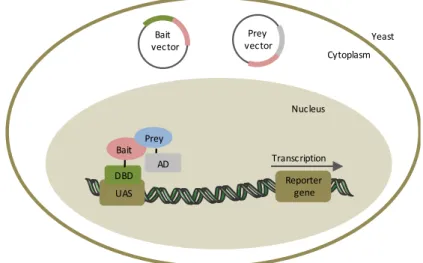
Figure A3.1. Schematic representation of YTH principle. ... 43
Figure A3.2. Schematic representation of a co-immunoprecipitation followed by mass spectrometry. ... 45
Figure B1.1. Effect of bioportides on PPP1 activity ... 98
Figure B1.2. Bioportides translocation into bovine and human spermatozoa (previous page) .... 100
Figure B1.3. Impact of the bioportides in bovine spermatozoa motility parameter ... 101
Figure B1.4. Molecular dynamics of PPP1R2 RVxF and PPP1R2 RVxF SC peptide ... 102
Figure B1.5. Schematic representation of the structure of PPP1CC complexed with PPP1R2 ... 105
Figure B2.1. GSK3 in human testis and sperm ... 127
Figure B2.2. Total and serine phosphorylated GSK3 levels in human sperm (previous page) ... 130
Figure B2.3. Subcelular localization of GSK3 and GSK3 in mature human sperm (previous page) ... 131
Figure B2.4. Yeast co-transformation of GSK3 and GSK3 interactors ... 143
Figure B2.5.Co-Immunoprecipitation of GSK3 from normospermic human sperm sample ... 144
Figure B2. 6. Venn diagram showing the overlap of GSK3⍺ and GSK3β interactomes ... 145
Figure B2.7. GSK3α sperm motility network ... 146
Figure B2.8. GSK3α testis network ... 147
Figure B2.9. GSK3β sperm motility network ... 148
Figure B2.10. GSK3β testis network ... 149
Figure B2.11. LRP6 and AKAP11 in human testis and sperm ... 151
Table of Tables
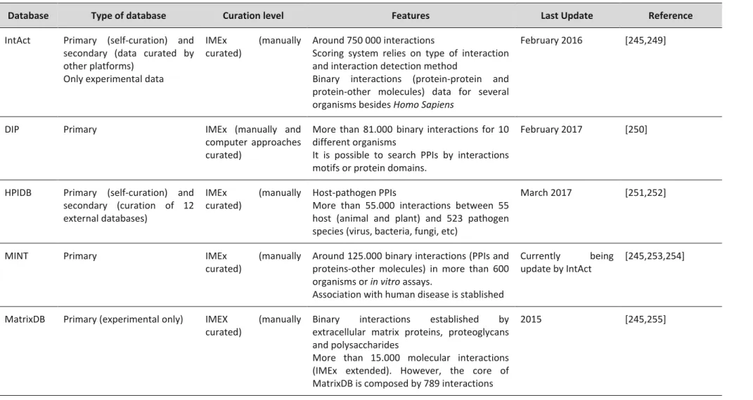
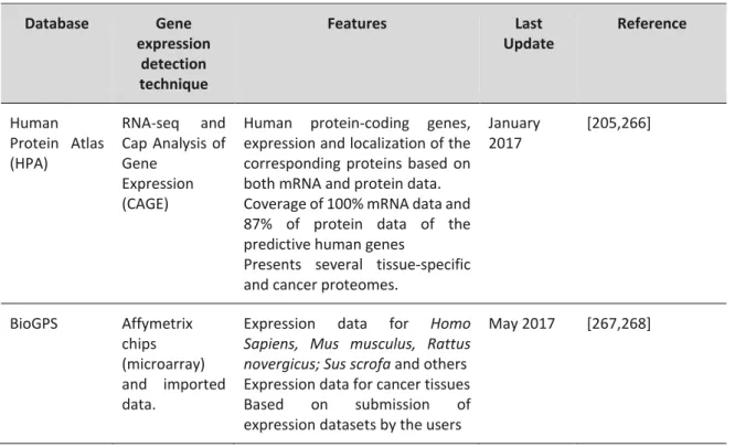
Table A3.1. Protein-protein interactions databases ... 49
Table A3.2. Tissue gene expression databases and repositories ... 51
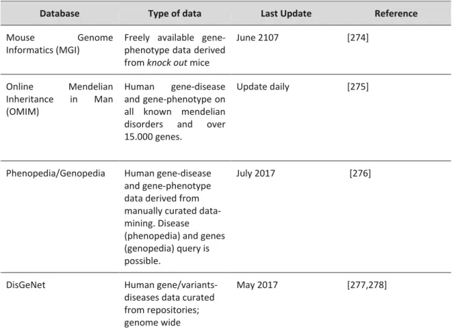
Table A3.3. Gene-disease association and animal model gene-phenotype databases ... 53
Table B1.1. Peptide sequence, peptide designation and length of the bioportides (BP) used in this study ... 92
Table B1.2. Aminoacids substitution studies of RVxF motif ... 102
Table B2.1. GSK3 human testis interactors ... 132
Table B2.2. GSK3 human testis interactors ... 135
Table B2.3. GSK3 human sperm interactors ... 137
Table B2.4. GSK3 human sperm interactors ... 138
Table of Supplementary Figures
Supplementary Figure B1.1. Effect of PPP1R2 on PPP1 activity ... 112
Supplementary Figure B1.2. Impact of bioportides on bovine spermatozoa viability ... 113
Supplementary Figure B2. 1. Expression and auto-activation of the reporter genes tests of pAS2-1-GSK3 and pAS2-1-GSK3 in AH190 yeasts ... 168
Supplementary Figure B2.2. Solubilizing effect of different lysis buffers for GSK3 and GSK3 .. 168
Supplementary Figure B2.3. Co-Immunoprecipitation of GSK3 from normospermic human sperm sample (previous page) ... 169
Supplementary Figure B2.4. The GSK3 interactome network ... 169
Supplementary Figure B2.5. The GSK3 interactome network (previous page) ... 170
Supplementary Figure B2.6. The GSK3 general male infertility network ... 170
Table of Supplementary Tables
Supplementary Table B1.1. Descriptive and statistical measures of the effect of bioportide in PPP1 activity
... 110
Supplementary Table B1.2. Inferential statistics of the effect of bioportide in PPP1 activity
... 110
Supplementary Table B1.3. Descriptive and statistical measures of the effect of bioportide in bovine sperm motility at 1 and 2 hours of incubation
... 111
Supplementary Table B1.4. Inferential statistics of the effect of bioportides on bovine sperm motility at 1 and 2 hours of incubation
... 112
Supplementary Table B2.1. Testis expression levels of GSK3 interactome
... 165
Supplementary Table B2.2. Testis and sperm related phenotypes/diseases/annotations of GSK3 interactome
... 165
Supplementary Table B2.3. Correlation coefficient between % of immotile sperm and GSK3 isoform expression and activation in human sperm
... 165
Supplementary Table B2.4. Top 5 of GeneOntology enrichment of GSK3 testis interactome
... 165
Supplementary Table B2.6. Top 5 of GeneOntology enrichment of GSK3 testis interactome
... 166
Abbreviations
Abs Absorbance
AD Activating domain
AKAP11 A kinase anchor protein 11
AKAPs A kinase anchor proteins
AKT RAC-alpha serine/threonine-protein kinase ARHGAP6 Rho GTPase-activating protein 6
ATP adenosine triphosphate
BP Bioportide
Ca2+ Calcium
cAMP Cyclic adenosine monophosphate
CatSper Cation channel of sperm
co-IP co-Immunoprecipitation
CPPs Cell penetrating peptides
DAG Diacylglycerol
DBD DNA binding domain
DNA Deoxyribonucleic acid
ESTs Expressed sequence tag
GSK3 Glycogen synthase kinase 3
GSK3 Glycogen synthase kinase 3 isoform alpha GSK3 Glycogen synthase kinase 3 isoform beta
H+ Hydrogen HCO3 Bicarbonate HP Homing peptides KI Knock in KO Knock out LDHC Lactate dehydrogenase C
LRP6 Low-density lipoprotein receptor-related protein 6 MAPKs Mitogen-activated protein kinase
Mn2+ Manganese
mRNA messenger Ribonucleic acid
Na Sodium
p17 17kDa protein
PBS Phosphate buffer solution
PDPK1 3-phosphoinositide-dependent protein kinase 1
PFA Paraformaldehyde
PGK2 Phosphoglycerate kinase 2
PIK3C Phosphatidylinositol 3-kinase
PPIN Protein-protein interactions network
PPIs Protein-protein interactions
PPME1 Protein Phosphatase Methylesterase 1
PPP1 Phosphoprotein phosphatase 1
PPP1CC2 Phosphoprotein phosphatase 1 catalytic subunit C2 PPP1R11 Phosphoprotein phosphatase 1 regulatory subunit 11 PPP1R2 Phosphoprotein phosphatase 1 regulatory subunit 2
PPP1R2P3 PPP1R2 pseudogene 3
PPP1R7 Phosphoprotein phosphatase 1 regulatory subunit 7 PPP2CA Phosphoprotein phosphatase 2 catalytic subunit A PPP3CC Phosphoprotein phosphatase 3 catalytic subunit C PRKA cAMP-dependent protein kinase catalytic subunit alpha
PRKC Protein kinase C
PRSS37 Probable inactive serine protease 37
PSA Prostate specific antigen
RAF-1 RAF proto-oncogene serine/threonine-protein kinase
RNA Ribonucleic acid
RNA-seq RNA sequencing
sAC Soluble adenylyl cyclase
Ser Serine residues
SERCAs Sarco/endoplasmic reticulum Ca2+-ATPase
SFK Src family kinase
Thr Threonine residues
Tyr Tyrosine residues
VOCCs L-type voltage-operated Ca2+ channels
YTH Yeast two-hybrid
A1. Male reproductive System
The male reproductive system has the purpose of producing, maturate and deliver the male gametes to the female reproductive tract. Anatomically, the male reproductive system is composed of two testes, a system of genital ducts (tubuli recti; efferent ducts; epididymis, deferent ducts; ejaculatory ducts and urethra), accessory glands (seminal vesicle, prostate and bulbourethral glands) and the penis. Functionally, spermatozoa or sperm must be produced (spermatogenesis) in the testis, matured in the epididymis and delivered to the female reproductive system by the penis. Moreover, the production of sexual hormones, specifically testosterone, occurs in the testis. In the next section, the morphological and functional characteristics of the Homo sapiens male reproductive system will be briefly described [1]. In Figure A1.1 an overview of the male reproductive system is presented.
A1.1. Testis – Where everything begins
Human testes are ovoid shape organs, localized outside the abdominal cavity and accommodated within the scrotum. This localization maintains the testes at an average temperature of 32ºC, optimal for its function. Within each lobule, there are 1 to 3 seminiferous tubules, the functional unit of the testis, and Leydig cells, which are responsible for the production of testosterone [2,3].
Seminal vesicle Ductus deferens Epididymis Testis Bladder Ureter Penis Bulbourethral glands Prostate Seminiferous tubule Sertoli cell Germ cells Blood vessel Leydig cell
Sertoli cell nucleus
A B
Figure A1.1. Overview of the male reproductive system. A. Components of the male reproductive system. The
bladder and the ureter are not part of the male reproductive system. B. Schematic cross-section of a testicular tubule, showing germ cells inserted in Sertoli cells. In the interstitium there are Leydig cells and blood vessels. Figures were produced using Servier Medical Art.
The seminiferous tubules are composed of germ cells line and Sertoli cells. Figure A1.1, describes the morphological structure of the seminiferous tubule. Germ cells will originate mature sperm through spermatogenesis. In Homo sapiens more than 200 million sperm are produced every day. Spermatogenesis can be divided in two distinct phases: (i) proliferative and meiotic phase; and (ii) haploid phase. The proliferative phase ensures the continuum of sperm production through a constant renewal of spermatogonia (the least mature germ cells). Consequently, spermatogonia can either undergo mitosis and duplicate itself or enter meiosis. By mitosis, spermatogonia yields primary spermatocytes and the latter, divides meiotically into two secondary spermatocytes. Finally, secondary spermatocytes divide into spermatids. Meiosis will reduce the chromosomal information into half. The haploid phase is characterized by the acquisition of crucial morphological structures necessary for fertilizing potential. It is divided in two sub-phases: spermiogenesis and spermiation. Spermiogenesis is defined as the process of transforming a round haploid spermatid into a highly specialized spermatozoon. At the organelle level, the Golgi complex will originate the acrosome [4]; the spermatid cytosol is merged into sperm head skeleton and, sperm axoneme is formed by outer dense fibers and fibrous sheath [5–9]. To accomplish such metamorphosis, transcription, translational and prost-translational modification increase drastically. Yet, by the end of spermiogenesis, transcription is ceased due to replacement of histones for protamines in sperm DNA, which results in highly packed DNA [10]. From this point on, sperm must rely on existing proteins to control its function. Spermiation refers to the detachment of fully differentiated sperm from the seminiferous epithelium and the journey through the lumen of the seminiferous tubules [11]. Also, during spermiation, the remnants of germ cells cytoplasm are rejected.
Sertoli cells provide structural support and nourishment of germ cells; perform phagocytosis of degenerating germ cells; allow spermiation of fully mature sperm, control germ cell proliferation and differentiation by paracrine secretion of regulatory proteins and create a unique microenvironment for the germ cell line (blood-testis barrier) [12–14]. Morphologically, Sertoli cells are pyrimid-like cells that involve partially the germ cell line. Around 18% of the seminiferous tubule is composed of Sertoli cells and the number of Sertoli cells positively correlates with sperm output. Adjacent Sertoli cells form tights junctions, creating a unique environment, isolating germ cells from the rest of the body. This is particularly important to isolate germ cells form the immune system [13].
The last type of cells present in testis are the Leydig cells. These cells lay between seminiferous tubules and contact directly with blood vessels. Beginning in puberty, Leydig cells increase the production of testosterone, in response to luteinizing hormone [15]. This stimulates the beginning
of spermatogenesis, through Sertoli cell signaling. Through the entire adulthood, spermatogenesis is controlled by constant feedback with the nervous system (hypothalamus and pituitary) [16].
A1.2. Epididymis – where everything starts to function
By the end of spermatogenesis, sperm is morphological complete but functionally immature. To be able to fertilize an oocyte, sperm must journey through the epididymis [17]. Epididymis is a highly-convoluted duct that connects the efferent ducts to the deferent ducts. It is localized in the posterior surface of the testis and in primates can have a length range from 1-7 meters [18]. Estimates of the time necessary for sperm to migrate through human epididymis are inconsistent. Rowley et al suggested a range of 1 to 21 days and Amann and Howards between 3-4 days [19,20]. The epididymis has five main roles: (i) transport of testicular sperm out of the testis; (ii) create a suitable environment for sperm maturation; (iii) promote progressive spermatozoa motility; (iv) prepare sperm for fertilization; (v) and sperm storage. Sperm goes through morphological and functional maturation in the epididymis.
Since testicular sperm are transcriptionally silent cells, the maturation of sperm through the epididymis depends on post-translational modification of pre-existing proteins and protein exchanges between the epididymal lumen and the sperm. Phosphorylation, glycosylation and proteolysis of sperm proteins are among the most common post-translational modification. Most proteins that suffer this process are linked to interaction between sperm and oocyte (zona pellucida and oocyte membrane) and acrosome reaction [21–23]. The role of phosphorylation on sperm maturation, particularly motility acquisition, is discussed further on section A2.
Epididymosomes, small extracellular vesicles secreted by the epididymis´s epithelial cells are the major players in the interchange of proteins between sperm and epididymal lumen. It was suggested by Cornwall that epididymosomes allow the safe delivery of proteins to sperm, escaping the proteolytic activity in the epididymal fluid [17]. Within the proteins delivered by epididymosomes, proteins involved in sperm motility, immunological protection and inhibition of capacitation are the more abundant [23].
A1.3. The spermatozoa – the final result
Sperm can be divided in two main structures: head and flagellum. These structures are surrounded by a unique plasma membrane. In the next section, we will focus on the morphological features of sperm head, connecting piece and plasma membrane composition (Figure A1.2). An extensive description of sperm flagellum can be consulted in section A2.
The human sperm head is spatula-shaped and its main components are the nucleus and the acrosome. These two structures are surrounded by a small amount of cytoplasm and cytoskeleton structures [24]. The acrosome lies anteriorly to the nucleus, and the cytoplasm lies in the narrow space between both structures and the plasma membrane. Within the nucleus, the DNA is highly compacted and its volume is considerably less than in somatic cells [24]. These results from: protamines highly compacted DNA and sperm only having half the genetic information of somatic cells. Still, approximately 4% of DNA is bound to histones and has been proved that the expression of such DNA is essential for early embryo development [25]. Surrounding the nucleus is the nuclear envelope. The unique feature of sperm nuclear envelope is that, contrary to somatic cells, it does not present nuclear pore complex [26]. Furthermore, the sperm nucleus is protected by a rigid structure formed by bonding of structural proteins, the perinuclear theca [5]. These features result in an optimal shaped nucleus for mobility and better protection of the genetic information. The acrosome covers half to two thirds of the sperm head. This organelle is derived from the Golgi
Figure A1.2. Schematic representation of the human sperm head and connecting piece. Human sperm head contains
anteriorly the acrosome and posteriorly the nucleus. The nucleus is surrounded by the nuclear envelope and the perinuclear theca. The connecting piece is composed by the capitulum and the segmented columns and connects the head to the flagellum.
complex and presents an inner acrosomal membrane adjacent to the nucleus and an outer acrosomal membrane adjacent to the plasma membrane. The acrosome houses several proteases, hidroglycolases and esterases, some which are sperm-specific (e.g acrosin), that upon specific zona
pellucida signaling are released by exocytosis (acrosome reaction). These proteases will create a
path for sperm penetration into the zona pellucida [27]. After acrosome reaction, the equatorial region of these organelle is exposed and is involved in the initial interaction between sperm and oocyte membrane [28].
Immediately behind the sperm head is the connecting piece. This structure functions as an anchor between the head and the tail. Consequently, it must be somewhat flexible. The connecting piece is formed by two main structures: the capitulum and the segmented columns. The capitulum inserts into the head and distally interacts with the segmented columns. Distally, the segment columns fuse with the other dense fibers of the flagellum [29,30].
In general, the sperm plasma membrane contains 70% phospholipids, 25% neutral lipids and 5% glycolipids [31]. The most abundant phospholipid of the plasma membrane is sphingomyelin. This lipid confers rigidity to bilayer membranes. Regarding neutral lipids, human sperm membrane contains very high amounts of cholesterol. Cholesterol is a plasma membrane stabilizer with the particularity of maintaining membrane fluidity [32]. Consequently, cholesterol is a key lipid for sperm permeability and motility. In human sperm plasma membrane, the only glycolipid is seminolipid [33], which is present only in mammalian sperm and Schwann cells [34]. It is believed that this molecule participates in maintenance of lipid diffusion barriers (see bellow) [33]. After epididymis transit, plasma membrane presents high cholesterol/phospholipid ratio. This results in plasma membrane stabilization which is beneficial to the journey in the female reproductive system.
Mammalian sperm plasma membrane is divided into five macrodomains: acrosome; postacrosome, equatorial segment; midpiece and principal piece. Each domain appears to have a unique composition and organization which reflects different properties and functions [35,36]. For example, the acrosomal macrodomain is highly fusogenic compared with the post acrosomal macrodomain. One hypothesis for this is that the acrosomal macrodomain is rich in phospholipids that form “cone shape holes” into the membrane, resulting in an unstable membrane [33]. Another question that arises is how these macrodomains are confined to a specific part of the sperm. Lipid diffusing barriers have been described in sperm, specifically in the posterior ring and annulus. The
posterior ring segregates the content of sperm plasma membrane of the head and tail and the annulus between the midpiece and principal piece [36].
A1.4. Accessory glands
After epididymal transit, sperm are pushed to the deferent ducts, ejaculatory ducts and finally urethra, upon stimulation. During this journey, the accessory glands (Figure A1.1) secret several fluids that will form the seminal plasma. Please note that the epididymal fluid and sperm cells represent around 3-5% of all semen volume [37]. Altogether, sperm and seminal plasma form the semen.
Secretion from the seminal vesicle is responsible for around 50% of the seminal plasma content and is alkaline and very rich in fructose. Fructose will be consumed by sperm cells producing energy and the alkaline environment will balance the acidic female reproductive tract fluids. This will result in stimulation of sperm motility. Fructose quantification is widely used as a marker for seminal vesicle function. However, some question have arisen regarding the fidelity of such marker. Since fructose is consumed by sperm, fructose concentration also depends on the rate of fructose consumption sperm and not exclusively on seminal vesicle function. Several roles for seminal vesicle secretion have been described. Promotion of sperm motility; increase stability of sperm DNA and suppression the immune system activity in the female reproductive system are some [38].
The next contribution to the seminal plasma are the secretions from the prostate which represent around 20-30% [37]. Prostatic fluid is rich in zinc (Zn2+), citric acid and prostate specific antigen (PSA). Moreover, it presents a slightly acidic pH [39]. It appears that Zn2+ stabilizes spermatic DNA and prevents DNA fragmentation, specifically by stabilizing chromatin [40] It further presents anti-microbial activity [39,41]. Citric acid maintains an acidic environment and since it is 100x more secreted by the prostate than by other accessory glands, it may be used as a prostate activity function biomarker. Finally, the PSA promotes liquefaction of semen and consequently stimulates sperm motility [39,42]. Similarly, to the epididymis, the prostate also secretes small vesicles, the prosteosomes, which may fuse with the sperm cell. Theses vesicles are rich in proteins and will transfer such proteins to sperm, by membrane fusing.
The last accessory glands are the bulbourethral glands which, upon sexual stimulation, secrete, to the urethra, an alkaline mucus-like fluid rich in glycoproteins. This fluid neutralizes the acidity of
pre-existing urine residues and provides lubrication for the penis during intercourse [43]. The bulbourethral contribution to the seminal plasma is very small (around 1%)[37].
Finally, the last step to accomplish fertilization is releasing the semen into the female reproductive system by ejaculation. Ejaculation is defined by the expulsion of the semen through the penis. Upon stimulation, the sympathetic nervous system activates the contraction of muscle in the epididymis, seminal vesicle, prostate and deferent ducts. Consequently, the semen is released to the urethra. The accumulation of semen in the urethra stimulates the contraction of muscles within the penis (bulbospongiosus and ischiocavernosus muscles) resulting in the release of semen [44].
A1.5. Male Fertility: infertility and contraceptives
Infertility is defined as the incapability of conceive after 12 months of unprotected intercourse [45]. In a recent study, Agarwal estimated that 15% couple’s worldwide are infertile, from which 50% have a male contribution and 20-30% are exclusively due to male factors [46]. Furthermore, idiopathic male infertility predominates as the major cause of infertility (50% of all male infertility cases) [47]. The fact that key mechanisms responsible for several sperm processes, such motility, are still not fully understood, may be the key to understand idiopathic cases of male infertility. Understanding sperm physiology is a step towards unveil and more important diagnose and treat idiopathic male infertility.
On the other hand, male contraceptives have not evolved as much as female contraceptives in the last decades. They fall in three main categories: vasectomy, male condom and withdrawal. In 2015, all together, male contraceptives represented 21% of contraceptive practice worldwide [48]. Vasectomy, is the blockage of the vas deferens and consequently passage of sperm. It is an invasive procedure that until recently was irreversible and has a very high effectiveness (97%-98%). Male condom is a physical barrier that prevents sperm cells to meet the oocyte. Most men refer discomfort using a condom. However, it is the only contraceptive that prevents sexual transmitted diseases and has an effectiveness of 85%. Finally, withdrawal, the pulling out of the penis from the vagina prior to ejaculation, is the least effective contraceptive (73%) [49].
Considering that the period of male contraceptive usage is increasing (earlier sexual initiation and latter fatherhood) and the choices available for men are limited, we believe that a new male contraceptive is necessary. Targeting the signaling pathways involved in sperm motility acquisition may be the future of male contraceptives. Using this approach, spermatogenesis is not disturbed,
ensuring that it is reversible, but keeps sperm from encountering the oocyte. Therefore, understanding the mechanism involved in sperm motility acquisition is key.
A2. Signaling mechanisms in mammalian sperm motility
aHuman spermatozoon is one of the most differentiated cell types and must leave the male body where is produced and achieve its goal in the female reproductive system [50]. In order to fertilize an egg, the sperm is formed in the testes, in a process called spermatogenesis. At the end of spermatogenesis, sperm are morphologically complete but functionally immature and incapable of fertilizing an egg. To be functional, sperm cells must undergo: (i) maturation in the epididymis; (ii) capacitation and (iii) acrosome reaction in the female reproductive system [51]. These events are co-dependent, since acrosome reaction does not occur if capacitation is impaired and capacitation depends on functional maturation of sperm in the epididymis. Motility acquisition is essential for human sperm function and ultimately male fertility. In 2011, Paoli defined sperm motility as a propagation of transverse waves along the flagellum in a proximal- distal direction producing an impulse that pushes the spermatozoon through the female genital tract [52].
Severe asthenozoospermia is one of the causes of male infertility which arises from the inability of the sperm cell to reach the oocyte [53]. Primary or activated motility is acquired throughout the journey in the epididymis. Although the exact mechanism behind motility acquisition is still far from being fully understood, specific signaling events are described in the literature as essential for this process [51,54]. Low-amplitude symmetrical tail movements characterize sperm activated motility and drive sperm in a straight line in a non-viscous media (seminal plasma) [55]. However, in fallopian tubes, sperm must acquire a specific type of motility, hyperactivated motility, which is characterized by high amplitude and asymmetric flagellar bends. Only this type of flagellar movement allows sperm to overcome dense mucus, detach from the oviductal epithelium and penetrate the egg’s protective vestments [56]. Curiously, in the viscous media hyperactivated sperm swim in a circular or figure-8 pattern [55,57]. Alterations in pH, specific molecules, and ion concentration changes are a few of the crucial events for stimulation of hyperactivated motility [58,59]. However, the cellular mechanism and signaling pathways responsible for this type of motility are not fully described.
To be motile, human sperm need a morphologically complete flagellum; be able to produce energy
aPublished review paper: Freitas MJ, Vijayaraghavan S, Fardilha M (2017) Signaling mechanisms in mammalian sperm
to power flagellar movement; and functional signaling pathways (to transduce external signals into internal signals). This review will discuss these three topics, but will mainly focus on the signaling pathways involved in human sperm motility regulation. For an in-depth review on sperm bioenergetics please see du Plessis et al. [60]
A2.1. Sperm flagellum - structure and function
The human sperm are composed of two main structures: head and flagellum (Figure A2.1). The head comprises the nucleus and the acrosome. The nucleus houses the genetic information to be delivered to the oocyte. Upon acrosome reaction, the acrosome integrity is disrupted and its content is released digesting the oocyte’s zona pelucida [61]. The flagellum contains the motile apparatus necessary for sperm motility [55,62] and is divided into four ultrastructures: connecting piece; midpiece; principal piece and end piece [55]. The connecting piece attaches the flagellum to the sperm head; the midpiece contains the sperm mitochondria; the principal piece and the end piece generate the flagellar waveform pattern motility [55,62,63]. The main structure of the flagellum is the axoneme, which is the sperm motility motor. This structure is well conserved throughout evolution, present in flagella from protozoans to humans [50,62]. The axoneme originates in the connecting piece and terminates in the end piece. Typically, the axoneme is composed of nine microtubules doublets and a central pair, designated a 9+2 structure. The nine microtubules doublets connect to each other by nexin links and connect to the central pair by projections, the radial spokes. The latter are responsible for positioning and spacing the microtubules doublets in a perfect circle around the central pair microtubule. Projecting from the microtubules doublets are the inner and outer axonemal dynein arms (classified according to their position in relation to the doublet microtubule). These proteins are key for motility, by promoting sliding of a microtubule doublet in relation to the adjacent. The flagellar beating pattern begins with a dynein from one doublet transiently interacting with the following doublet. In the presence of ATP, axonemal dynein “walks” towards the base of the flagellum, forcing the adjacent microtubule doublet to slide down. Since microtubules are attached to the connecting piece, this movement encounters resistance, leading to the bending of the flagellum. At the end, the dynein detaches from the adjacent microtubule. To obtain a flagellum waveform movement and consequently motility, this process has to occur on one side of the axoneme and be inactive on the opposite site. Hence, the flagellar beat appears to be based on an “on-and-off” switch of the axonemal dynein arms, in specific points in the axoneme [50,55,62,63].
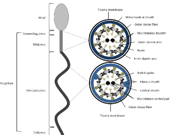
Figure A2.1. Schematic representation of human spermatozoon and flagellum structure. Human sperm is divided into
two parts: head and flagellum. The flagellum is further divided into four structures: connecting piece; midpiece; principal piece and endpiece. A cross-section shows that the flagellum structure differs between midpiece and principal piece. In midpiece, plasma membrane and mitochondrial sheath surround the outer dense fibers. Within outer dense fibers, is the axoneme, composed of the microtubule doublets associated with the dyneins arms (inner and outer), radial spoke and microtubule central pair. Nexin connects adjacent microtubule doublet. In the principal piece, plasma membrane and fibrous sheath surround the outer dense fibers. In two opposing microtubule doublets, the outer dense fibers are replaced by to longitudinal columns of fibrous sheath.
In mammalian sperm, between the axoneme and the plasma membrane there are several accessory structures, such as the mitochondrial sheath, outer dense fibers and fibrous sheath [50,55]. In the midpiece, the axoneme is surrounded by outer dense fibers and the mitochondrial sheath, while in the principal piece the axoneme is surrounded by outer dense fibers and fibrous sheath. The end piece has no accessory structures between the axoneme and the plasma membrane [62]. The mitochondrial sheath is composed of individual mitochondria coiled helically around the axoneme. In humans, the midpiece length is about a dozen mitochondrial turns [64]. The outer dense fibers have a petal-like shape, are directly above the axoneme microtubules doublets and diminish in diameter from base to tip of the principal piece [65]. The outer dense fibers appear to be responsible for maintaining the passive elastic structure and recoil of the flagellum and to protect the axoneme against shearing forces [66]. In the principal piece, the fibrous sheath confers
flexibility, shape and plane to the flagellar beat [67]. It also supports and ensures compartmentalization of signaling proteins that regulate motility, capacitation, and hyperactivation. In the principal piece, two opposing outer dense fibers are replaced by fibrous sheath projections [65] (Figure A2.1).
The regulation and propagation of this “on-and-off” signal and the conversion into flagellar bending appears to reside in the control of the ATPase activity of axonemal dynein arms. Although this process is not fully understood, alterations in pH, ATP availability, calcium concentration and phosphorylation of key proteins appears to modulate axonemal dynein arms activity and consequently sperm motility. The process of ATP production and the signal pathways that control axonemal dynein activity will be discussed in the next topics [68].
A2.2. Energy for motility - Oxidative phosphorylation vs glycolysis
One of the key requirements for sperm motility is energy availability. ATP is the fuel used by axonemal dynein ATPases within the flagellum [69], and active protein modifications, such as phosphorylation, also depend on ATP. Thus, it is not surprising that sperm requires exceptionally high amounts of ATP when compared to somatic cells [70]. Consequently, a constant and adequate supply of ATP is crucial [69]. In spite of the efforts [69,71,72], a long-standing debate exists on the metabolic pathway responsible for sperm motility bioenergetics: oxidative phosphorylation in mitochondria, glycolysis in the flagellum and head, or both.
In mammalian sperm, oxidative phosphorylation occurs in mitochondria, which are exclusively located in the midpiece. A mature mammalian spermatozoon contains approximately 72-80 mitochondria [73] and in theory can produce more than 30 ATP molecules per glucose molecule [74]. Since midpiece is localized at the anterior end of the flagellum the transport of ATP to the all length of the flagellum must be efficient. Ford et al believe that the model of flux transfer chains proposed by Dzeja and Terzic in 2003, is able to transport the ATP produced in mitochondria through the entire flagellum [72,75]. It was indeed shown that an increase in human sperm motility requires a parallel increase in mitochondrial activity [52,71,76]. Also, the use of specific inhibitors for the mitochondrial electron transport chain and ATP synthase decreases drastically human sperm motility [74]. Moreover, high mitochondrial activity levels increase the success of in vitro fertilization rate [69]. These studies suggest that human sperm motility correlates with
mitochondrial functional status. Further, mitochondrial activity is negatively correlated with morphological alterations in the midpiece, which appears to reinforce the role of mitochondrial ATP production in sperm motility [71].
In spite of the reports supporting the role of mitochondria in sperm motility, its contribution to flagellar beat can be questioned. Since mitochondria are localized in the midpiece, it has been argued if ATP diffusion and carrier systems are able to supply ATP throughout the entire length of the flagellum (about 50µm in humans) [72,74]. Also, some authors argued that if ATP produced in the mitochondria fuels motility, the levels of reactive oxygen species produced during the electron transport chain would be harmful to DNA integrity [77]. However, both enzymatic (e.g. superoxide dismutase and glutathione peroxidases) and non-enzymatic antioxidants (e.g. glutathione and ascorbic acid) present in human sperm and seminal plasma appear to control the levels of ROS activity [77–79].
A growing hypothesis for the source of ATP (or at least part of the ATP) in sperm is the glycolytic pathway. Glycolysis is the process by which glucose is converted into pyruvate. During this process, energy is released in the form of ATP and NADH, with a rate of 2 ATP molecules per glucose. When human sperm are deprived of glucose (the starting unit of glycolysis) or when glycolysis is blocked, ATP content and protein tyrosine phosphorylation decreases. Consequently, sperm exhibits decreased motility [74,80–83]. Mukai and Okuno proved that even when mitochondria function is conserved, mouse sperm motility decreases when glycolysis is impaired. Moreover, a sperm-specific lactate dehydrogenase (LDHC) accounts for 80-100% of the LDHC activity in human sperm and is anchored to the fibrous sheath along the length of the flagellum, representing a local ATP production closer to the site of ATP consumption [83]. Also, Odet et al showed that a disruption in mouse sperm-specific Ldhc resulted in impaired fertility due to immotile sperm [84]. Furthermore, sperm-specific LDHC presents a low Km for pyruvate and a high Km for lactate, suggesting a higher affinity of LDHC for pyruvate and consequently a preference for the glycolytic energy pathway. It is noteworthy to mention that although most mammals rely, at least partially, on glycolysis for motility, the bull seems to be an exception. Oxidative phosphorylation in bull sperm appears to be the only source of ATP [85].
A third possibility for ATP availability in human sperm is a cooperation and dependence between oxidative phosphorylation in mitochondria and glycolysis in the flagellum. This hypothesis is supported by the different energetic subtracts of the reproductive tract fluids [60,70]. It appears
that mammalian sperm switch between metabolic pathways depending on oxygen availability and glucose, pyruvate, lactate, sorbitol, glycerol, and fructose concentration in the fluid [74,81,86–89]. For example, in the human female reproductive tract, glucose, pyruvate and lactate are found in the range of 0.5-3.2mM, 0.1-0.2-mM and 4.9-10.5mM, respectively. Sperm must adapt its bioenergetic metabolism according to the metabolites available from the epididymis until the fallopian tubes [90].
A2.3. Signaling pathways in sperm motility
Sperm leaving the testes are immotile and acquire motility throughout the epididymis journey. Sperm is virtually devoid of transcription and translation due to highly condensed DNA and lack of endoplasmatic reticulum [91]. Since gene expression cannot be accounted for functional alterations in sperm, activation or inhibition of specific signaling pathways and protein post-translational modifications must be involved. The interaction between sperm and the environment created by the epididymis and the female reproductive tract are essential to trigger sperm motility.
Several signaling pathways have been described as having a role in mammalian sperm motility. In the next section, the most relevant signaling pathways and messengers involved in sperm motility acquisition in the epididymis and hyperactivated motility in the female reproductive tract will be described.
A2.3.1. Sperm motility in the male reproductive system – A journey through the epididymis After spermatogenesis, sperm is morphologically complete but functionally immature. When entering the epididymis, a long convoluted tubule that connects the testis to the vas deferens, human sperm is incapable of fertilizing an oocyte. The epididymis is roughly divided into three regions: caput, corpus, and cauda. The caput is adjacent to the testis and the caudal portion adjacent to the vas deferens [12]. Only during the journey through the epididymis the sperm acquire fertilization ability. Epididymal maturation involves the interaction of sperm with proteins that are synthesized and secreted into the epididymis in a region-dependent manner [17]. Most of studies concerning epididymis function are carried out in rodent models, due to the limited availability of human epididymal tissues at reproductive ages, the impossibility of mimicking the epididymis environment in vitro and the difficulty to manipulate the human epididymis experimentally. The exact mechanism behind sperm motility acquisition in the epididymis is still
unknown [55].
One of the first described signaling events responsible for sperm motility acquisition within the epididymis is the control of Phosphoprotein Phosphatase 1 (PPP1, also known as PP1) activity in sperm (Figure A2.2). PPP1CC2 (also known as PP12), a testis-enriched sperm-specific PPP1 isoform is distributed throughout the flagellum, midpiece and posterior region of the head [92] suggesting a role in motility and acrosome reaction [93,94]. In 1996 Smith et al described, for the first time, the association between PPP1 activity and sperm motility. In caput sperm PPP1 activity is high and sperm is immotile. Conversely, in caudal sperm PPP1 is inactive and sperm are motile [95,96]. In the following years, several studies attempted to unveil the signaling pathways responsible for PPP1 activity modulation in sperm, through the epididymis journey. PPP1 regulatory subunit 2, PPP1R2 (also known as Inhibitor-2) is a PPP1 inhibitor [97]. In sperm, PPP1R2 localizes throughout the principal piece, midpiece and posterior and equatorial regions of the head. Former studies described PPP1R2 activity in human sperm and that some of the sperm PPP1 population is bound to PPP1R2 and therefore inactive [98]. When phosphorylated at threonine 73, human PPP1R2 is unable to bind to PPP1, rendering its activity [99]. Glycogen synthase kinase 3 (GSK3) is the kinase responsible for PPP1R2 phosphorylation. Interestingly, GSK3 activity has been extensible correlated with sperm motility regulation both in cauda and caput (bovine, mouse and macaque models) [100– 102]. GSK3 is 6 times more active in caput than in caudal sperm and its activity is correlated negatively with sperm motility [100,102]. Moreover, GSK3 appears to have an isoform-specific function on sperm motility. When GSK3alpha is knockout there is a decrease in sperm motility and metabolism, while GSK3beta conditional knockout is fertile [103].
Recently, Koch et al showed that the Wnt signaling can be partially responsible for GSK3 activity regulation in epididymal sperm. In corpus and caudal epididymis, Wnt signaling proteins are released in epididymosomes from epididymal principal cells. In the epididymis lumen Wnt proteins bind to Low-density lipoprotein receptor-related protein 6 receptor (LRP6), activating it. In turn, LRP6 induces GSK3 inhibition, which leads to decreased PPP1R2 phosphorylation [104]. Synergistically, GSK3 activity can be modulated by Phosphoprotein phosphatase 2A (PPP2 also known as PP2A). Dudiki et al demonstrated that in caput sperm demethylated and phosphorylated PPP2 isoform alpha (PPP2CA) is active. Consequently, it dephosphorylates GSK3 in serine residues rendering active. Subsequently, PPP1R2 threonine 73 is phosphorylated and the inhibitor becomes inactive, resulting in active PPP1 and immotile sperm. On, caput sperm, methylation of PPP2CA increases due to a decrease in Protein phosphatase methylesterase 1 (PPME1) activity. In these
conditions, PPP2CA becomes inactive resulting in increased GSK3 serine phosphorylation and thus, its inactivation. Subsequently, PPP1R2 is active and inhibits PPP1, leading to motile sperm [105] (Figure A2.2).
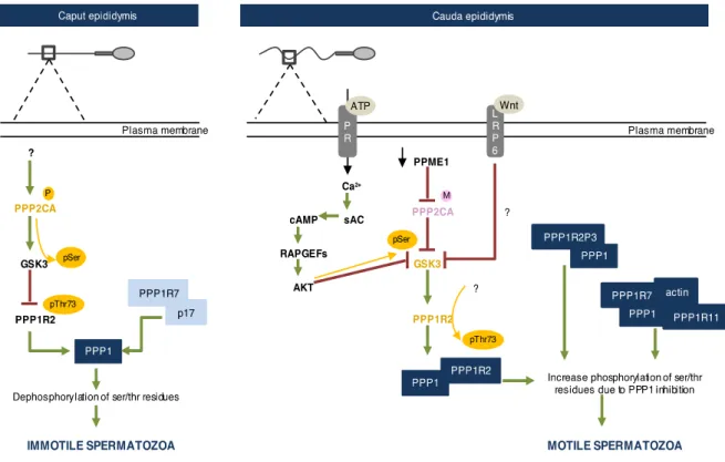
Figure A2.2. Schematic representation of the signaling events required for sperm motility acquisition in the epididymis.
In caput epididymis, PPP2CA is phosphorylated and consequently active, which in turn dephosphorylates GSK3 at serine residues, rendering it active. GSK3 phosphorylates PPP1R2 at thr73 which inhibits the interaction between PPP1R2 and PPP1 resulting in active PPP1. PPP1R7 is bound to p17, which leads to free and active PPP1. Active PPP1 results in dephosphorylation of key residues and consequently immotile sperm. In cauda epididymis, PPME1 activity decreases increasing PPP2CA methylation, resulting in inhibition of PPP2CA. Consequently, GSK3 serine phosphorylation increases leading to GSK3 inhibition. Also, Wnt binds to LRP6 receptor which promotes GSK3 inhibition by an unknown mechanism. Moreover, ATP binds to purinergic receptors (PR), resulting in calcium influx. Calcium activates sAC, which produces cAMP activating Rap guanine nucleotide exchange factor (RAPGEFs). The latter activates AKT that phosphorylates GSK3 at serine residues inactivating it[106]. GSK3 is inhibited, which leads to decrease Thr73 PPP1R2 phosphorylation (the phosphatase responsible is unknown). Consequently, PPP1R2 binds PPP1. Also, PPP1 is bound to PPPP1R2P3 and in a complex with PPP1R7, actin and PPP1R11. Thus, PPP1 activity is inhibited and ser/thr phosphorylation of key residues increases leading to motile sperm. P: Phosphorylation; M: Methylation. Green arrows: activation. Red arrows: inhibition. Yellow arrows: phosphorylation. In yellow: phosphorylated proteins. In pink: methylated proteins.
Moreover, in 2013, Korrodi-Gregório et al identified a new PPP1R2 isoform in human sperm, PPP1R2P3 (also known as inhibitor 2-like) [98]. This isoform has the unique feature of threonine 73 being replaced by proline avoiding GSK3 phosphorylation. Korrodi-Gregório et al hypothesized that PPP1R2P3 is only present in caudal motile sperm, representing a constitutively inhibitor of PPP1, and therefore responsible for the process of sperm motility acquisition along the epididymis journey [98] (Figure A2.2).
Besides PPP1R2, PPP1 regulatory subunit 7 (PPP1R7) and PPP1 regulatory subunit 11 (PPP1R11), other two PPP1 inhibitors are present in sperm, suggesting a synergetic mechanism for PPP1 activity control [107]. PPP1R11 (also known as I3) is a human homolog of the mouse Tctex5, a protein associated with male infertility due to impaired sperm motility. On mouse sperm, PPP1R11 is localized in the head and principal piece of the flagellum, the same subcellular localization of PPP1 [108]. In rat liver cells, PPP1R7 (also known as sds22) inhibits PPP1, and in rat testis, it associates with PPP1CC2. In caput, bovine sperm PPP1R7 and PPP1CC2 do not interact. Instead, PPP1R7 is associated with a 17kDa protein (p17) [109], resulting in active PPP1. Conversely, in mouse caudal sperm, PPP1R7, PPP1R11, actin and PPP1 form a complex catalytically inactive [107] (Figure A2.2). Although PPP1 plays a crucial role in keeping motility at check in caput sperm, its substrates are still unknown. Besides PPP1, a sperm-specific isoform of calcineurin (PPP3CC) appears to be involved in epididymal maturation. Upon ablation of PPP3 and regulatory subunit PPP3R2, male mice are infertile due to impaired hyperactivation and penetration of zona pelucida. Phenotypically, sperm without PPP3CC presents an inflexible midpiece. When sperm is hyperactivated, the bending capacity of the midpiece increases, however, PPP3CC null sperm are incapable of exhibiting this increase. Interestingly, inhibition of PPP3CC with specific inhibitors, results in a quick phenotype (5 days) alteration from normal to inflexible midpieces. After one week of halting drug administration, the sperm are completely recovered and fertility is restored [110].
Since for sperm motility dephosphorylation must be shut down, it is not surprising that phosphorylation must increase. It is well known that the soluble adenylyl cyclase/Cyclic adenosine monophosphate/cAMP-dependent protein kinase (sAC/cAMP/PRKA; cAC/cAMP/PKA) signaling pathway affects positively sperm motility. Although the sAC/cAMP/PRKA signaling is mostly associated with hyperactivated motility [58], its involvement in sperm motility acquisition is unquestionable (see below) [111,112]. In 2013, Vadnais et al, proposed a cross talk between the GSK3/PPP1R2/PPP1 and sAC/cAMP/PRKA pathways during motility acquisition in the epididymis [106] (Figure A2.2). In Figure A.2. the main signaling pathways involved in motility acquisition in the epididymis are represented.
A2.3.2. Sperm motility in the female reproductive system
During unprotected intercourse, millions of sperm are deposited in the female reproductive tract, more specifically in the vagina. From there on sperm must swim until they reach the oocyte in the fallopian tube. Although sperm is already motile when ejaculated, hyperactivated motility must be acquired to overcome all the filters and traps imposed by the female reproductive tract.
Interestingly, it is the unique female enviroment that triggers the signaling pathways essential for sperm hyperactivated motility [113]. In the past years, many efforts have been made to unravel the role of key messengers and signaling pathways involved in hyperactivated motility.
Firsts messengers - Calcium, bicarbonate, and progesterone
In sperm, calcium (Ca2+) plays a central role in events preceding fertilization, specifically, motility, chemotaxis and acrosome reaction. The relevance of Ca2+ on eukaryotic cell physiology is reflected in the several Ca2+-depended enzymes, intracellular Ca2+ stores and Ca2+ channels [114] . Human sperm is no exception. The most described role of Ca2+ in human sperm motility is the activation of the soluble adenylyl cyclase (sAC). Moreover, inhibition of Ca2+ signaling is associated with male subfertility [115]
In human sperm, mean basal Ca2+ is kept around 100nM-200nM, while in the extracellular medium varies between 1-2mM [116]. This gradient concentration is accomplished by a Ca2+-ATPase pump, which promotes Ca2+ efflux with ATP consumption [117,118]. A low resting Ca2+ concentration is what keeps human sperm in a basal motility state in the caudal portion of the epididymis and vas deferens. However, in the female reproductive tract, Ca2+ concentration must increase to induce hyperactivated motility. The female reproductive system controls the increase in Ca2+ concentration in the sperm through clues in specific places, and menstrual cycle phase [119].
The influx of Ca2+ into human sperm is promoted by several mechanisms: increase in membrane permeability [120]; depolarization [121]; inhibition of the Ca2+-ATPase pump; activation of voltage-dependent calcium channel (VOCCs). Yet the main known mechanism for Ca2+ influx into sperm is the CatSper (cation channel of sperm), identified in 2001 by Ren et al [122]. This channel, located at the principal piece of the flagellum, is the only constitutively active Ca2+ conductance present in
human sperm, responds weakly to voltage alterations and is pH-sensitive [122,123]. Moreover, null mice for CatSper1 are infertile [122]. Human CatSper activation is triggered mainly by extracellular progesterone (see below), prostaglandins [124] and an alkaline environment (created by increasing HCO3- concentrations) [125]. Curiously, mouse CatSper is activated by neither progesterone nor
prostaglandins. This suggests a species-specific Ca2+ influx process, possibly to avoid cross-species
fertilization [124]. Although it is not located to the sperm’s head, CatSper appears also to be involved in the acrosome reaction by increasing Ca2+ concentration [126]. Further, Brenker et al
concluded that a range of small odorant molecules present in the female reproductive tract activates CatSper, resulting in chemotaxis of the sperm towards the oocyte [127].
Although the process of Ca2+ influx is essential for sperm motility, it is established that the human sperm has Ca2+ stores. The most promising candidates for Ca2+ stores in human sperm are the acrosome, the nuclear membrane and the cytoplasmic droplet [128]. Interestingly, it appears that in the sperm flagellum there are no Ca2+ stores, suggesting that the stores are important on processes such as acrosome reaction, rather than in motility. Moreover, the presence in human sperm of sarcoplasmic/endoplasmic reticulum calcium ATPases (SERCAs), channels that transport Ca2+ from the intracellular medium to Ca2+ stores in somatic cells, further reinforces the presence and functional importance of Ca2+ stores [128,129].
Progesterone is probably the most potent activator of capacitation of human sperm [130]. It is produced by the cumulus oophorus cells that surround the oocyte. At nanomolar concentration range, progesterone induces Ca2+ influx and promotes extensive phosphorylation through the
activation of several kinases, such as PRKA [131], Protein kinase C (PRKC), Mitogen-activated protein kinases (MAPKs) and Phosphatidylinositol 4,5-bisphosphate 3-kinase (PIK3C, PI3K) [132,133]. Phenotypically, progesterone increases the number of motile sperm, induces hyperactivated motility and acrosome reaction, and appears to be involved in sperm chemotaxis towards the oocyte [133–138].
In somatic cells, progesterone acts through classic nuclear progesterone receptor and regulates gene expression. Conversely, sperm is transcriptionally silent and the effect of progesterone on sperm physiology is far too quick to be explained by gene expression [139]. In 2011, Strünker et al and Lishko and et al concluded that progesterone activates the CatSper channel [124,125]. As sperm leave the epididymis and mixes with the prostatic seminal vesicle fluid, the bicarbonate (HCO3-)
content increases [140]. Reaching the female reproductive system, sperm encounters an acidic environment, which should reduce motility. Yet, the basic pH of the seminal plasma neutralizes the acidic pH and allows sperm motility [141] and the semen is deposited closely to the uterus cervix so that sperm can quickly move out of the vagina [56,142]. Within the uterus, the rich HCO3– alkaline environment is essential for sperm hyperactivated motility [143]. Curiously, throughout the menstrual cycle, HCO3– concentrations varyfrom 35nM at the follicular phase to at least 90nM at ovulation, potentiating fecundation [142]. Sperm-specific Na+/ HCO
3- co-transporters mediate the
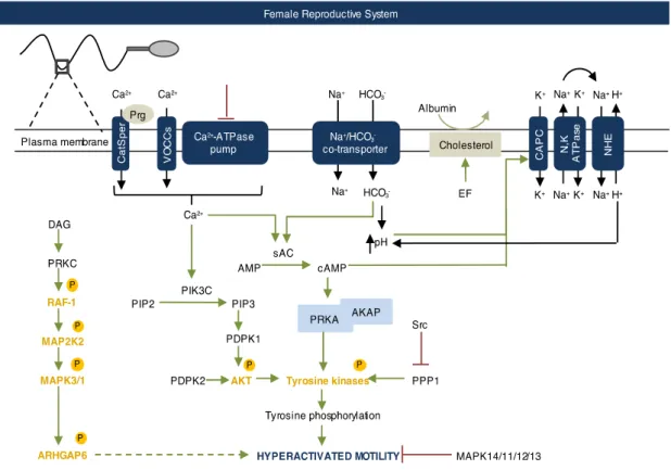
influx of HCO3- and, as a result, there is an increase on sperm pH and depolarization [144]. Though,
to achieve complete depolarization, there must be a Na+ and K+ influx, Na+ is transported by the
Na+/ HCO
3- co-transporters and K+ influx is mediated by Calcium-activated potassium channels and
intracellular alkalinization and cAMP, which hints a HCO3- indirect regulation of K+ [141]. Within the
sperm, HCO3- activates factors that exchange phospholipids within the bilayer plasma membrane.
Consequently, cholesterol is vulnerable to albumin, which is the most abundant protein on the female reproductive system, and the main cholesterol acceptor. Albumin can decrease up to 40% of the sperm cholesterol content and this leads to an increase on membrane fluidity [145,146]. Depolarization, intracellular alkalinization and increased membrane fluidity promote influx of Ca2+.
The Na,K-ATPase pump is a membrane protein found in all eukaryotes [147]. By using the energy released from ATP hydrolysis the Na, K-ATPase pump promotes the efflux of three molecules of Na+
and influx of two molecules of K+ [148]. Two subunits compose the Na,K-ATPase protein, the alpha
and beta subunits. In several species, including human, the alpha4 sbunit presents the most restricted expression. It is present in sperm principal piece only in mature sperm of males in sexual maturity. Besides the Na,K-ATPase alpha4 subunit, only subunit alpha1 is present in sperm [147]. Knockout studies revealed that the Na,K-ATPase alpha4 subunit is crucial for sperm physiology, since alpha4 subunit KO is completely sterile. Knockout sperm presents reduced primary and hyperactivated motility, bent flagellum, increased intracellular Na+ and cell plasma membrane
depolarization [149]). Mcdermott et al reinforce the role of Na,K-ATPase alpha4 subunit on human male fertility by showing that an overexpression of this protein in mouse testis results in an increased total motility (among other parameters of sperm movement) [150]. Although the exact mechanism underlying the role of ATPase in sperm physiology is not fully characterized, Na,K-ATPase alpha4 isoform appears to regulate intracellular H+. Since is unlikely that the Na,K-ATPase
transports H+, its ability to regulate intracellular H+ arises from its effect on the activity of a Na+/H+
exchanger (NHE). NHE uses the Na+ gradient established by the Na,K-ATPase to extrude H+ in
exchange for the influx of Na+ [151] and, consequently there is an increase in the intracellular pH
[151] (Figure A2.3). In bovine sperm, Jimenez et al demonstrate that Na,K-ATPase activity is up-regulated during capacitation. Also, when Na,K-ATPase activity is impaired, the intracellular decrease in Na+ and plasma membrane depolarization that typically accompany sperm capacitation
are inhibited [152]. Ouabain, a cardiac glycoside produced in adrenal glands, is a Na,K-ATPase inhibitor which may have a physiological role in fertilization.[153–155].
Signaling pathways in hyperactivated motility
It appears that all the processes that occur in the female reproductive system increase the Ca2+ and