In vitro design of a novel lytic bacteriophage
cocktail with therapeutic potential against
organisms causing diabetic foot infections
Joa˜o J. Mendes,
1,2Clara Leandro,
1Carla Mottola,
3Raquel Barbosa,
1Filipa A. Silva,
1Manuela Oliveira,
3Cristina L. Vilela,
3Jose´ Melo-Cristino,
4Andrzej Go´rski,
5Madalena Pimentel,
1,6Carlos Sa˜o-Jose´,
1,6Patrı´cia Cavaco-Silva
1,7and Miguel Garcia
1 Correspondence Joa˜o J. Mendes jmendes@technophage.pt Received 3 January 2014 Accepted 27 May 2014 1TechnoPhage, S.A., Lisbon, Portugal
2Internal Medicine Department, Santa Marta’s Hospital/Central Lisbon Hospital Center, Lisbon, Portugal
3
Interdisciplinary Center of Research in Animal Health, Faculty of Veterinary Medicine of the University of Lisbon, Lisbon, Portugal
4Institute of Microbiology, Faculty of Medicine of the University of Lisbon, Lisbon, Portugal 5Ludwik Hirszfeld Institute of Immunology and Experimental Therapy, Polish Academy of Sciences,
Wroclaw, Poland
6Department of Microbiology and Immunology, Faculty of Pharmacy, University of Lisbon, Lisbon, Portugal
7
Centro de Investigac¸a˜o Interdisciplinar Egas Moniz (CiiEM), Superior Institute of Health Sciences Egas Moniz, Monte de Caparica, Portugal
In patients with diabetes mellitus, foot infections pose a significant risk. These are complex infections commonly caused by Staphylococcus aureus, Pseudomonas aeruginosa and Acinetobacter baumannii, all of which are potentially susceptible to bacteriophages. Here, we characterized five bacteriophages that we had determined previously to have antimicrobial and wound-healing potential in chronic S. aureus, P. aeruginosa and A. baumannii infections. Morphological and genetic features indicated that the bacteriophages were lytic members of the family Myoviridae or Podoviridae and did not harbour any known bacterial virulence genes. Combinations of the bacteriophages had broad host ranges for the different target bacterial species. The activity of the bacteriophages against planktonic cells revealed effective, early killing at 4 h, followed by bacterial regrowth to pre-treatment levels by 24 h. Using metabolic activity as a measure of cell viability within established biofilms, we found significant cell impairment following bacteriophage exposure. Repeated treatment every 4 h caused a further decrease in cell activity. The greatest effects on both planktonic and biofilm cells occurred at a
bacteriophage : bacterium input multiplicity of 10. These studies on both planktonic cells and established biofilms allowed us to better evaluate the effects of a high input multiplicity and a multiple-dose treatment protocol, and the findings support further clinical development of bacteriophage therapy.
INTRODUCTION
Diabetes mellitus affects an estimated 171 million patients worldwide (Wild et al., 2004) and has become a major world epidemic. Even with the best preventative care, 9 %
of patients will develop a diabetic foot infection (DFI), which brings the consequent risk of amputation (Lavery et al., 2006). Qualitative and quantitative aspects of wound micro-biology are critical determinants of the wound outcome. Gram-positive micro-organisms are the first to colonize and acutely infect breaks in the skin, whereas chronic wounds develop a more complex polymicrobial microbiology, including aerobic Gram-negative rods (Lipsky et al., 2004).
Abbreviations: AB, alamarBlue; BT, bacteriophage therapy; DFI, diabetic foot infection; IM, input multiplicity; NCBI, National Center for Biotechnology Information; p.i. post-infection.
These micro-organisms aggregate in communities encased within extracellular polymeric substances on the wound surface. Such an entity is defined as a biofilm, and shows increased resistance to immunological and antimicrobial attack (Percival et al., 2012). In current clinical practice, DFI treatment includes debridement and systemic antibiotics (Lipsky et al., 2004). The increased incidence of antibiotic-resistant bacterial strains, such as meticillin-antibiotic-resistant Sta-phylococcus aureus and pan-drug-resistant, non-fermenting, Gram-negative bacilli, threatens the efficacy of antimicrobial therapy (Mendes et al., 2012). Thus, it is necessary to identify new therapeutic strategies for DFIs.
Bacteriophages are viruses that consist of a genome con-tained within a protein coat and that specifically infect bacteria. In contrast to filamentous bacteriophages, the multiplication of tailed bacteriophages and release of the newly formed virus particles always involves lysis of the host bacterial cell. However, among tailed bacterio-phages, some may not immediately follow this lytic pathway. The genome of these so-called temperate bacteriophages may instead reside in the host cell (integrated in the bacterial chromosome or in a plasmid-like form in the cytoplasm) and be propagated for several bacterial generations without lysis. In contrast, strictly lytic phages do not have this option and usually undergo the lytic pathway once inside the bacterial host (Ansaldi, 2012). Bacteriophage therapy (BT) is the use of lytic bacteriophages to reduce or elimi-nate pathogenic bacteria. BT has become a broadly relevant technology for veterinary, agricultural and food microbio-logical applications; however, the treatment of human infections with BT attracts the greatest interest (Kutter et al., 2010).
The use of bacteriophages as antibacterial agents for suppurative infections began shortly after the discovery of bacteriophages. Bruynoghe and Maisin first demonstrated BT, using bacteriophages to treat S. aureus skin infections (Bruynoghe & Maisin, 1921). However, following the discovery and general application of antibiotics, interest in the therapeutic uses of bacteriophages waned. Recently, the increase in antibiotic-resistant bacterial strains has reinvi-gorated enthusiasm about these bacteria-specific viruses (Chopra et al., 1997). This interest is particularly true in cases in which bacteriophages can be applied externally (topical application), as is the case for DFIs.
The development of an effective BT is a multistep process consisting of: (i) bacteriophage isolation and assessment for antibacterial activity against specific bacterial strains; (ii) bacteriophage characterization and screening for un-desirable traits; (iii) in vitro posology and dosage regimen design; (iv) pre-clinical animal efficacy and toxicology studies; and (v) regulated human clinical trials. Although the use of bacteriophages to treat DFIs is promising, difficulties in any of these steps can hinder widespread clinical application (Abedon, 2010).
Recently, we demonstrated the antimicrobial activity and wound-healing capability of a topically delivered bacteriophage
suspension against wounds chronically infected with chronic S. aureus, Pseudomonas aeruginosa and Acinetobacter bau-mannii in two animal models of diabetes mellitus (Mendes et al., 2013). In the current study, we present a character-ization of the bacteriophages used in this previous study. We examined their spectrum of activity, genetic and morpho-logical structures, and activity against planktonic cells and established biofilms. Collectively, the findings justify the posology and dosage regimen used in the animal studies.
METHODS
Bacterial strains.The S. aureus 743/06, P. aeruginosa 433/07 and A. baumannii 1305/05 host strains were isolated from human clinical samples that were collected and identified in hospitals in the Lisbon area. The three strains were characterized previously as biofilm producers (Mottola et al., 2013). Bacterial clinical isolates used for bacteriophage host-range investigation included S. aureus (n5132), P. aeruginosa (n593) and A. baumannii (n5103) from wound speci-mens. Of these isolates, 44 were from DFIs. The epidemiology, clinical details and specific microbiology of our collection of DFI isolates have been described previously (Mendes et al., 2012). All isolates were stored in tryptone soy broth (TSB; Biokar Diagnostics) with 15 % glycerol (w/v) at 270uC until needed. For the experiments, single bacterial colonies were grown in TSB at 37uC. After a 24 h incu-bation, the bacterial cells were suspended in saline and adjusted to McFarland’s scale 0.5 (bioMe´rieux), producing a final working suspension of approximately 5.06108
c.f.u. ml21.
Bacteriophage isolation, amplification and purification. S. aureus F44/10 and F125/10, P. aeruginosa F770/05 and F510/08, and A. baumannii F1245/05 bacteriophages were isolated from envir-onmental water samples from the Lisbon area. Standard methods for bacteriophage isolation (Adams, 1959) were employed for all five bacteriophages using the host strains described above. The obtained bacteriophage plaques were purified by repeated single plaque isolation to ensure that each contained only one type of bacteriophage. To produce bacteriophage stocks in sufficient quantities for the experiments, a previously described protocol of amplification, concentration by high-speed centrifugation and purification on a CsCl gradient (Miller, 1987) was used for all five bacteriophages. Briefly, a final lysate of each bacteriophage was centrifuged at 10 000 g for 20 min at 4uC. The pellet was discarded, and the supernatant fraction was concentrated overnight at 8000 r.p.m. (JA-14 rotor; Beckman Coulter). The bacteriophage pellet was resuspended in SM buffer (5.8 g NaCl l21, 2 g MgSO4.7 H2O l21, 50 ml 1 M Tris/HCl, pH 7.5). This concentrated bacteriophage suspension was loaded onto a discontinuous CsCl gradient and centrifuged at 30 000 r.p.m. for 5 h at 4uC in a Beckman L-90 ultracentrifuge with an SW41Ti rotor (Beckman Coulter). The banded bacteriophage particles were collected and thoroughly dialysed against SM buffer. Final bacterio-phage titres were determined using double agar overlay plaque assays (Kropinski et al., 2009). Purified bacteriophages were stored at 4uC and further diluted in SM buffer to achieve a working suspension of approximately 261010p.f.u. ml21prior to the assays.
Morphology of bacteriophages.The morphology of each of the five bacteriophages was analysed by transmission electron microscopy at the Fe´lix d’He´relle Reference Center for Bacterial Viruses, Laval University, Que´bec, Canada. Briefly, a 200-mesh Formvar carbon-coated copper grid (Pelco International) was deposited face down on 10 ml staining suspension (2 % uranyl acetate, pH 7.0, for all bac-teriophages except for F770/05, which was stained with 2 % phos-photungstic acid, pH 7.0). After 30 s, 10 ml bacteriophage suspension
was mixed with the stain. After 2–3 min, the grid was deposited face up on blotting paper. The grid was dried for 5 min and then observed at 80 kV using a JEOL 1230 transmission electron microscope. These data were integrated with the genomic analysis, and the bacteriophages were classified according to the Ackermann (2009) classification.
Genomic analysis of bacteriophages. The DNA of all five bacteriophages was isolated using a standard phenol/chloroform extraction and DNA precipitation protocol (Sambrook et al., 1989). The purified nucleic acid was sent to Macrogen for commercial sequencing. In brief, pyrosequencing of the sample DNA was performed using a GS FLX Titanium General Library Preparation kit (Roche 454 Company) according to the manufacturer’s instruc-tions. The assembly of quality-filtered reads was performed using Genome Sequencer De novo Assembler software (Newbler) version 2.5.3. An extensive bioinformatics evaluation was conducted to analyse the sequences and identify regions of similarity with entries in databases, which yield clues about structure and function. Each genome sequence was scanned using the National Center for Biotechnology Information (NCBI)BLASTNandBLASTXbioinformatics tools (http://blast.ncbi.nlm.nih.gov/Blast.cgi). Prediction of ORFs was performed by integrating the results obtained by the programs GeneMark.hmm (http://exon.gatech.edu/genemark/eukhmm.cgi) and MetaGeneAnnotator (http://metagene.cb.k.u-tokyo.ac.jp). Protein homo-logy searches were performed with theBLASTPprogram (http://blast. ncbi.nlm.nih.gov/Blast.cgi?PAGE=Proteins) using the NCBI non-redundant protein sequence database. The genome sequences were deposited in the patent division of GenBank (specific patent nos: WO2010090542 and WO2012036580).
Bacteriophage host range. The five bacteriophages were tested against a panel of clinical isolates using the bacteriophage spot-test procedure (Armon & Kott, 1993). Briefly, 3 ml top-0.7 % tryptone soy agar (TSA; Biokar Diagnostics) was added to 200 ml overnight culture of each clinical isolate and poured over TSA. The agar was allowed to solidify, after which 5 ml each bacteriophage suspension (approx. 108p.f.u.) was spotted on the bacterial lawn of each different isolate. The drop was allowed to dry, and the plates were incubated overnight at 37uC. Specific bacteriophage-sensitive isolates showed clear areas where the bacteriophage suspensions had been spotted. Bacteriophage activity against planktonic cells. To determine the activity of the bacteriophages against planktonic cells in vitro, a kinetic time–kill assay (NCCLS, 1999) was performed using a modified protocol. Briefly, 1 ml host bacterial suspension (56108
c.f.u.) was diluted in 9 ml TSB, yielding a final concentration of 56107c.f.u. ml21. For single-bacteriophage studies, 100 ml (56109p.f.u.) specific bacteriophage was added, yielding a final concentration of 56108p.f.u. ml21[input multiplicity (IM) of bacteriophage : bacterium of 10]. For combination studies, 100 ml (56109 p.f.u.) each bacteriophage suspension was added, resulting in a final concentration of 56108 p.f.u. ml21(IM of 10) for each of the bacteriophages. An additional kinetics assay was performed for P. aeruginosa 433/07, in which 10 ml (56108
p.f.u.) bacteriophage F770/05 suspension was added (yielding an IM of 1), alone or in combination with the bacteriophage F510/08 at an IM of 10. Control experiments were performed in parallel using bacteriophage buffer instead of a bacteriophage suspension. All mixtures were incubated at 37uC with agitation, and 100 ml aliquots were collected at 0, 1, 3, 5 and 24 h post-infection (p.i.). Bacterial quantification was performed using a 10-fold serial dilution method (Murray et al., 2003). All experiments were conducted in triplicate. The results are presented as the mean±SD and are expressed as log-transformed values [log (c.f.u. ml21)] over time.
Combined bacteriophage activity against established biofilms. The activity of the bacteriophages against established biofilms was examined using a modification of previously described protocols
(Cerca et al., 2005; Pettit et al., 2005). Briefly, 1 ml of each of the host bacterial suspensions (56108
c.f.u.) was diluted in 9 ml TSB, and 100 ml of this dilution (56106
c.f.u.) was added to a 96-well flat-bottomed polystyrene microtitre plate (Orange Scientific) and incubated at 37uC for 24 h to allow biofilm formation. After incubation, the planktonic bacteria were removed carefully with a sterile pipette. The number of biofilm cells at 24 h has been demonstrated previously to be approximately 107c.f.u. per well for all bacterial species (Mottola et al., 2013). Next, 150 ml bacteriophage suspension (IMs of 10 and 100) diluted in TSB was added to the wells. The following bacteriophage suspensions were used for each bacterium: for S. aureus, a 1 : 1 combination of F44/10 and F125/10; for P. aeruginosa, a combination of F770/05 and F510/08 at a 1 : 10 ratio; and for A. baumannii, F1245/05 alone. Biofilms treated with TSB alone served as positive controls in measurements of cell metabolic activity (see below).
The microplates were incubated at 37uC for 4 or 24 h. At each time point, the wells were processed according to a previously described protocol (Pettit et al., 2005) using alamarBlue (AB; Thermo Scientific), and their absorbance at 570 and 600 nm was measured using a SpectraMax 340PC microplate reader (Molecular Devices). A second assay was performed in which, after biofilm formation, planktonic bacteria were removed from the wells and replaced with a bacteriophage suspension every 4 h over a 24 h incubation period. In the positive-control group, planktonic bacteria were removed from the wells and replaced with TSB every 4 h. These plates were then processed as described previously (Pettit et al., 2005).
Biofilm susceptibility experiments were performed a minimum of three times. All results are presented as the percentage variation of AB±SD. This value was calculated using the manufacturer’s formula,
with one exception: the medium-only negative control in the formula was replaced by a more robust negative control that consisted of medium plus bacteriophage at each IM (i.e. IMs of 10 and 100). Strong antimicrobial suppression was defined as a¢50 % reduction in AB compared with the positive control.
Statistical analysis.For all datasets, comparisons between groups were performed using two-tailed Student’s t-tests, and values of P,0.05 were considered significant. All data were entered into a spreadsheet program (Excel; Microsoft) for statistical analysis. Analytical statistics were performed using Analyse-it, version 2.21 Excel 12+ (Analyse-it Software), a statistical add-in program for Excel.
RESULTS
Bacteriophage features
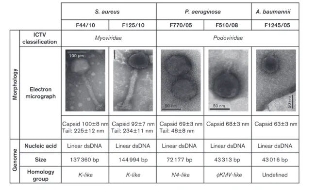
After purification, the selected bacteriophages were initially characterized according to plaque morphology. The S. aureus F44/10 and F125/10, P. aeruginosa F770/05 and F510/08, and A. baumannii F1245/05 bacteriophages pro-duced clear lytic plaques ranging from 1.5 to 5 mm in diameter. Plaques produced by the bacteriophages F770/05, F510/08 and F1245/05 were surrounded by growing opaque halo zones. The morphological and genomic characteristics of the five bacteriophages are presented in Fig. 1.
Morphology
To classify the purified bacteriophages based on their virion morphology, we used transmission electron micro-scopy. The staphylococcal bacteriophages F44/10 and F125/
10 appeared to be composed of a contractile tail and an isometric head, with a baseplate structure also discernible at the tip of the F44/10 tail. These features, along with their genomic properties (see below), allowed us to classify F44/ 10 and F125/10 as members of the family Myoviridae. The Pseudomonas bacteriophages F770/05 and F510/08 and the Acinetobacter bacteriophage F1245/05 had short tails and were classified as members of the family Podoviridae. The family Podoviridae consists of different subgroups. Although there are certain morphological similarities between the bacteriophages F510/08 and F1245/05 and the wKMV-like group (Chang et al., 2011; Lammens et al., 2009), definite morphological assignment of F1245/05 could not be performed due to the uncharacteristic morphology of these virion particles.
Genomic analysis
The bacteriophages were characterized at the genomic level by determining and analysing their genome sequences. The bacteriophages F44/10 and F125/10 had the largest genomes and hence a greater number of putative genes and ORFs, which is in agreement with the characteristic features of viruses belonging to the family Myoviridae (Lavigne et al., 2009). The genomes of the bacteriophages F44/10 and F125/10 displayed high similarity (up to 98 % nucleotide sequence identity, 80–90 % genome coverage) to those of a group of highly related staphylococcal
myoviruses, which includes bacteriophages K (O’Flaherty et al., 2004), A5W (GenBank accession no. EU418428) and GH15 (Gu et al., 2012). The bacteriophages F510/08 and F770/05 shared high sequence identity (up to 98 % nucleo-tide sequence identity, 83–98 % genome coverage) with Pseudomonas wKMV-like and N4-like viruses, respectively (Ceyssens et al., 2010). Examples of wKMV-like viruses are the bacteriophages wKMV and LUZ19 (Ceyssens et al., 2006; Lavigne et al., 2003) and of N4-like viruses are LIT1 and LUZ7 (Ceyssens et al., 2010). The bacteriophage F1245/05 presented no significant similarity at the DNA level with any known bacteriophage in the databases, except for a few short segments with up to 4 % nucleotide sequence identity and 81 % genome coverage.
The deduced products of the predicted genes of all bacteriophages were compared with sequences in the NCBI non-redundant protein sequence database using
BLASTP. No significant similarity with known virulence or toxin proteins or with elements typically associated with lysogeny (integrases, repressors and antirepressors) could be found. Finally, the protein similarity searches did not reveal potential exopolysaccharide depolymerase genes. Bacteriophage host range
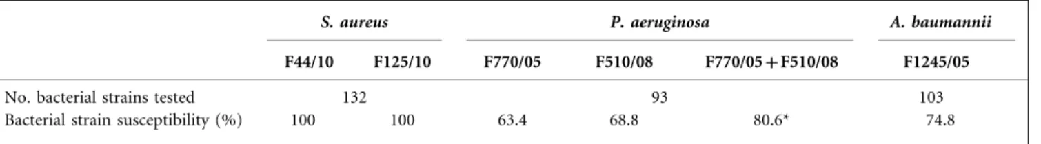
To gain insight into the host range of selected bacter-iophages, the susceptibility of three panels of clinical isolates of S. aureus (n5132), P. aeruginosa (n593) and A.
S. aureus P. aeruginosa
Podoviridae Myoviridae
K-like K-like N4-like φKMV-like
A. baumannii F44/10 ICTV classification Morphology Genome Electron micrograph
Nucleic acid Linear dsDNA Linear dsDNA Linear dsDNA Linear dsDNA Linear dsDNA 137 360 bp 100 mm 50 nm 50 nm 50 nm 144 994 bp 72 177 bp 43 313 bp 43 016 bp Size Undefined Homology group Capsid 100±8 nm
Tail: 225±12 nm Tail: 234±11 nmCapsid 92±7 nm Capsid 69±3 nm Capsid 68±3 nm Capsid 63±3 nmTail: 48±8 nm
F125/10 F770/05 F510/08 F1245/05
Fig. 1. Morphological and genomic characteristics of the bacteriophages used for BT. Five bacteriophages previously shown to successfully treat infections in vivo were characterized using transmission electron microscopy. Representative images are shown. The genomes were sequenced by pyrosequencing and analysed extensively using BLASTN,BLASTX, GeneMark.hmm, MetaGeneAnnotator andBLASTP. ICTV, International Committee on the Taxonomy of Viruses.
baumannii (n5103) was tested for each species-specific bacteriophage. There was a degree of variability in the host range of each bacteriophage (Table 1). All tested staphylo-coccal strains were susceptible to both S. aureus bacter-iophages (F44/10 and F125/10). In contrast, when examined individually, the P. aeruginosa bacteriophages F770/05 and F510/08 lysed only 63.4 and 68.8 % of the tested isolates, respectively. However, when these results were considered together, we observed that 80.6 % of the P. aeruginosa isolates were infected by at least one of the bacteriophages, whereas 51.6 % were susceptible to both bacteriophages. Finally, of the tested A. baumannii strains, 74.8 % were susceptible to the bacteriophage F1245/05.
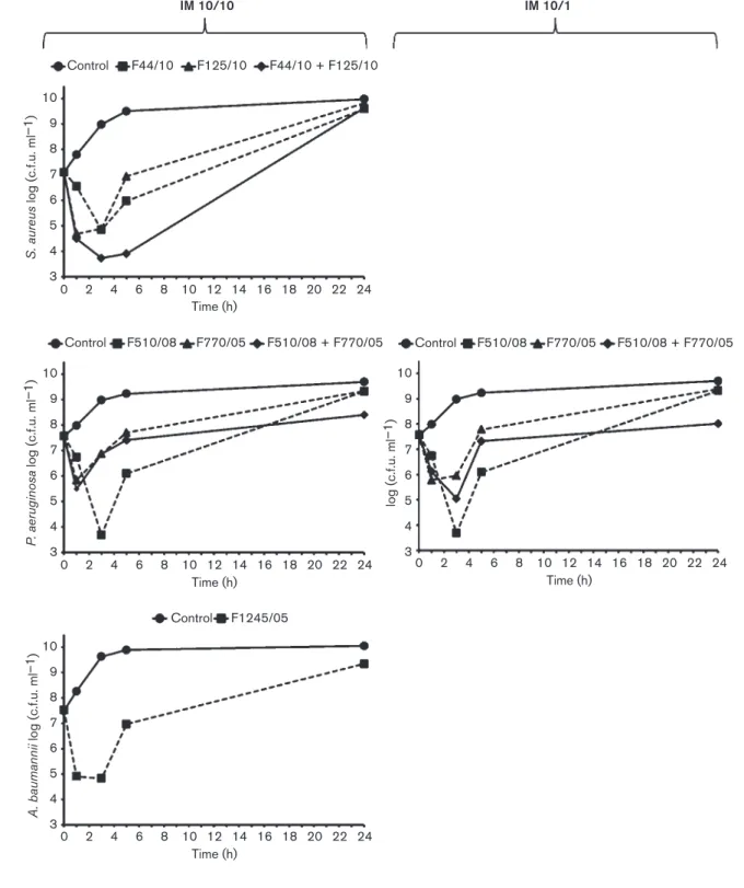
Bacteriophage activity against planktonic cells To evaluate the activity of the selected bacteriophages against planktonic cells, liquid cultures of the different bacterial hosts were exposed to the corresponding bacter-iophages, both individually and in combination, and cell growth/viability was monitored over time with constant agitation. The time–kill curves are presented in Fig. 2. S. aureus 743/06, when challenged with either F44/10 or F125/ 10 at an IM of 10, showed impaired growth, with reductions in cell counts of 2.3±0.3 and 2.2±0.2 log (c.f.u. ml21), respectively, at 3 h p.i. However, after 24 h, the cultures recovered to near-control levels. The reduction in the number of viable cells observed at 3 h was significantly enhanced when the two bacteriophages were used in combination [3.4±0.2 log (c.f.u. ml21); P,0.01]. Nevertheless, there was no difference in the recovery of growth at 24 h.
At an IM of 10, the P. aeruginosa bacteriophage F510/08 caused a 3.9±0.4 log (c.f.u. ml21) reduction in the viability of P. aeruginosa 433/07 at 3 h p.i. This reduction was more modest [0.7±0.4 log (c.f.u. ml21)] for F770/05 at the same IM and time point. When the two bacteriophages were combined, the kill curve was not different from that of F770/05 for the first 5 h; however, the combination provided a statistically significant reduction relative to the control at 24 h [1.3±0.3 log (c.f.u. ml21); P,0.01]. When the IM of F770/05 was reduced to 1, combined with F510/08 at an IM of 10, the initial 3 h reduction was more pronounced [2.5±0.4 log (c.f.u. ml21); P,0.01]. Similarly, this combination caused a statistically significant reduction [1.7±0.3 log (c.f.u. ml21); P,0.01] relative to the control
at 24 h. A. baumannii 1305/05 suffered an initial 2.7±0.2 log (c.f.u. ml21) reduction at 3 h after single-bacteriophage (F1245/05) challenge. Although this bacterial strain recov-ered by 24 h, it did not reach the control levels of viability. Activity of bacteriophage combinations against established biofilms
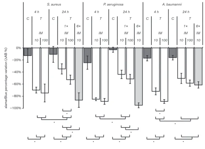
We also studied the ability of the bacteriophages to eliminate cells in established biofilms by treating biofilms with species-specific bacteriophage combinations. AB, which quantita-tively measures cell metabolic activity using an oxidation– reduction indicator that changes colour in the presence of metabolically active cells, was used to measure cell viability in biofilms with and without treatment. The viability of cells within a biofilm is one of the most important aspects when evaluating the effectiveness of antimicrobial agents; there-fore, we used a quantification method based on metaboli-cally active cells, as determined by AB. This assay is a reliable and reproducible method for evaluating biofilm suscept-ibility and is considered to be preferable over the viable plate-count method, as it is very difficult to recover all surviving adherent bacteria as single cells using the latter method (Pettit et al., 2005). The AB-based assay has been used to identify antimicrobials with enhanced efficacy against certain clinically important bacterial biofilms (Pettit et al., 2005, 2009).
Fig. 3 shows the percentage of AB reduction in control and treated biofilms at 4 and 24 h using different IMs and frequencies of application. At 4 h, the tested bacteriophage preparations strongly reduced the cell viability of all bacterial hosts, independently of the IM. There was only a statistically significant difference between IMs for A. baumannii; the higher IM resulted in a greater reduction in metabolic activity (71.9±5.8 vs 88.7±3.1 %; P,0.01). At 24 h, after a one-time bacteriophage preparation application, the cell viability of all bacterial strains was less suppressed than at 4 h but still significantly different from that of the control. At 24 h, there were no statistically significant differences between IMs except for S. aureus, for which the higher IM resulted in a greater reduction in cell viability (34.8±8.5 vs 52.6±7.7 %; P,0.01). In experi-ments using multiple bacteriophage treatexperi-ments, a greater reduction in cell viability was observed compared with the reduction following one-time bacteriophage treatment.
Table 1. Susceptibility of wound bacterial isolates to candidate bacteriophages for BT
S. aureus P. aeruginosa A. baumannii
F44/10 F125/10 F770/05 F510/08 F770/05+F510/08 F1245/05
No. bacterial strains tested 132 93 103
Bacterial strain susceptibility (%) 100 100 63.4 68.8 80.6* 74.8
Control 10 9 8 7 6 S. aureus log (c.f.u. ml –1) 5 4 3 22 20 18 16 14 12 Time (h) 10 8 6 4 2 0 24 F44/10 F125/10 IM 10/10 IM 10/1 F44/10 + F125/10 Control 10 9 8 7 6 P. aeruginosa log (c.f.u. ml –1) 5 4 3 22 20 18 16 14 12 Time (h) 10 8 6 4 2 0 24 F510/08 F770/05 F510/08 + F770/05 Control 10 9 8 7 6 A. baumannii log (c.f.u. ml –1 ) 5 4 3 22 20 18 16 14 12 Time (h) 10 8 6 4 2 0 24 F1245/05 Control 10 9 8 7 6 log (c.f.u. ml –1 ) 5 4 3 22 20 18 16 14 12 Time (h) 10 8 6 4 2 0 24 F510/08 F770/05 F510/08 + F770/05
Fig. 2. Time-kill curves of the target bacteria during planktonic growth when challenged with their specific bacteriophages (alone or in combination). Bacterial strains were grown in TSB with constant agitation and with or without bacteriophages. Growth was monitored and quantified by calculating cfu/ml at 0, 1, 3, 5, and 24 hours. In the left panel (input multiplicity, IM 10/ 10), assays were performed on S. aureus 743/06, P. aeruginosa 433/07, and A. baumannii 1305/05 in which the specific bacteriophage suspensions were added to provide an IM of 10. In the right panel (IM 10/1), an additional assay was performed on P. aeruginosa 433/07 in which the bacteriophage F770/05 suspension was added to provide an IM of 1, alone or in combination with the bacteriophage F510/08 at an IM of 10.
This trend was found for S. aureus and P. aeruginosa but not for A. baumannii.
DISCUSSION
Effective bacteriophage preparations for therapeutic pur-poses require careful design through a multistep research process of bacteriophage characterization, cocktailing and dosing. This process includes in vitro studies, such as those presented here, and in vivo studies, which have been published previously (Mendes et al., 2013). Ideally, the characterization of bacteriophages for BT should be as thorough and complete as possible. However, in certain cases, it may be more practical to minimize this process and to focus the characterization on particular traits that are the most desirable for a specific application. Combining different bacteriophages in the same preparation (mixtures of two or more bacteriophages within a given formulation)
frequently results in a broader spectrum of antibacterial activity and/or lytic efficacy and may allow targeting of bacteria under different conditions or in different environ-ments (Chan & Abedon, 2012). Finally, in vitro experi-ments such as those described in this work are useful for evaluating the direct interaction between a drug and bac-teria, which enables the selection of candidate bacteriophages. These studies also provide valuable information for the determination of optimal posology (Abedon & Thomas-Abedon, 2010).
Integration of the information emerging from the mor-phological and genomic analyses revealed that the bacter-iophages used here were all tailed bacterbacter-iophages (order Caudovirales), with two belonging to the family Myoviridae and three to the family Podoviridae. Genome sequence analysis did not identify any known genes related to lysogeny or traits that might enhance the virulence of the target bacteria, which is an important observation
S. aureus P. aeruginosa A. baumannii
4 h C T IM 10 100 0% –20% –40% –60%
alamarBlue percentage variation (
∆AB %) –80% –100% * * * * * * * * * * * * * 24 h C T IM 1× IM 10 6× 10 100 4 h C T IM 10 100 24 h C T IM 1× IM 10 6× 10 100 4 h C T IM 10 100 24 h C T IM 1× IM 10 6× 10 100
Fig. 3. Analysis of the activity of the bacteriophages against bacterial biofilms. Bacteria were grown for 24 h to establish biofilms and the bacteriophages were then added. The following bacteriophage suspensions were used for each bacterium: for S. aureus, a 1 : 1 combination of F44/10 and F125/10; for P. aeruginosa, a combination of F770/05 and F510/08 at a 1 : 10 ratio; and for A. baumannii, F1245/05 alone. Cell metabolism was quantified with AB and is reported as the percentage reduction relative to growth in untreated controls. C, negative control; T, treated with bacteriophage; 1!, one-time bacteriophage suspension application, 6!, bacteriophage suspension application every 4 h for 24 h. *P,0.01 (square brackets indicate the comparisons between different groups).
regarding the safe use of bacteriophages. Another import-ant selection criterion for bacteriophages for BT is their host range, which should be as broad as possible, par-ticularly including clinically prevalent bacterial species (Gill & Hyman, 2010). In this study, members of family Myo-viridae exhibited the broadest spectrum of lytic activity, whereas viruses of the family Podoviridae exhibited a narrower spectrum, particularly the pseudomonal bacter-iophages. The spectrum of activity of the staphylococcal bacteriophages was relatively broad as expected, given their high relatedness to bacteriophages K and A5W, both of which have been described previously as polyvalent bacteriophages (O’Flaherty et al., 2005; GenBank accession no. EU418428). Nevertheless, the host ranges of the Pseu-domonas and Acinetobacter bacteriophages were remarkable compared with those of other species-specific bacterio-phages (Merabishvili et al., 2009; Popova et al., 2012). The overall morphology, genomic characterization and host-range results suggested that these bacteriophages are very good candidates for BT. However, care must be taken when generalizing these results, because the bacterial clinical isolates used for the bacteriophage host-range investigation reflect only the microbiological profile of diabetic foot ulcers in a particular geographical area, and these vary worldwide. Also the sensitivity of the spot test must be taken into account. Whilst the use of high bacteriophage titres (108p.f.u. per spot) for host-range analysis is routine when considering bacteriophages for BT (Kutter, 2009), it should be noted that the use of lower titres may reduce host-range. In our host-range investigation, the use of bacteriophage stock dilutions up to 103 p.f.u. per spot yielded differences from the presented results by up to 23 % (unpublished data).
Time–kill curves provide detailed information about antimicrobial efficacy against planktonic bacteria as a function of time. These curves are often used to study the antibacterial effect of single and combination drug compounds and dosing regimens before in vivo efficacy studies (NCCLS, 1999). In the current study, following bacteriophage exposure, all bacteria had an initial bacterial reduction to a nadir between 1 and 3 h p.i., followed by regrowth that was noticeable after 5 h and even more pronounced after 24 h. The Pseudomonas bacteriophage combination resulted in a significantly greater reduction in bacteria compared with the reduction stimulated by most active single bacteriophage 24 h after bacteriophage exposure. However, the decrease was insufficient to be considered as a synergistic effect, defined as a¢2 log (c.f.u. ml21)-fold decrease by a combination compared with the most active single agent (NCCLS, 1999). In the Pseu-domonas combination study, when an IM of 1 was used for the Pseudomonas F770/05 bacteriophage instead of an IM of 10, we observed a greater initial bacterial reduction after 3 h, but similar results were obtained at 24 h. This interaction was not specifically analysed in our study, and there is no obvious explanation for this, but clearly further studies would be of interest.
The study presented here has certain limitations. First, only a single bacterial inoculum was used. This value was care-fully selected based on several lines of evidence. A higher inoculum (107 c.f.u. ml21) was used than the normal 105 c.f.u. ml21 inoculum used in previous time–kill studies testing antibiotics (NCCLS, 1999) because we wanted to mimic a worst-case scenario, similar to that found in wounds (Loc-Carrillo et al., 2012). In a previous epide-miological study (Mendes et al., 2012), microbiological products (aspirates, biopsies and swabs collected using the Levine method) of clinically infected foot ulcers in patients with diabetes were found to have a maximum bioburden of 107c.f.u. (g tissue)21(or cm22of ulcer area). Additionally, the most recent study using a previously optimized rodent model (Mendes et al., 2013) tested this bacteriophage cocktail on infected wounds with a known mean wound bioburden of 7.54±0.19 log (c.f.u.) per ulcer.
Secondly, the IM in nearly all experiments was 10 (fixed IM). The final IM chosen was selected based on the ‘multiplicity of 10 rule’, which states that if the goal is a significant reduction in bacterial density, then one should strive for in the order of 10 bacteriophages adsorbed to the average bacterium (Abedon, 2009; Kasman et al., 2002). Previous studies on infected animal and human burn tissue have concluded that low-titre bacteriophage administra-tion (IMs lower than 10) is unlikely to be successful (Goode et al., 2003; Kumari et al., 2010). Furthermore, increasing the IM increases the success of BT by reducing bacterial numbers.
Thirdly, we observed regrowth in planktonic cells exposed to bacteriophages within 24 h. This observation may be indicative of the development of resistance, as in vitro resistance is frequent in both BT and antibiotic therapy. For example, a study (O’Flynn et al., 2004) previously found in vitro resistance frequencies of 1026–1024 for single-phage treatments and 1026 for double-phage or triple-phage cocktails against Escherichia coli O157 : H7. Similarly, resistance to fusidic acid can readily be selected from an initial high inoculum, with a mean frequency of 1026–1028. This resistance has not limited the antibiotic’s topical use and does not appear to be a clinical problem (Sahm et al., 2013; Turnidge & Collignon, 1999). However, these observations do not imply in vivo resistance. According to certain studies, the rate of development of resistance to bacteriophages is approximately 10-fold lower than the rate of the development of antibiotic resistance (Carlton, 1999). Nonetheless, as observed here, in vitro studies show that bacteriophage resistance can evolve within hours, independently of the use of bacteriophage combinations. However, the evolution of bacteriophage resistance in vitro does not seem relevant to in vivo scenarios, in which bacteria replicate more slowly and are challenged by more difficult environmental conditions. A previous study (Capparelli et al., 2007) found a mean resistance frequency of 1.261028for S. aureus treated with
bacteriophages in vitro. However, the researchers were unable to isolate any bacteriophage-resistant S. aureus
strains in vivo. Indeed, even though the resistance of bacteria to the bacteriophage cocktails used here was not specifically studied, we previously found that the presence of residual bacteria did not globally hinder planimetric or histological improvement (Mendes et al., 2013). In the current study, the greatest reduction in bacterial counts occurred at 3 h, and regrowth was observed at 5 h, which enabled us to conclude that the best time to give a ‘boost’ application of bacteriophage would be between these two time points.
In a previous study (unpublished data), we found that the plaques of the bacteriophages F770/05, F510/08 and F1245/ 05 were surrounded by growing opaque halo zones, which could be related to the presence of a virion-associated exopolysaccharide depolymerase (Cornelissen et al., 2011). This and related enzymes have been found to enhance the biofilm-eradicating activity of bacteriophages compared with non-depolymerase-producing bacteriophages (Hughes et al., 1998). Based on genomic analysis, none of our bacteriophages seemed to produce any obvious extracellular polysaccharide or exopolysaccharide depolymerase. However, because bacteriophages that do not produce depolymerases have also been used in biofilm elimination (Chibeu et al., 2012), we sought to investigate the effect of bacteriophage combinations on the viability of target bacterial cells in pre-formed biofilms at 4 and 24 h. Here, assays using an IM of 10 produced nearly identical results as assays using an IM of 100, with two exceptions. First, we observed different results between an IM of 10 and an IM of 100 after 4 h for A. baumannii; however, this discrepancy may have arisen because only one bacteriophage was used. When previous experiments used a combination of two bacteriophages, the IM doubled, producing synergistic results (Abedon & Thomas-Abedon, 2010). Secondly, differences between IMs were observed after 24 h for S. aureus. This result may have occurred because the receptor for the bacterio-phage F44/10, which we speculate to be N-acetylglucosa-mine in the cell-wall teichoic acid, is very frequent (relative to other receptors) in both live cells and bacterial debris. This property means that active bacteriophages may adsorb to fragments of lysed cells (debris) instead of live cells, at a higher rate. This phenomenon may ultimately lead to injection of the genetic material in a suicidal manner, eliminating the bacteriophage from the system (Rabinovitch et al., 2003). Increasing the IM amplifies the probability of bacteriophage–bacterium interaction, resulting in a true cell infection. Moreover, in vivo, a bacteriophage dose suffi-ciently in excess of the target bacterium population (IM ¢100) should be given to account for bacteriophage loss, dilution (associated with absorption and distribution), decay and/or inefficiencies of bacteriophage adsorption to bacteria (e.g. inefficiencies in penetration into biofilms in vivo).
It is well known that bacterial regrowth occurs after biofilms have been exposed to antibiotics (Kussell et al., 2005). One possible way to limit this regrowth is through multiple dose applications. Our results using multiple dose
applications, as opposed to single-application dosing, are similar to the results observed in Georgia, where BT is the current standard of clinical care, and in Poland, where BT is used as an experimental treatment under a compassion-ate-use regulatory provision (Abedon et al., 2011; Kutter et al., 2010; Mie˛dzybrodzki et al., 2012). These results were also corroborated experimentally in previously published animal studies (Capparelli et al., 2007). This implies that a significant proportion of the bacteria in biofilm do not have genotypic resistance but rather some form of phenotypic resistance, which is reversible by the modifica-tion of the causal environmental factors. Various equally valid and non-mutually excluding theories have been pre-sented that could explain the possible coexistence dynamics of bacteriophages and susceptible bacteria: numerical refuge, physiological refuge and shielding by bacterial debris. The numerical refuge theory (Chao et al., 1977) predicts that simple mass-action interactions between bacteriophages and sensitive and resistant bacteria deter-mine the stability of the population. Thus, in our study, when new bacteriophages were added (creating a higher bacteriophage density), a decline in the number of sensitive cells resulted. The physiological refuge hypothesis (Lenski & Levin, 1985) postulates that during certain stages of bacterial life cycles sensitive bacteria may become trans-itorily resistant to bacteriophage infection. In the present study, fresh medium was then added. This altered the life cycle of the present sensitive bacteria (e.g. from stationary to logarithmic), thereby potentially converting them from a temporarily resistant state into a susceptible state. Finally, the shielding by bacterial debris theory (Rabinovitch et al., 2003) predicts that active bacteriophages adsorb onto fragments of lysed cells (debris) and inject their genetic material in a suicidal manner, thus discounting from the system as a bacteriophage. In the present study, as new bacteriophages were added, dead cells were removed, thus reducing non-productive binding as described. None of these observations was noted for A. baumannii, perhaps because this was the only case in which we used a single bacteriophage, limiting the importance of non-heritable mechanisms in the reduction in resistance induced by mutation.
In conclusion, we prepared, purified and characterized bacteriophage cocktails with a broad spectrum of activity against S. aureus, P. aeruginosa and A. baumannii strains that commonly cause DFIs. The complementary studies on both planktonic cells and established biofilms allowed us to better evaluate the effects of a high IM (¢10) and a multiple-dose treatment protocol (every 4 h for 24 h). We believe that this work takes an important step towards the future clinical application of BT.
ACKNOWLEDGEMENTS
This study was supported by TechnoPhage S.A. and Tecnifar. Additionally, the studies on established biofilms were conducted with the financial support of the Foundation for Science and
Technology (PTDC/SAU-MIC/122816/2010 – ‘Biofilms in diabetic foot: microbial virulence characterization and cross-talk of major isolates’). C. M. holds a Foundation for Science and Technology PhD fellowship (SFRH/BD/72872/2010), and M. O. is a researcher in the Foundation for Science and Technology ‘Cieˆncia 2007’ programme. These funding bodies did not have any role in the design of the experiments; in the collection, analysis and interpretation of data; in the writing of the manuscript; or in the decision to submit the manuscript for publication.
REFERENCES
Abedon, S. T. (2009). Kinetics of phage-mediated biocontrol of bacteria. Foodborne Pathog Dis 6, 807–815.
Abedon, S. T. (2010).The ‘nuts and bolts’ of phage therapy. Curr Pharm Biotechnol 11, 1.
Abedon, S. T. & Thomas-Abedon, C. (2010). Phage therapy pharmacology. Curr Pharm Biotechnol 11, 28–47.
Abedon, S. T., Kuhl, S. J., Blasdel, B. G. & Kutter, E. M. (2011).Phage treatment of human infections. Bacteriophage 1, 66–85.
Ackermann, H.-W. (2009).Phage classification and characterization. In Bacteriophages Methods and Protocols, pp. 127–140. Edited by M. R. J. Clokie & A. M. Kropinski. Clifton, NJ: Humana Press.
Adams, M. H. (1959).Methods of study of bacterial viruses: isolation of bacterial viruses. In Bacteriophages, pp. 447–449. New York: Interscience.
Ansaldi, M. (2012).Cell biology perspectives in phage biology. Front Biosci (Elite Ed) 4, 1823–1829. .
Armon, R. & Kott, Y. (1993).A simple, rapid and sensitive presence/ absence detection test for bacteriophage in drinking water. J Appl Bacteriol 74, 490–496.
Bruynoghe, R. & Maisin, J. (1921).Essais de the´rapeutique au moyen du bacteriophage. C R Soc Biol 85, 1120–1121 (in French). Capparelli, R., Parlato, M., Borriello, G., Salvatore, P. & Iannelli, D. (2007).Experimental phage therapy against Staphylococcus aureus in mice. Antimicrob Agents Chemother 51, 2765–2773.
Carlton, R. M. (1999). Phage therapy: past history and future prospects. Arch Immunol Ther Exp (Warsz) 47, 267–274.
Cerca, N., Martins, S., Cerca, F., Jefferson, K. K., Pier, G. B., Oliveira, R. & Azeredo, J. (2005). Comparative assessment of antibiotic susceptibility of coagulase-negative staphylococci in biofilm versus planktonic culture as assessed by bacterial enumeration or rapid XTT colorimetry. J Antimicrob Chemother 56, 331–336.
Ceyssens, P. J., Lavigne, R., Mattheus, W., Chibeu, A., Hertveldt, K., Mast, J., Robben, J. & Volckaert, G. (2006). Genomic analysis of Pseudomonas aeruginosa phages LKD16 and LKA1: establishment of the phiKMV subgroup within the T7 supergroup. J Bacteriol 188, 6924–6931.
Ceyssens, P. J., Brabban, A., Rogge, L., Lewis, M. S., Pickard, D., Goulding, D., Dougan, G., Noben, J. P., Kropinski, A. & other authors (2010).Molecular and physiological analysis of three Pseudomonas aeruginosa phages belonging to the ‘‘N4-like viruses’’. Virology 405, 26–30.
Chan, B. K. & Abedon, S. T. (2012).Phage therapy pharmacology phage cocktails. Adv Appl Microbiol 78, 1–23.
Chang, K. C., Lin, N. T., Hu, A., Lin, Y. S., Chen, L. K. & Lai, M. J. (2011). Genomic analysis of bacteriophage wAB1, a wKMV-like virus infecting multidrug-resistant Acinetobacter baumannii. Genomics 97, 249– 255.
Chao, L., Levin, B. R. & Stewart, F. M. (1977).A complex community in a simple habitat: an experimental study with bacteria and phage. Ecology 58, 369–378.
Chibeu, A., Lingohr, E. J., Masson, L., Manges, A., Harel, J., Ackermann, H. W., Kropinski, A. M. & Boerlin, P. (2012). Bacterio-phages with the ability to degrade uropathogenic Escherichia coli biofilms. Viruses 4, 471–487.
Chopra, I., Hodgson, J., Metcalf, B. & Poste, G. (1997).The search for antimicrobial agents effective against bacteria resistant to multiple antibiotics. Antimicrob Agents Chemother 41, 497–503.
Cornelissen, A., Ceyssens, P. J., T’Syen, J., Van Praet, H., Noben, J. P., Shaburova, O. V., Krylov, V. N., Volckaert, G. & Lavigne, R. (2011). The T7-related Pseudomonas putida phage w15 displays virion-associated biofilm degradation properties. PLoS ONE 6, e18597. Gill, J. J. & Hyman, P. (2010).Phage choice, isolation, and preparation for phage therapy. Curr Pharm Biotechnol 11, 2–14.
Goode, D., Allen, V. M. & Barrow, P. A. (2003). Reduction of experimental Salmonella and Campylobacter contamination of chicken skin by application of lytic bacteriophages. Appl Environ Microbiol 69, 5032–5036.
Gu, J., Liu, X., Lu, R., Li, Y., Song, J., Lei, L., Sun, C., Feng, X., Du, C. & other authors (2012).Complete genome sequence of Staphylococcus aureus bacteriophage GH15. J Virol 86, 8914–8915.
Hughes, K. A., Sutherland, I. W. & Jones, M. V. (1998). Biofilm susceptibility to bacteriophage attack: the role of phage-borne polysaccharide depolymerase. Microbiology 144, 3039–3047. Kasman, L. M., Kasman, A., Westwater, C., Dolan, J., Schmidt, M. G. & Norris, J. S. (2002).Overcoming the phage replication threshold: a mathematical model with implications for phage therapy. J Virol 76, 5557–5564.
Kropinski, A., Mazzocco, A., Waddell, T., Lingohr, E. & Johnson, R. (2009).Enumeration of bacteriophages by double agar overlay plaque assay. In Bacteriophages Methods and Protocols, vol. 1. Isolation, Characterization, and Interactions (Methods in Molecular Biology) series, vol. 501, pp. 69–76. Edited by M. Clokie & A. Kropinski. New York: Humana Press, Springer Science + Business Media.
Kumari, S., Harjai, K. & Chhibber, S. (2010).Topical treatment of Klebsiella pneumoniae B5055 induced burn wound infection in mice using natural products. J Infect Dev Ctries 4, 367–377.
Kussell, E., Kishony, R., Balaban, N. Q. & Leibler, S. (2005).Bacterial persistence: a model of survival in changing environments. Genetics 169, 1807–1814.
Kutter, E. (2009).Phage host range and efficiency of plating. Methods Mol Biol 501, 141–149.
Kutter, E., De Vos, D., Gvasalia, G., Alavidze, Z., Gogokhia, L., Kuhl, S. & Abedon, S. T. (2010). Phage therapy in clinical practice: treatment of human infections. Curr Pharm Biotechnol 11, 69–86. Lammens, E., Ceyssens, P. J., Voet, M., Hertveldt, K., Lavigne, R. & Volckaert, G. (2009).Representational difference analysis (RDA) of bacteriophage genomes. J Microbiol Methods 77, 207–213.
Lavery, L. A., Armstrong, D. G., Wunderlich, R. P., Mohler, M. J., Wendel, C. S. & Lipsky, B. A. (2006).Risk factors for foot infections in individuals with diabetes. Diabetes Care 29, 1288–1293.
Lavigne, R., Burkal’tseva, M. V., Robben, J., Sykilinda, N. N., Kurochkina, L. P., Grymonprez, B., Jonckx, B., Krylov, V. N., Mesyanzhinov, V. V. & Volckaert, G. (2003).The genome of bacteriophage wKMV, a T7-like virus infecting Pseudomonas aeruginosa. Virology 312, 49–59.
Lavigne, R., Darius, P., Summer, E. J., Seto, D., Mahadevan, P., Nilsson, A. S., Ackermann, H. W. & Kropinski, A. M. (2009). Classification of Myoviridae bacteriophages using protein sequence similarity. BMC Microbiol 9, 224.
Lenski, R. E. & Levin, B. R. (1985).Constraints on the coevolution of bacteria and virulent phage: a model, some experiments and predictions for natural communities. Am Nat 125, 585–602. Lipsky, B. A., Berendt, A. R., Deery, H. G., Embil, J. M., Joseph, W. S., Karchmer, A. W., LeFrock, J. L., Lew, D. P., Mader, J. T. & other authors (2004).Diagnosis and treatment of diabetic foot infections. Clin Infect Dis 39, 885–910.
Loc-Carrillo, C., Wu, S. & Beck, J. P. (2012).Phage therapy of wounds and related purulent infections. In Bacteriophages in Health and Disease. pp. 185–202. Edited by P. Hyman. Cambridge, USA: CAB International.
Mendes, J. J., Marques-Costa, A., Vilela, C., Neves, J., Candeias, N., Cavaco-Silva, P. & Melo-Cristino, J. (2012). Clinical and bacterio-logical survey of diabetic foot infections in Lisbon. Diabetes Res Clin Pract 95, 153–161.
Mendes, J. J., Leandro, C., Corte-Real, S., Barbosa, R., Cavaco-Silva, P., Melo-Cristino, J., Go´rski, A. & Garcia, M. (2013).Wound healing potential of topical bacteriophage therapy on diabetic cutaneous wounds. Wound Repair Regen 21, 595–603.
Merabishvili, M., Pirnay, J. P., Verbeken, G., Chanishvili, N., Tediashvili, M., Lashkhi, N., Glonti, T., Krylov, V., Mast, J. & other authors (2009).Quality-controlled small-scale production of a well-defined bacteriophage cocktail for use in human clinical trials. PLoS ONE 4, e4944.
Mie˛dzybrodzki, R., Borysowski, J., Weber-Da˛browska, B., Fortuna, W., Letkiewicz, S., Szufnarowski, K., Pawełczyk, Z., Rogo´z˙, P., Kłak, M. & other authors (2012).Clinical aspects of phage therapy. Adv Virus Res 83, 73–121.
Miller, H. (1987). Practical aspects of preparing phage and plasmid DNA: growth, maintenance, and storage of bacteria and bacterio-phage. Methods Enzymol 152, 145–170.
Mottola, C., Mendes, J., Cavaco-Silva, P., Melo-Cristino, J. & Oliveira, M. (2013).Relevance of inoculum size on biofilm formation by dia-betic foot bacterial isolates. In Portuguese Congress of Microbiology and Biotechnology 2013. 6–8 December, Aveiro Portugal.
Murray, P., Baron, E., Jorgensen, J., Pfaller, M. & Yolken, R. (2003). Manual of Clinical Microbiology, 8th edn. Washington, DC: American Society for Microbiology.
NCCLS (1999). Methods for Determining Bactericidal Activity of Antimicrobial Agents: approved guideline M26-A. Wayne, PA: National Committee for Clinical Laboratory Standards.
O’Flaherty, S., Coffey, A., Edwards, R., Meaney, W., Fitzgerald, G. F. & Ross, R. P. (2004). Genome of staphylococcal phage K: a new lineage of Myoviridae infecting Gram-positive bacteria with a low G+C content. J Bacteriol 186, 2862–2871.
O’Flaherty, S., Ross, R. P., Meaney, W., Fitzgerald, G. F., Elbreki, M. F. & Coffey, A. (2005).Potential of the polyvalent anti-Staphylococcus bacteriophage K for control of antibiotic-resistant staphylococci from hospitals. Appl Environ Microbiol 71, 1836–1842.
O’Flynn, G., Ross, R. P., Fitzgerald, G. F. & Coffey, A. (2004). Evaluation of a cocktail of three bacteriophages for biocontrol of Escherichia coli O157 : H7. Appl Environ Microbiol 70, 3417–3424. Percival, S. L., Hill, K. E., Williams, D. W., Hooper, S. J., Thomas, D. W. & Costerton, J. W. (2012). A review of the scientific evidence for biofilms in wounds. Wound Repair Regen 20, 647–657.
Pettit, R. K., Weber, C. A., Kean, M. J., Hoffmann, H., Pettit, G. R., Tan, R., Franks, K. S. & Horton, M. L. (2005). Microplate Alamar blue assay for Staphylococcus epidermidis biofilm susceptibility testing. Antimicrob Agents Chemother 49, 2612–2617.
Pettit, R. K., Weber, C. A. & Pettit, G. R. (2009).Application of a high throughput Alamar blue biofilm susceptibility assay to Staphylococcus aureus biofilms. Ann Clin Microbiol Antimicrob 8, 28.
Popova, A. V., Zhilenkov, E. L., Myakinina, V. P., Krasilnikova, V. M. & Volozhantsev, N. V. (2012).Isolation and characterization of wide host range lytic bacteriophage AP22 infecting Acinetobacter bauman-nii. FEMS Microbiol Lett 332, 40–46.
Rabinovitch, A., Aviram, I. & Zaritsky, A. (2003).Bacterial debris – an ecological mechanism for coexistence of bacteria and their viruses. J Theor Biol 224, 377–383.
Sahm, D. F., Deane, J., Pillar, C. M. & Fernandes, P. (2013).In vitro activity of CEM-102 (fusidic acid) against prevalent clones and resistant phenotypes of Staphylococcus aureus. Antimicrob Agents Chemother 57, 4535–4536.
Sambrook, J., Fritsch, E. & Maniatis, T. (1989).Molecular Cloning: a Laboratory Manual, 2nd edn. Cold Spring Harbor, NY: Cold Spring Harbor Laboratory Press.
Turnidge, J. & Collignon, P. (1999).Resistance to fusidic acid. Int J Antimicrob Agents 12 (Suppl. 2), S35–S44.
Wild, S., Roglic, G., Green, A., Sicree, R. & King, H. (2004). Global prevalence of diabetes: estimates for the year 2000 and projections for 2030. Diabetes Care 27, 1047–1053.