Cite this: Chem. Soc. Rev., 2014, 43, 5326
Amyloid-based nanosensors and nanodevices
Charlotte A. E. Hauser,*aSebastian Maurer-Strohband Ivo C. Martins*cSelf-assembling amyloid-like peptides and proteins give rise to promising biomaterials with potential applications in many fields. Amyloid structures are formed by the process of molecular recognition and self-assembly, wherein a peptide or protein monomer spontaneously self-associates into dimers and oligomers and subsequently into supramolecular aggregates, finally resulting in condensed fibrils. Mature amyloid fibrils possess a quasi-crystalline structure featuring a characteristic fiber diffraction pattern and have well-defined properties, in contrast to many amorphous protein aggregates that arise when proteins misfold. Core sequences of four to seven amino acids have been identified within natural amyloid proteins. They are capable to form amyloid fibers and fibrils and have been used as amyloid model structures, simplifying the investigations on amyloid structures due to their small size. Recent studies have highlighted the use of self-assembled amyloid-based fibers as nanomaterials. Here, we discuss the latest advances and the major challenges in developing amyloids for future applications in nanotechnology and nanomedicine, with the focus on development of sensors to study protein–ligand interactions.
1. Introduction
Nanotechnology offers exciting opportunities for the development of advanced biomaterials. Nano-sized biological entities and chemically defined compounds can be used as building blocks to engineer devices and substrates with desired physico-chemical and biomimetic properties.1–9This has already promoted major break-throughs leading to new nanomaterials and nanodevices.1,3,7,10
The majority of the current materials exploited for nanotechnology is based on classical polymeric systems, on which a multitude of publications has been issued in recent years and reviewed elsewhere.9,11–19The main objective of this article is to foster a greater understanding of the less exploited amyloid-based materials. The term amyloid, referring to the Greek word for starch, was first coined by German botanist Matthias Schleiden for starch-like material in plants and later applied to deposited aggregates in the brain by German pathologist Rudolph Virchow. Virchow was originally convinced that these brain deposits were from starch or cellulose origin.20 Our emphasis in this review is on amyloids, which are structures that self-assemble from protein and peptide monomers into cross-beta structures and are defined by a characteristic fiber diffraction
pattern. Pioneering work on the determination of the ordered structure of amyloid fibrils by X-ray fiber diffraction analysis was done by Louise Serpell and colleagues.21–24Since amyloids are generally derived from natural proteins, they have a low immunogenic and inflammatory potential, due to their intrinsic biocompatibility.2,8,25–28Earlier studies have demonstrated that highly ordered amyloid cross-beta structures are associated protein-specific amyloid core sequences of four to seven amino acids.29–32 The core sequences are able to self-assemble into
structurally highly similar fibrils that resemble the fibrils derived from the entire protein. Therefore, they are willingly used as model systems, because they simplify the studies on amyloido-genesis due to their substantially shorter size.29–31,33,34Based on the existing structural knowledge, it would theoretically be possible to create novel peptides that form amyloids, simply by using Nature’s diverse toolbox. In practise, this has been proven to be challenging. Although prediction tools are available, the prediction of the potential of a given peptide sequence to form amyloid fibers is not satisfactory. Thus, continuous improve-ments are needed. In particular, we want to point out that not every reported amphiphilic peptide that forms b-sheets by self-assembly is evidentially an amyloid structure.35,36Known amyloid peptide motifs can be combined with lipids, nucleic acids, sugar moieties and other building blocks. In addition, synthetic moieties will expand chemical versatility and functionalization potential. In particular, amyloid core sequences or very short amyloid-like peptides containing only a few amino acids have been exploited for technical applications. The most inspiring feature of these novel materials and devices is their short size. The current level of proficiency for understanding the mechanism
aInstitute of Bioengineering and Nanotechnology (IBN), 31 Biopolis Way,
The Nanos, #04-01, Singapore 138669, Singapore. E-mail: [email protected]
bBioinformatics Institute (BII), Agency for Science Technology and Research (A*STAR),
30 Biopolis Street, #07-01, Matrix 138671, Singapore. E-mail: [email protected]
cInstituto de Medicina Molecular, Faculdade de Medicina, Universidade de Lisboa,
Av. Prof. Egas Moniz, 1600-049, Lisbon, Portugal. E-mail: [email protected]; Fax: +351 217 999 477; Tel: +351 217 999 476
Received 21st February 2014 DOI: 10.1039/c4cs00082j
www.rsc.org/csr
REVIEW ARTICLE
Published on 30 April 2014. Downloaded by Universidade de Lisboa on 21/03/2016 12:50:37.
View Article Online
of self-assembly of amyloid-like peptides and proteins at the nanoscale is rather limited. Nonetheless, the existing knowledge already allows the design of defined nanomaterial and nanodevices in dimensions down to atomic level accuracy. For example, the structure of amyloid fibers from the protein transthyretin (TTR) has recently been determined at an unprecedented 0.5 Å atomic-resolution.37Already earlier, other structures of amyloid fibers, close to atomic-resolution, had been reported.24,31,32,37–43 The nano-range dimensions constitute a key advantage, since their scale roughly matches the size of biological molecules, allowing promising developments.10,44,45 It encompasses the study and monitoring of individual biomolecules under biologically relevant conditions.45–47It is now possible to track a biomolecule, such as a protein, in its biological context, for example when it interacts with a ligand inside a cell.47,48 Developing this further by employing biocompatible materials, while simultaneously lowering the threshold at which protein– ligand interactions are detectable, will lead to further advances. We discuss the possibility of using amyloid-based nanomaterials as tools to detect protein–ligand interactions and cover future applications in nanotechnology and related fields, such as bio-nanoelectronics.
1.1. Major requirements of novel biomaterials
The rapidly expanding field of nanotechnology requires devices that achieve high levels of complexity and control of micro-environment on a molecular scale.49 The current state-of-the-art in micro- and nanofabrication technology allows the construction of nanodevices in very small dimensions and the creation of different topologies.50–59Multitudes of techniques are now used for the construction process, although the field has not yet reached sufficient maturity. Commonly used methods require modification of a substrate via several processes such as thin film deposition (an additive process to the substrate), pattern transfer lithography (also an additive process to the substrate), selective etching (an ‘‘erosive’’ process of the substrate) and injection-moulding (which can be applied onto silicon, glass or polymer substrates).60–65 The disclosed methods can also be combined. Using micro-channels, through which a flow of a given substance can be passed, add an additional dimension to the devices by allowing the flow speed, the liquid composition, and other parameters to be modified in a controlled and well-established manner. All this put together has led to the popular lab-on-a-chip (LOC) concept, where the major goal is to build a completely automated and sensitive biosensor system on a small portable chip.66 Biological nanostructures, in particular self-assembling peptide nanostructures, have been shown to exhibit promising features that make them suitable for the fabrication of LOCs and microfluidic devices. Different to nanomaterial such as carbon-based nanotubes or silicon nanowires, the self-assembling peptides are used under mild fabrication conditions. Further-more, the peptide fiber fabrication process is much faster, less costly and does not require a clean-room.67–71 Ideally, scaled-down systems can perform as many operations as those currently conducted in a standard-size laboratory, but with lower sample volumes, shorter analysis times and reduced costs. All the
mentioned systems can be applied for the accurate detection and quantification of biological molecules in small samples which will have an impact on drug discovery strategies, on food and water quality, on research-related applications and, finally, on clinical diagnostics. There is no doubt that nanotechnology offers smart tools that will circumvent complex and bulky infrastructure in the laboratory and that future advances will rely on such fast and easy-to-use LOC devices. However, to guarantee successful developments, further technological improvements are needed, such as lower threshold levels at which biological species are detected. This will enable sensitive detection of rare biochemical analytes and influence the signal to noise ratio, as will be discussed in the following sections.
1.2. Recent progress of the lab-on-a-chip system
Successfully engineered highly sensitive ‘‘lab-on-a-chip’’ devices are a major goal in the development of microfluidics biosensors.63,72
Three major steps are required to proceed in a controlled fashion within the confined space of the microchip, namely: (i) the handling of the species in terms of fluidics, (ii) the occurrence of the biological phenomena under study and (iii) the sensing (i.e., the detection) of the occurrence of the biological phenomena. Regarding the first step, the handling of the species, it comprises the actual design of the chip, in combination with the flow channels schematics, the engineering of the flow paths and the method(s) of controlling the flow parameters. There is no question that this is the key step of any approach aiming to construct a lab-on-a-chip biosensor. The second step, the detection of the biological event, is somewhat simpler, since different approaches can be followed. The most successful procedure involves the use of optical and electrochemical sensors and will be discussed later on. Concerning the last step, the sensing, the biological event under study can range from simply determining and quantifying the presence of a biomolecule (such as a nucleic acid or a protein) up to the development of cell-chips for the analysis of protein–ligand interactions. For a perfect chip, the first stage (fluidics) is essentially independent from the next two steps and easily adaptable to different biological problems under study. Furthermore, shortening or merging the last two steps, i.e., the biological event step and the detection step, is an important move forward, given the possibilities it opens in terms of simplification of the chip design. Regarding self-assembled peptide nanostructures, the diphenylalanine (FF) dipeptide and its aromatic dipeptide analogue, the tert-butyl dicarbonate Boc-FF, have been extensively investigated for the development of LOC and microfluidic devices.68–70Depending on the fabrication
process, different nanostructures, such as nanotubes, nanowires and nanoparticles can be created. The main advantage of the FF peptide structures that makes them suitable material for nano-fabrication is their strong mechanical properties, their resistance to proteolytic, thermal and chemical manipulations. Their stability under liquid conditions, when using them in organic or aqueous environments, and the ease of overcoming the low innate conductivity of these peptide nanostructures by applying
them as etching masks or doping them with conductive material, suggests their usefulness for nanosensor developments.68–70
1.3. Commonly used nanomaterials
The most commonly used nanomaterials are carbon-based materials, such as carbon nanotubes. Also metal nanoparticles and quantum dots are widely exploited nanomaterials.73–78 Nanomaterials applied to electrochemical or optical immunoassays have been utilized in the detection of biomolecule activity.8,9,79,80 Carbon nanomaterials, such as carbon-based nanotubes and nano-fibers, have been amply discussed in the context of their aptness for the development of nanosensors. They all feature a number of characteristics, such as biocompatibility, chemical functionality and conductivity, which are suitable for the detection of biological interactions. They are especially useful for ultrasmall volumes and very low concentrations of solutes. In particular, carbon nanotubes have been investigated for the detection of biomolecules, namely cytochrome c, hydrogen peroxide and nicotinamide adenine dinu-cleotide, demonstrating their great potential for the development of detection kits and other similar approaches.81,82Recently, carbon
nanotubes have been reported to induce inflammation in in vivo applications, in particular lung inflammation and fibrosis.83 Hence, their practicability might be somewhat limited.
Nanoparticles are giving alternatives for the study of biomolecules but they also have some drawbacks. A recent investigation observed strong interactions of nanoparticles with proteins from biological fluids, derived from living systems, showing that the protein-coated nanoparticles were critically affected in their mode of action and pathophysiology.84In spite of this, nanoparticles are frequently used as labels, markers or probes, to report on the presence and activity of a given biological molecule. They can be composed of colloidal metals (gold or silver) in conjugation with a bioactive molecule (DNA, protein, antibody). In such a set-up, the biomolecule provides specificity for the analyte to be investigated, while the metal enables the interaction to be detected. Quantum dots represent a variation of the colloidal metal-based nanoparticles. Here, the metal is sub-stituted by inorganic crystals (group II–VI or III–V elements) or silica, with the same final objective, to be successfully employed in different biomedical applications.
It is anticipated that nanoparticles and carbon-based nano-materials will continue to play a major role in the developing field of nanotechnology. However, there is an ongoing search for novel materials in order to overcome the restrictions that they inherently possess. Briefly, regarding carbon-based nano-materials, such as nanotubes, it is important to keep in mind that these structures, besides their inflammation potential, are essentially composed of one or more concentric tubes of graphite. As a matter of fact, these tubes are relatively fragile. The same problem applies for standard carbon nanoparticles. Their resistance to the surround-ing environment and their ability to allow for chemical reactions to occur in the proximity is comparatively reduced. The fragility is certainly an additional specification that is inconvenient for many applications. There is clearly a demand for other materials that overcome these limitations. Ideally, novel materials should effort-lessly integrate with the current technologies while also providing novel features that can be exploited for innovative applications.
One of the possibilities is the use of amyloid-based nanomaterials. Amyloids are generated during amyloidogenesis. They can be observed when peptides and proteins aggregate. The amyloid fiber structures have been extensively studied and characterised, as earlier mentioned, but the mechanism of amyloidogenesis is still unclear. Since only the final structures, the amyloid fibers, will be employed as nanomaterial, the precise control over each step during fiber formation is less important. Given the fibers’ well-established prop-erties, the fabrication of fascinating nanomaterials from amyloids for nanotechnology research is easy to imagine.
2. Amyloidogenesis
The use of amyloids as nanomaterials to probe interactions between biomolecules is highly beguiling.1–9It implies that the size of the amyloid nanomaterial matches the dimensions of the molecules under analysis. Thus, using amyloid nanomaterials for analytical purposes should provide the maximum possible degree of miniaturization and simultaneously minimize the use of reagents and samples. Ideally, amyloid nanomaterials with unique chemical and physical properties, as to size, composition, conductivity, magnetic properties, mechanical strength, light absorbing and emitting properties, should be engineered without any problems. Assuming that the amyloid nanomaterials fulfil all the desired criteria, they should hit the mark of detecting nanoscale protein–ligand interaction and selecting low level biomolecule analytes. Simultaneously, they should be adaptable for the analysis of other biomolecules with minimal change in the biosensor design. A further requirement for relevant amyloid biosensor nanomaterials is their resistance to the surrounding milieu. Thus, when chemical reactions take place in the immediate vicinity of the nanomaterial, they should not affect it. Furthermore, the possibility to chemically modify the amyloid nanomaterial for a specific function without affecting the chemical and mechanical stability is of utmost importance. For some applications, it is also desirable to have amyloid nanomaterial that self-assembles in a well-established and ordered manner to provide distinct topographies. Thus, it is of particular interest to define strategies for controlled self-assembly and surface patterning when using amyloids. In fact, amyloid fibrils exhibit exactly these features. Under most biological conditions amyloids are not strongly affected by the environment and can be chemically modified for a specific function. For this reason, amyloid materials are becoming attractive tools in nanotechnology research. However, uncertainties about amyloids’ biological activity and potential toxicity have hampered further use of amyloid fibrils in nano-technology. In order to give more clarity and to avoid this misconception, the state-of-the-art is briefly discussed in this section, highlighting our current understanding of amyloidogenesis and the different structural entities involved.
2.1. Amyloidogenesis: a highly organized protein–peptide aggregation process
The mechanism of amyloidogenesis is still a mystery, but remains of foremost interest manifested by its implication in
the onset of debilitating degenerative diseases, such as Alzheimer’s, Parkinson’s, ALS, Creutzfeld–Jakob and diabetes type II disease, besides many others.85–88It is less known that amyloids are also
involved in non-pathological events. These amyloids, also named ‘‘functional amyloids’’, seem to exert regular biological functions. Functional amyloids are found within all type of species, from fungi and bacteria to insects and mammalians. Hence, it is very likely that the amyloid structure provides an important structural feature in the morphogenesis of a species. Peptide hormones are for example stored in secretory granules in an amyloid-like cross-beta-sheet conformation.89 It has been proposed that reversible amyloid-like nanofibrils are the inherent structural forms of stored peptide hormones.90 Other examples for functional amyloids in humans are amyloid fibers that serve as templates for the biosynthesis of melanin.91–93Furthermore, it has been shown that human serum albumin, lysozyme and other native proteins have a predisposition under certain situations to give way to amyloid fibril structures.94,95It is conceivable that the transition of a non-fibrillar protein to a self-aggregated amyloid fibrillar state does not necessarily only play a role in disease pathogenesis but also has a key function in signalling, communication and memory-storage. Examples for the latter events have been reported for bacteria in their formation of biofilms as a way to communi-cate.96–98 For prion amyloids it was demonstrated that they might be essential for the storage of memory in the brain.99 There exists a variety of hypotheses, aiming to explain the process of amyloid formation with the misfolding theory as the most prominent example.100–102As a matter of fact, there is confusion regarding how initiation and progression of the process occurs, whether it involves exclusively larger poly-peptides or proteolytically degraded smaller-sized core sequences that steer the event.103The adopted view of a disordered nature of the process of amyloidogenesis and the variety of species found in solution could explain some of the reluctance in nanotechnology developments to use amyloid fibrils. Therefore, we would like to stress the fact that at least all final amyloid structures, although being unrelated in respect to sequence, function and species, have the same distinct X-ray fiber diffrac-tion pattern.21,104,105 The characteristic diffraction pattern that serves as the hallmark for amyloid designation is pointing to an orderly arranged end structure that assumingly could be explained by a highly organized self-assembly process. The complexity of controlling the amyloid formation process is most likely due the early stages where heterogeneous populations of precursors and oligomeric states appear next to each other. Any efforts employing amyloids as nanomaterials and taking advantage of their self-assembling properties require an under-standing of amyloids’ properties and of the structural features of the species found during amyloid fibril formation. It is impor-tant to keep in mind that amyloidogenesis is not a simple two state process. A multitude of intermediate species can be found between the initial monomeric peptide–protein sequences and the final mature amyloid fibril. The variety of species found has led to a number of different notations for these intermediates. In the case of the amyloid beta peptide, intermediate species have been designated as oligomers, protofibrils, annular assemblies
and diffusible ligands. For simplicity, in this article, the inter-mediate species will be referred to as oligomers/protofibrils, which facilitates the description while it still reflects the variety of species observable. In this view, amyloidogenesis can be described as an ordered process where monomers lead to oligomers/protofibrils that then convert themselves into the mature amyloid fibrils, as described in Fig. 1.35,106–108For the sake of clarity, throughout the text, the schematic drawings of amyloidogenesis processes depict monomer arrangements describing inter-molecular interactions. We also opted for employing mostly a depiction of anti-parallel beta-sheet arrange-ments. This necessary simplification of the amyloidogenesis processes allows, hopefully, for the reader to more easily under-stand the key messages in each drawings.
For the application of amyloid fibers in nanotechnology it is important to consider the kinetics of amyloid fibril formation and the dependency on nucleation seeds.109–114 In short, the presence of pre-assembled oligomers/protofibrils, which can act as nucleation seeds, greatly accelerates the process. Pre-assembled oligomers/protofibrils eliminate or shorten the lag phase during which monomers slowly self-assemble into higher order aggregates. Monomers rapidly proceed into mature fibrils in a very fast manner via monomer addition to the nuclei. Hence, any amyloid-based nanotechnology approach must factor into account the possibility that multiple oligomeric species may simultaneously be present and that the kinetics of the process can vary due to the initial conditions.
2.2. Amyloid fibrils: highly ordered cross-beta structures Several key findings helped to shape our current understanding of amyloid fibril structure.35,85,100,101,105,107,108,115–118Transient aggregation was observed in several proteins, and even in inclusion bodies, suggesting an ordered simultaneous formation and re-solubilisation of aggregates. Spectroscopy studies identified two major types of aggregates: aggregates, where the formation of beta-sheets is involved and, to a lesser extent, aggregates that retain a native conformation. Interestingly, both can eventually evolve into amyloid fibrils invariably rich in cross-beta structure, as elucidated in Fig. 2.
It is noteworthy to mention that the formation of beta-sheet rich aggregates does not automatically imply the generation of amyloid fibrils. Amyloid fibrils however typically contain a high content of beta-sheet structures and require the formation of beta-sheet rich precursor aggregates.117 The self-assembly process (during which conformational change(s) occur) ends in the typical beta-sheet enriched amyloid fibril structure. Detailed characterization of amyloid fibril structure suggests that fibrils are in a cross-beta arrangement, where tightly packed beta-sheets orientate themselves perpendicularly to the fiber elongation axis. Intra- and intermolecular hydrogen-bonding, salt bridges, solvent effects as well as stacking of the sheets with potentially occurring aromatic side-chains are most likely the main factors that stabilize the mature fibril structure. The rigid arrangements could explain the stability of amyloid fibers in aqueous solutions and support well their use in nanotechnology.
2.3. Amyloid toxicity: pathological fibrils precursors versus physiological mature fibrils
The use of amyloids as nanomaterials was partially delayed due to several misconceptions. A very common misunderstanding relates to the toxicity of amyloid fibrils. It is a result of their initial characterization as toxic species in studies, focussing on the debilitating degenerative amyloid diseases, such as Alzheimer’s and Parkinson’s disease among others. Since fibril
formation was first observed in these diseases, it was initially erroneously assumed that the fibrils were the cause of toxicity. In fact, in later studies the precursor intermediates of the fibrils were classified as the cause of toxicity, while the mature fibrils in physiological conditions were found to be relatively benign and innocuous.96This widespread misperception has certainly hindered the exploration of the promising technological applica-tions of amyloid fibrils. Another commonly found misconception relates to the view of amyloid fibrils to exert no function. Since for a long period of time no clear physiological role for amyloid fibrils was found, it was initially assumed that these are not likely to perform any function, constituting what could be called a ‘‘wrong turn’’ in the protein folding pathway. In fact, amyloid fibrils perform a number of physiological roles, such as serving as extracellular matrix materials of bacteria, as essential amphipathic materials of fungi and bacteria, as protective envelops of fish and insect eggs, and as the essential nanowire component of spider’s
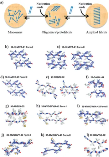
Fig. 2 Different amyloid fibril structures. Amyloid fibrils can self-assemble
both into a (a) parallel as well as an anti-parallel arrangement (shown in panel (a) of Fig. 1). The schematic drawings of parallel and anti-parallel amyloid monomer arrangements depicted here are used as simplified examples to facilitate the understanding of the main text. (b–l) Crystal structures of several amyloid Ab segments, shown in projection down the fiber axes, with the Ab segments packed as pairs of interdigitated b-sheets (the basic unit of the fibril) and displaying b-sheet structures composed of parallel strands (e, g, h, l) and of antiparallel strands (b–d, f, i–k) (adapted
with permission).117
Fig. 1 Main features of amyloidogenesis. (a) Amyloidogenesis is a highly
ordered nucleation-dependent process, where monomers give rise to intermediate structures (oligomers/protofibrils) that later give rise to the mature amyloid fibril. It is important to clarify that the schematic drawing of an antiparallel amyloid monomer arrangement is only used as a showcase, since there are parallel monomer arrangements possible as well. (b) Irregular protein aggregates and (c) amyloid fibrils are clearly
distinct. Reprinted with permission.35(d) Mature amyloid fibrils are invariably
rich in b-turn structure (adapted with permission),106in contrast to other
irregular protein aggregates, showing (e) quasi-crystalline amyloid fibrils
with highly ordered cross -beta structures (adapted with permission).108
(f) In most amyloidogenic sequences that are associated with diseases, for example the Alzheimer’s-related amyloid beta peptide 42 sequence, the toxicity of amyloid fibrils is found to be associated with the fibril precursor aggregates (oligomers/protofibrils) and not with the mature amyloid fibrils
(adapted with permission).107
silk. Furthermore, functional amyloids have vital functions like skin pigmentation in mammals, including humans, where an amyloidogenic sequence plays a key role as a vital scaffold for melanin biosynthesis,91–93depicted in Fig. 3.
The formation of melanin in the human melanosome occurs via the interplay of small reactive molecules, precursors of melanin, and an amyloid sequence. In brief, the melanin precursor (MP) is activated by a tyrosinase (T) resulting in the activated melanin precursor (AMP) which then interacts with the amyloidogenic sequence PmeI17 (PmeI) to form mature melanin. The amyloid sequence PmeI drives a fast amyloid formation process: mature melanin is formed with PmeI serving as a template that organizes the melanin precursors and accelerates the covalent polymerization of these molecules into the mature melanin molecule.91–93 Amyloidogenic protein sequences have been suggested to play other physiological roles, namely involving fibrin and tissue-type plasminogen activator. Fibrin results from thrombin-mediated proteolysis of fibrinogen. Fibrin is primarily involved in blood clotting and platelet activation mechanisms, regulated by tissue-type plasminogen activator, which is a regulator of fibrin clot lysis.119,120 Interestingly, several amyloidogenic peptides, namely amyloid beta-peptide and fibrin-derived amyloidogenic sequences interact with tissue-type plasminogen activator,120 suggesting additional roles for amyloidogenic
sequences in the modulation of the activity of other biomolecules. Furthermore, amyloid conformers of endostatin were reported to possess anti-angiogenic activity.121–123 All these findings clearly indicate that amyloidogenesis plays an important physiological role in different organisms, including humans.
3. Amyloid fibrils in nanotechnology
The variety of physiological functions of amyloid fibrils exploited by Nature reveals their harmless potential for nano-technology. Furthermore, the stability and reduced cytotoxicity of the mature pathological fibril found in degenerative diseases
(when compared to the fibril precursor, the protofibril) is an important information. In addition, their recognized diversity and the fact that they are well-characterized in terms of structure and formation explain the current interest in these structures. Several applications are under scrutiny, with the most promising ones using amyloids as potential nanowires for electronics, as functional templates and liquid crystals, for the production of amyloid-based gels in cell adhesion and wound-healing, and as drug delivery systems.
3.1. Amyloid-based drug delivery
The conjugation of peptides–proteins with drug compounds is a useful strategy to create long-acting drug depots and to overcome some of the limitations related to the use of the individual drugs alone, such as premature enzymatic degrada-tion or limited blood circuladegrada-tion and bioavailability.124Amyloid fibrils of insulin have been shown to possess a large void space able to accommodate molecular iodine. Iodine molecules seem to be solvent accessible and can eventually be released. Thus, insulin-based amyloid fibrils may potentially provide a model for the design of protein-based drug delivery media.125In case
that the drug by itself self-assembles to amyloid structures, as was found for gonadotropin-releasing hormone (GnRH) analogues, it is possible to formulate such amyloids as stable drug depots that offer a controlled release of the active drug from the amyloid conjugate.126This concept was demonstrated for a family of short-and long-acting analogues of GnRH,126 Verified by electron microscopy the long-acting GnRH-analogues formed amyloid-like fibrils and manifested a wide variety of morphologies. Moreover, the duration of action of release depended on the ability of the GnRH analogue amyloids to slowly release active peptide, supporting the role of amyloids as drug depots. These findings were supported by in vitro and in vivo tests, which also showed that these amyloid fibrils were not toxic to dermal cells. Based on the current understanding of the amyloidogenesis process and on experimental data currently available,89,126a generic amyloid-based drug delivery system is depicted in Fig. 4.
Recent studies support the use of amyloids as drug-delivery systems. The therapeutic antibody-derived candidacidal deca-peptide (killer deca-peptide, KP) was shown to spontaneously and reversibly self-assemble into fibril-like structures.127Since this self-assembled amyloid state may avoid proteolysis but still will slowly release the active dimeric peptide form over time, it can also serve as a drug delivery system. Recently, another micellar nanocontainer delivery and release system has been designed employing a peptide–polymer conjugate of hybrid molecules which self-assembles into micelles comprising a modified amyloid peptide core surrounded by a poly-ethylene glycol (PEG) corona.128 In the peptide–PEG hybrid system studied,
enzymatic degradation, using alpha-chymotrypsin, leads to selective cleavage close to the PEG–peptide linkage. Then the break-up of the micelles and the release of free active peptides follow. The trade-off between possible amyloid toxicity and drug delivery potential seems to favour the later, encouraging further research along this concept. Also, the FF dipeptide,
Fig. 3 Physiological relevant amyloid fibril formation. Amyloids were
initially considered the pathological outcome of a misfolding event, but it is now clear that amyloid fibrils can have physiological functions, such as in melanin synthesis in the human melanosome. Briefly, the melanin precursor (MP) is activated by a tyrosinase (T) resulting in the activated melanin precursor (AMP), which interacts with the amyloidogenic
sequence PmeI17 (PmeI) leading to mature melanin.91–93
assembled to microtubes, has been explored for effectively deliver drugs. Using rhodamine as a model drug, it was shown that the FF microtubes released the drug in a steady-state profile, following first-order kinetics. Applying the microtubes to erythrocytes and fibroblast cells, had no impact on cell viability, demonstrating the potential of these carriers to deliver drugs at a constant rate.129
3.2. Amyloid nanowires and nanotubes
Since amyloids tend to self-associate into organized fibrils, the first nanotechnology applications investigated were related to the design of nanowires and nanoelectronic materials. This approach was very successful. Using peptides combined with
metals or enzymes, it was possible to produce artificially engineered peptide/protein-based nanowire structures. Reches and Gazit first demonstrated that the diphenylalanine dipeptide structural motif, also found in b-amyloid, could form rigid nanotubes that, after reduction of ionic silver within the nano-tubes and proteolysis of the peptide results in the formation of nanowires.130 Furthermore, Gazit and coworkers were able to control the challenging task of an aligned arrangement of FF nanotubes on self-assembled arrays by the vapour deposition technique, as shown in Fig. 5.131
With this attractive technology, the number, length and density of the nanostructures can be carefully tuned by simply adjusting the amount of peptide building blocks in the gas phase. Thus, self-assembling peptides can be employed as promising nanomaterials for the construction of ultracapacitors to store energy.131In future, it is conceivable that on the basis of this technology large-scale arrays of arranged peptide nanotubes or nanowires will be fabricated which then can be utilized as high-surface-area electrodes for energy storage applications, for microfluidic devices and for smart surfaces with favourable self-cleaning abilities due to their highly hydrophobic properties.
Several other groups have investigated FF nanotubes for different nanofabrication purposes exploiting their strong stiffness and their stability under defined conditions, as for example Castillo-Leo´n, Svendsen and colleagues. They looked into the electrical properties of FF nanotubes,132and explored applications, using FF nanotubes as etching material,133or for the fabrication of silicon wires134and as electrode material for biosensor development, in particular for the fabrication of PEDOT nanowires.135Recently, Reches and colleagues were able to self-assemble the aromatic FF dipeptides and its Boc-protected analogue into either tubular or spherical structures and obtain various morphologies from these self-assembling peptides, which complement the classical amyloid fibril structure, as shown in Fig. 6.136
Briefly, when both structures were put together, they co-assembled to interesting necklace or beaded string structures under reversible and stable conditions.136Using this strategy, one can imagine a large variety of novel, complex mixed structures. Decoration of these co-assembled chains with biological entities, such as enzymes, antibodies and other bioactive moieties or with chemical cues opens up enormous opportunities for sensor, drug delivery and tissue engineering applications. Hence, employing self-assembling peptides will also support the exploration of other morphologies that go beyond and complement the well-established fibril structure. In addition to that, a novel direction has been taken by using FF dipeptides for the fabrication of biomimetic artificial motors, as described in Fig. 7.137
Here, the biomimetic hybrid motor system contains a metal–organic framework (MOF) together with the FF peptide. MOFs made of metal ions and bridging organic ligands are nanoscale porous materials that have been proposed as motor systems due to their rigidity, density and distinct pore size. The FF peptides assembled in the highly ordered pore array of the MOFs. When the assembled peptide structures got released from the MOF, a swimming motion was initiated due to a large
Fig. 4 Amyloid fibrils as slow drug release systems. In a simple setting,
amyloid monomers (blue arrows) may entrap the drug (D) within the mature amyloid fibrils during the amyloidogenesis process. Afterwards, this conjugate may be safely transported to the vicinity of the drug molecular target (T). The drug can then be slowly released to perform its functional binding to its molecular target.
surface tension difference around the MOF. The authors envision new applications towards smart autonomous motors that might mimic swimming bacteria, and with integrated recognition sites, might harvest targets, such as toxic chemicals. Scheibel and
colleagues showed that N-terminal and middle region (NM) amyloid fibrils of wild-type Sup35p, a prion determinant of S. cerevisiae, were stable under varying harsh conditions. Most importantly, they manipulated the fibril length according to the assembly conditions, making amyloid fibrils ranging from 60 nm to several hundred micrometers in length.48Moreover, by mutating one lysine to a cysteine, the resulting sequence assembled to amyloid fibrils that were able to covalently bind to gold nano-particles. When placed on gold electrodes and further chemically treated with silver and gold, a hybrid gold–silver nanowire of a diameter of 100 nm was produced that could effectively conduct an electrical current. This is the first example of a regular solid metal wire fabricated from amyloid peptides–proteins that could properly be called a nanoscale electrical circuitry, i.e., an amyloid nanowire. Herland and colleagues produced an electronically active lumines-cent nanowire by combining insulin-derived amyloid fibrils with the PFF polymer. This could be employed as an active layer in a light emitting diode with quantum efficiency greater than a similar construct without the amyloid species included.138–140 Subse-quently, Hamedi and colleagues reported the formation of amyloid fibrils, using insulin coated with polymer alkoxysulfonate (PEDOT). This resulted in the construction of a functional electrochemical transistor operating at low voltages between 0 and 0.5 V.141Further
advances will require small sized circuits and a number of different possible applications to be investigated. Exploring the limits of the technology, Malisauskas and colleagues managed to form ultrathin silver wire of 1 nm diameter and up to 2 mm in length using lysozyme amyloid fibrils with hollow channels filled with silver.142 Barrau and colleagues used amyloid nanowires in
Fig. 5 Self-assembling peptides are promising nanomaterials for the
construction of ultracapacitors to store energy. (a) Self-assembling aromatic dipeptide nanotubes (ADNTs) can be produced and vertically aligned via the depicted vapour deposition technique being composed of (b) individual single peptide nanotubes stabilized via (c) classical stacking interactions between aromatic moieties of the peptides. (d–f) Top-view of scanning electron microscopy images of (d) uncoated, (e) ADNT-coated and (f) carbon-nanotube-coated electrodes showing the deposited material. (g, h) Ultracapacitors based on the ADNT-coated electrodes can be fabricated as shown by (g) current density measurements of ADNT-coated (red line), carbon-nanotube-ADNT-coated (black line) and unADNT-coated (blue line) carbon electrodes combined with (h) capacitance density tests of the
ADNT-coated carbon electrodes (adapted with permission).131
Fig. 6 Complex peptide-based structures can be formed via the co-assembly
of two simple peptides, the diphenylalanine peptide and its tert-butyl dicarbonate (Boc) protected analogue. The distinct architectures range from tubular struc-tures to beaded strings to nanospheres that are stable, but also reversible
structures under specific conditions (adapted with permission).136
Fig. 7 Self-assembling amyloidogenic peptides can be used for the
development of molecular motors. (a) Combining a metal–organic framework (MOF) and self-assembling FF peptides allows the building of a ‘‘cage cell’’, (b) where energy can be stored. In addition, the controlled release of self-assembling FF peptides (c, d), located inside nanoscaled pores of the energy-storing-cell, can generate motion. (e) A MOF ‘‘mini boat’’ with a narrow opening makes the chemical motor motion more efficient. (f) Optical image of the moving MOF ‘‘mini boat’’. (g) Comparison of the velocity and lifetime of the movement of the free particle (out of the boat) with the loaded boat. This strategy paves the way for new applications using molecular self-assembling
peptide systems (adapted with permission).137
organic photovoltaic devices to enhance the transport properties (with fibrils acting as a template for donor–acceptor materials). These promising results greatly expand the promising applications of amyloid-based nanowires143 and merit further investigations
into the potential uses of amyloid-based nanowires.
3.3. Functional templates and the use of amyloids for nanosensing
It is possible to functionalize amyloids with ligands such as fluorophores, antibodies, enzymes or other tags that are suitable for the desired application. A variety of bioactive molecules can be employed. In Fig. 8, the concept of the amyloid-based immobilization of active enzymes is illustrated.
Accordingly, nanostructured films from amyloid fibril-forming hen egg lysozyme could be generated in a scalable macroscopic approach, as depicted in Fig. 9.144
The fabricated films made from self-assembling amyloid protein fibrils showed high rigidity up to 5–7 GPa and a well-defined order that encouraged the alignment of fluorophores.144
This approach allowed producing well-ordered amyloid films, functionalized with fluorophores and was easily distinguishable from non-functionalized films. It demonstrates that amyloidogenic molecules support the alignment of otherwise unstructured components (such as fluorophores), which in turn gives rise to new methods for the bottom-up construction of highly organized multifunctional materials.
Other breakthrough designs have made use of bifunctional protein nanowires for the development of highly sensitive immunoassays.10,145 One exemplary approach, applied to the detection of pathogen Yersinia pestis, is shown in Fig. 10.
Briefly, two version of the amyloidogenic yeast amyloid protein Sup35p were designed, one fused with protein G (Sup35p-G), a protein well-known for its ability to bind to different mammalian immunoglobulins, the other fused with another protein, the MPH (methyl-parathion hydrolase) enzyme (Sup35p-MPH). The two versions of hybrid Sup35p molecules were co-incubated, resulting in amyloid fibrils with two func-tionalities, i.e. the ability of protein G to interact with protein G antibody and the enzymatic activity of MPH. Interestingly, these mixed Sup35p-MPH–Sup35-G amyloid fibrils (which can also be understood as protein nanowires) exhibited a high Sup35p-MPH to Sup35p-G ratio. A dramatic improvement in the detection threshold was achieved, when the G protein section of the Sup35p-MPH–Sup35-G amyloid fibrils was allowed to interact with G protein antibody. The interaction was measured via the MPH activity. This approach led to a 100-fold enhancement of the detection sensitivity towards the pathogen Yersinia pestis.10,145 With the aim to obtain an ultra-sensitive molecular biosensor, an auto-biotinylated bifunctional protein nanowire (bFPNw) was designed, based on the same principle and employing also Sup35. Protein G and a biotin acceptor peptide (BAP) were linked to this de novo designed structure.145The rationale for these auto-biotinylated bFPNws is that they could easily be adapted to existing diagnostics approaches involving antigen–antibody complexes and enzyme-based detections in a standard biotin–avidin detection system.
Theoretically, this could greatly enhance the sensitivity of immune-biosensing, which was indeed observed for Yersinia pestis detection. A 2000- to 4000-fold increase in sensitivity compared to classical immunoassays was achieved. Thus, the auto-biotinylated self-assembled bFPNw molecular biosensors demonstrate the potential use of self-assembling protein nano-wires in biosensing. In addition, the same group also showed that pesticides can be detected via a similar approach.146A new type of fluorescent molecular biosensors for the detection of the pesticide MP was developed by construction of a green fluores-cent protein (GFP) mutant fused to methyl parathion hydrolase (MPH). In this setup, MPH is used to detect the pesticide in a pH dependent reaction that releases H+. Linking MPH to the GFP mutant allows to detect the enzyme activity by monitoring pH induced changes in GFP fluorescence. The authors took this approach to the next level by fusing the MPH-GFP mutant to Sup35protein nanowires to further increase the response sensitivity of the molecular biosensor. This approach of fusing a classical protein biosensor structure to self-assembling protein nanowires, thereby increasing the response, is a promising new avenue of research. This same strategy may lead to other sensitive molecular biosensor systems.
Following on a similar line, Mezzenga and co-workers managed to fabricate interesting new biodegradable nanocomposite materials, in particular a graphene-based amyloid nanosensing device inspired originally by collagen structures.25,147 For the graphene–amyloid
Fig. 8 Amyloid sequences can be combined with active biomolecules B,
namely proteins, to (a) generate hybrid molecules with multi-functionalities. One such example is (b) joining enzymatic activity, provided by the active enzyme biomolecule B, with the stability and resistance to proteolysis provided by the amyloid fibril, represented by the blue arrow. Furthermore, in this set-up, the amyloid fibril may also provide the option of immobiliza-tion on a surface.
fibril composites, the globular milk protein b-lactoglobulin was used. The protein forms well-defined amyloid fibrils.148–151
This protein is another example for an amyloid fibril that not necessarily has to be toxic. On the contrary, b-lactoglobulin from milk is in daily use by millions of people, when their diet includes the consumption of yoghurt. Combining natural occurring amyloid nanofibers with graphene that is incompatible with biological entities, but shows exceptional mechanical and electronic properties, can create exciting novel hybrid materials.
These materials are conductive, can be degraded by enzymes and feature enzyme-sensing properties together with shape-memory. Hence, one can imagine that these hybrid graphene-based nano-composite materials will give rise to inspiring electronic, micro-mechanical and biological devices. The methodology followed to produce the graphene-based nanocomposite nanomaterial is described in detail in Fig. 11.
Baldwin and colleagues showed that SH3 amyloid forming sequences linked with soluble cytochrome b562 sequences resulted in the formation of stable fibrils. Importantly, the cytochrome section remained fully functional and was able to bind metalloporphyrins, the biological ligand of cytochrome,
Fig. 9 Well-structured and highly ordered protein films can be produced
from self-assembling amyloid peptides. (a) Amyloidogenic sequences can self-assemble into amyloid fibrils that are able to form nanostructured amyloid films by condensation of the fibrillar structures. (b) Atomic force micrograph of the mature amyloid fibrils from which the (c) free-standing protein film is derived. The film is stable and visible by scanning electron microscopy. (d, e) Optical images of plasticizer containing lysozyme amyloid films under crossed polarizers shows low transmission through the protein film when the objective polarizer is parallel to the fibril alignment in the film (left) and maximal transmission at an angle of 45 degrees (right), demonstrating that the fibrils are therefore perfectly aligned in the film plane. (f) Fluorescence intensity emitted from nano-structured films containing aligned fluorophores. Emission occurs either through a polarizing filter that emits light while the film rotates 360 degrees (filled blue squares) or in the absence of the polarizer (green open squares) showing that self-organizing protein nanofilms can be used to fabricate highly organized and perfectly aligned fluorophores-containing amyloid nanofilms. (g) This is observed in the fluorescence microscopy image of non-functionalized (left) and functionalized (right) fibril film (adapted
with permission).144
Fig. 10 Amyloids can increase the threshold of current antibody-based
detection of biomolecules. (a–d) Amyloid sequences can be employed to build a sensitive and easily operable immunoassay based on bifunctional protein nanowires composed of the yeast amyloid protein Sup35p genetically fused with protein G and an enzyme (methyl-parathion hydrolase, MPH). The approach takes advantage of the self-assembling, seeding and nucleation properties of the amyloidogenic sequences. (e–f) In the final bifunctional nanowire, the Sup35 acts as the skeleton arraying a few copies of protein G molecules with a large number of enzyme molecules. It demonstrates a high ratio of enzyme to protein G molecules that provokes a dramatic increase of the enzymatic signal when protein G binds to an antibody target. (g) Compar-ison of the sensitivity of bifunctional protein nanowires, commercial ELISA reagent, and MPH-protein G chimera for Yersinia pestis F1 antigen detection. At low concentrations of the antigen, a roughly 100-fold enhancement of the sensitivity can be achieved, when comparing the detection limit of a classical ELISA assay with the detection limit reached by the bifunctional protein
nanowires (threshold below 2 ng mL 1) (adapted with permission).10,145
supporting similar approaches for applications in nanotechnology.152 Liu and co-workers also managed to incorporate cytosine, a nucleobase, into the side chain of the amyloid peptide HHQALVFFA. This has opened a new avenue of possibilities via the joint use of amyloid sequences and nucleobases.153
Recently, Mu¨nch and colleagues reported that peptides derived from HIV-1 glycoprotein gp120 that self-assembled to amyloid-like fibrils could be useful as gene delivery vehicles. They demonstrated, as exemplified in Fig. 12, that artificial nanofibers received from a 12-mer peptide, called enhancing factor C (EF-C), were efficient agents for retroviral ex vivo transduction.154 Interestingly, the positively charged peptide nanofibers captured the viral particles for viral infection in a much more efficient way than semen-derived fibrils as well as RetroNectin, a commercially derived ‘‘gold standard’’ for enhanced retroviral gene transfer. Furthermore, the EF-C fibrils were stable at room temperature or 4 1C for at least two weeks without compromising infection-enhancing activity. They also displayed high stiffness. Positive net charges can easily be introduced into peptide sequences by choosing the correctly charged amino acid resi-dues which enables the nanofibrils to capture nucleic acids, such as DNA or RNA and promotes the attachment and fusion to target cellular membranes. Thus, novel gene transfer vehicles from short amyloid-like peptides could offer interesting new avenues. Rao and co-workers employed bovine insulin to make amyloid fibrils functionalized with poly(vinyl alcohol) (PVOH) resulting in a PVOH-amyloid composite stiffer than the amyloid-free version although it contained only 0.6% (weight) of peptide material.155This validates and serves as a proof-of-concept of the use of amyloid fibrils in films of polymeric materials to vary their mechanical/morphological properties.
The design of biologically functional surfaces was per-formed by Grass and colleagues employing transthyretin (TTR) amyloid fibrils as modulating biomaterial for promoting and exploiting cell adhesion, migration and differentiation in vitro.3Briefly, they have shown that TTR peptide sequences
combined with a biological cell adhesion motif sequence self-assembles in aqueous solution to form amyloid fibrils. These fibrils are bioactive with cells being able to interact specifically with the RGD group displayed on the fibril surface. These exciting findings points to a number of possible variations, where de novo engineered amyloid-containing surfaces can be created to exert specific interaction properties with a wide variety of cell types. Moving forward on this subject, Kasai and colleagues revealed that another amyloidogenic peptide, the A208 peptide, that contains N-terminal sequences of mouse laminin 1,156is able to form amyloid gels. Laminin 1 is involved in cell adhesion, migration, neurite outgrowth, tumor meta-stasis, and angiogenesis.143In fact, it is this amyloid fibrillar
form that promotes cell attachment via the bioactive laminin section. Other studies with insulin-based amyloid peptides also supported this approach.157Summarized, these results clearly suggest a role for multifunctional peptide fibrils as bioadhesives for tissue regeneration and engineering.
Amyloids may also have a role in cell differentiation, namely with IKVAV peptide, another very promising peptide for amyloid
applications.158–163 The peptide IKVAV corresponds to the neurite-promoting laminin epitope, which, when constituted into nanofibrils has been shown to induce very rapid differentia-tion of cells into neurons, at a better rate then soluble or peptide or laminin itself.156,158Hauser et al. have rationally designed a class of ultrashort aliphatic peptides containing 3–7 amino acids that have an innate ability to self-assemble to amyloid-like helical nanofibers. These fibers ultimately form hydrogels. Due to their small size these peptides are attractive, since they offer a cheap production alternative to other protein or peptide-based structures with longer sequences.106,164In a recent study,165the integrin-recognition motif RGD was conjugated to the hexamer peptide Ac-LIVAGKC (LK6C) and afterwards cysteine-mediated
disulfide bonds were introduced to the nanofibers assembled from the hexamer. The bioactive RGD motif visibly supported
Fig. 11 Graphene material and self-assembling amyloid peptides can
generate structures that are able to sense enzyme activity. (a–d) Schematic representations show the fabrication of free-standing films of amyloid fibrils–graphene composites. (a) Self-assembly of b-lactoglobulin into amyloid fibrils is accomplished through (b) electrostatically driven co-aggregation of amyloid fibrils and graphene oxide. (c) The binding of broken amyloid fibrils on graphene surfaces during reduction of graphene oxide forms (d) layers of amyloid fibrils and graphene nanosheets hybrid nanocomposites. (e) Transmission electron microscopy and (f) atomic force microscopy images of reduced graphene dispersions at pH 2 with a 1 : 8 graphene–amyloid fibril ratio demonstrating the formation of stable amyloid–graphene films that can remain conjugated and stable for several weeks. (g) The hybrid nanocomposite films can be used to develop a biosensor with enzyme activity as depicted in the inset. When incorporat-ing a desired protein, namely pepsin at 1%, it can detect the difference
between folded and unfolded protein (adapted with permission).147
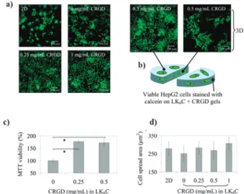
cell viability and spreading in a true 3D distribution.165These cross-linked 3D matrices made from amyloid nanofibers can easily be envisioned for further applications delivering thera-peutics and other compounds, as depicted in Fig. 13. In a recent
paper, Loo et al. showed that lysine-containing ultrashort amyloid-like peptides can be used as hydrogels for wound dressings to address unmet clinical needs in the treatment of partial thickness burns. The nanofibrous hydrogels accelerated wound closure in a rat model for partial thickness burns. Furthermore, the amyloid hydrogels effected earlier onset and completion of autolytic debridement compared to the control, used as a standard-of-care.166The ultrashort aliphatic amyloid peptides were further explored for localized injectable anti-cancer therapy. The peptides were functionalized with platinum anti-cancer drugs and oxaliplatin-derived hybrid peptide hydro-gels with up to 40% drug loading were tested for localized breast cancer therapy in a mouse model. Stably injected gels showed significant tumor growth reduction and a better tolerance com-pared to the free platinum drug.167
In peptide constructs such as those formed by a bioactive peptide plus an amyloid peptide, a major advantage for appli-cations would be that the kinetics of amyloidogenesis can be adjusted via subtle changes in the amyloid peptide sequence. Hence, for a particular clinical use, peptide sequence might be altered, while the nanofibrils bioactivity is maintained.159 Mutation of the peptide sequence by including more hydro-philic and bulky amino acids effectively slows down gelation through self-assembly of the nanofiber network. The ability to
Fig. 12 Amyloid fibrils can be employed as enhancers of retroviral
transduc-tion increasing gene transfer in mice models. (a–d) Amyloid fibrils, formed from a self-assembling 12 amino acids peptide named enhancing factor C (EF-C), are transduction enhancers. (a) Inoculating HeLa cells (blue) with viral particles with the MLV glycoprotein labelled with yellow fluorescent protein (MLV-YFP, green) in the absence (top line of the panel) of rhodamine-labelled fibers (Rho-EF-C, red) or in the presence of the labelled fibers shows that the presence of the fibers, which are not cytotoxic, enhances viral replication. (b) Quantitative comparison of the fibril effect in the increase of the virions rate of fusion with cells. (c) EF-C fibrils increase lentiviral transduction of 293T cells independently of the viral glycoprotein. (d) EF-C fibrils mediated lentiviral gene transfer is effective with several different cell types, namely human glioblastoma (U87MG), endocrine pancreatic tumour (BON), myeloid KG-1, peripheral blood mononuclear (PBL) and haematopoetic (CD34) stem cells. Importantly, EF-C fibrils can be immobilized (e–g) and allow efficient gene transfer into mice (h–j). (e) Z-stack images of immobilized Rho-EF-C fibrils only (top left), MLV-YFP only (bottom left) and immobilized Rho-EF-C fibrils exposed to virus (right; the upper image shows both channels, merged, and the lower images, separately). (f) EF-C fibrils coating facilitates lentiviral infection in a degree similar to an established standard method based on (g) RetroNectin (RN) which, (h) if put together possess an additive effect to each other. (i) EF-C fibril coating also prompts lentiviral gene transfer to mouse cells. (j) Treating bone marrow cells with the EF-C amyloid fibrils and transducing them with a GFP-labeled lentiviral vector before transplanting the cells into recipient mice results in a high success rate with a number of GFP-positive cells in peripheral blood, demonstrating that EF-C amyloid fibrils facilitate retroviral gene transfer
(adapted with permission).154
Fig. 13 Amyloidogenic peptides serve as novel three-dimensional (3D)
substrates suitable for tissue engineering applications. (a) HepG2 cells were seeded either directly onto wells (2D control) or onto gels (3D)
containing LK6C (an amyloidogenic peptide) with different concentrations
of conjugated integrin recognition motif RGD (CRGD). Cells proliferated in a 3D environment within the gel while the RGD motif enhanced cellular attachments. (b) After 4 days in culture the cells remained viable as evidenced by the calcein incorporation assay. (a) Visual inspection of HepG2 cultured cells suggested that CRGD-functionalized amyloidogenic peptide gels increased cell viability, as further demonstrated (c)
quantita-tively via the MTT cell viability assay. (d) Using 3D-LK6C in absence or
presence of CRGD and comparing it with the 2D-control gave no significant difference on the cell spreading area. Overall, this suggests new pathways for cell culture and tissue engineering by jointly exploring the 3D patterns created by different amyloidogenic peptides and the ability to functionalize them with several biologically relevant molecules (adapted
with permission).165
modify gelation kinetics in self-assembling systems without disrupting bioactivity is an important aspect for the development of injectable therapies in regenerative medicine. Feasible strategies for novel therapeutic approaches using self-assembling peptide nanofibers for angiogenesis and cardiac repair can be envisioned when reading the study of Lin et al., 2012.168Peptide nanofibers
mixed with vascular endothelial growth factor (VEGF) were injected into the heart of rats or pigs. Interestingly, the increased densities of arteries and arterioles after injection pointed to neovasculariza-tion. Furthermore, endogenous myofibroblasts and cardiomyocyte-resembling cells were newly recruited. These and other studies might encourage the use of amyloid nanofibers for future clinical translational applications, for injectable therapies such as cardiovascular repair and other regenerative treatments.
4. Challenges and future directions
The potential of proteins or peptides to form irregular protein aggregates or highly structured amyloid fibrils, although ultimately present in the polypeptide chain, varies strongly.35,108,169,170The
rational design of a de novo amyloid sequence, unrelated to any known amyloidogenic protein, is extremely challenging due to the complexity of the formation of amyloid fiber structure, including intermediate structures. Although the final structural properties of amyloid fibrils are highly similar, it is so far still impossible to predict the amyloid propensity from a given peptide sequence that is for example able to form beta-sheet structures. The reason for this dilemma is comprehensible in view that the mechanism of amyloid formation is far from being understood. Hence, it is not simply possible to assume that all existing or imaginable self-assembling peptide systems that are able to form beta-sheets or beta-turns are necessarily forming amyloid fibrils. The question remains which sequence of a self-assembling peptide monomer qualifies most likely to the formation of proper amyloid fibrils. Thus, we suggest referring best to the unequivocally and meticulously elaborated structural data set that were gained by Serpell and colleagues from natural occurring proper amyloid fibrils via X-ray analysis.21–24 The reported typical cross-beta diffraction data have given so far the most unifying answer in terms of characterization of all known amyloid structures. The cross-beta diffraction pattern generally exhibits a longitudinal 4.7–4.8 Å spacing and an equatorial spacing of around 10 Å. The term ‘‘cross-beta’’ refers to both diffraction signals, the longitudinal and the equatorial spacing, that form a typical cross pattern. Ideally, this should be taken as the gold standard to define and unmistakably classify amyloid fibrils. Several factors must be weighed in for the successful design of novel functional amyloid-based nanomaterials. The main aspects to take into consideration are described in Fig. 14.
Evidently, an important aspect is the analysis of the amino acid sequence of the amyloidogenic peptide or protein in question. The process of amyloid formation has similarities with the folding process, in the sense that it is highly ordered and ultimately governed by the primary protein sequence. In the case of hexapeptides, ideally, this should allow to use
simple scoring matrices with information regarding only primary structures that predict the amyloid propensity.108 The finding that the amyloid fibrils precursors (but not the fibrils themselves) are the cause for toxicity led to the careful characterization of the process of amyloidogenesis, i.e., the evolution from the peptide or protein monomers stage, to the precursor intermediates (where a multitude of species such as oligomers and protofibrils may be present), to, finally, arrive at the mature amyloid fibril stage.35,107,108,115,169,170It is generally agreed that a key mechanism to fibril formation is the nuclea-tion event.115,169,171Initially, there is a lag phase, during which monomers self-assemble into higher order aggregates, which form the seeding nucleus. The process will then further
Fig. 14 Main factors to consider for the assembly of amyloid fibers.
Amyloidogenic peptides and proteins that are suitable for amyloid fiber formation can be used for the insertion of new functions into the mature amyloid fibrils. Several factors are pivotal to successfully engineer amyloid fibers. To predict the amyloid propensity and functionality of a newly designed peptide sequence is a major hurdle. Modifying or adding func-tionality to an already known amyloid sequence might severely impact its amyloid propensity and functionality. Controlling the speed and moment of fibrilization/nucleation is another factor to be considered. Once these obstacles are surpassed, determining the engineered fibrils’ putative toxi-city, their stability in different environments and their ability to perform the designed function are the final steps for successful manufacturing novel amyloid-based biomaterials. The schematic drawing of an antiparallel amyloid monomer arrangement is only used as a showcase, since there are parallel monomer arrangements possible as well.
progress in an extremely fast manner by the addition of other nuclei or monomers, resulting in the formation of protofibrils and amyloid fibrils. However, the kinetics and amyloid morphology of related and almost identical peptides, such as the amyloid beta peptide 40 (Abeta40) and the amyloid beta peptide 42 (Abeta42), involved in Alzheimer’s disease and differing in only two amino-acids, can be significantly different.169 Therefore, it would give major advantages for amyloid-based nanotechnology to predict the amyloid propensity. This will be discussed in the following, together with the issues regarding the control of the amyloido-genesis process and of the eventual problem of fibril toxicity.
4.1. Predicting amyloid propensity and peptide design The issue of predicting the potential of peptide sequences that form amyloid fibers is a hot and problematic research topic. There have been continuously improvements by different groups over the years. The approaches range from special physico-chemical property win-dows to position-specific sequence matrices, explicit structural models, and combinations thereof.108,116,172–185Many methods base their prediction on the potential of hexapeptides to form amyloid fibres. This characteristic length has been found to cover several of the core motifs for amyloid fiber formation. Of course, fiber forming properties are not restricted to hexameric peptides alone. In fact, amyloid core sequences span sizes from four to seven amino acids, as earlier discussed.
Currently, the sequences known to form amyloid fibers are still relatively narrow in terms of amino acid coverage for each position and partially dominated by well-studied peptide families with high internal similarity. Consequently, sequence-based prediction of effects of selected point mutations to increase or decrease amyloid propensity for peptide design should work best for peptide scaffolds with sufficient related positive and negative known examples. Structural methods on the other hand are less affected by peptide family bias. These methods suffer from giving just a partial success rate of accurately describing all acting molecular forces in fiber structures. This includes solva-tion and entropic effects as well as limitasolva-tions in timescales for conformational sampling through simulations.
Although approaches combining sequence, physico-chemical property and structural terms tend to perform best overall, there is sufficient diversity among available methods that could result in divergent predictions in specific cases. However, another critical aspect for successful peptide design of amyloid fibers is the distinction of ordered amyloid fibers versus unordered amorphous beta aggregation, as discussed earlier. The delicate balance can be explored by combined use of prediction tools like Tango and Waltz.186Tango is based on physico-chemical
prin-ciples of beta-sheet formation and well-suited for distinguishing soluble from hydrophobic aggregating sequences.174Waltz, on the other hand, is specialized on the specific ordered amyloid fibers108which can be formed both by polar and hydrophobic peptides. Together, they elegantly complement each other. For example, Tango could be used on Waltz predictions to filter candidates which may be too hydrophobic and form fast amorphous aggregates instead of fibers.
Ultimately, peptide candidates predicted from any in silico approach still need to be experimentally characterized for amyloid fiber formation and further tested for suitability as nanomaterial. The expected hit rate of predicted peptides to actually form fibers currently lies around 50–80%. However, if one were to attempt to cover the whole sequence space by testing for example all possible hexapeptides, a peptide of length 6 and an alphabet of 20 amino acids would allow 64 000 000 possible sequences. Therefore, computational prediction appears indispensable to narrow down and identify a manageable number of candidates for experimental testing.
Interestingly, in the case of hexapeptides it turns out that nature did not sample the sequence space completely. For example, in the human proteome (IPI v3.5 nr100) only about 14% of all theoretically possible hexapeptides actually are found to occur naturally. Such peptide sequence space under-sampling of longer peptides is also seen in all other proteomes from eukaryotes to bacteria187and hence appears to be mainly a matter of the limited time that DNA-based organisms had so far for their evolution. At the same time, a strong biophysical phenomenon such as amyloid fiber formation should also have an impact on the fitness of an organism and on natural selection as was shown for beta aggregation and diseases.188 Consequently, peptide design efforts should not neglect the huge pool of peptides that do not naturally occur as it could be the source of yet to be discovered strongly amyloid forming peptides. A prime example for an unnatural amyloid system is the class of ultrashort aliphatic peptides. All peptides of this class do not correspond to any known sequence of a natural protein, but were rationally designed.118,164 Other peptide systems that adopt typical beta-sheet structures have been developed, for example by J. Collier, A. Aggelli, D. Pochan and others, discussed in a recent review.36
4.2. Controlling nucleation-dependent amyloidogenesis Despite significant differences in symptoms or functions as well as in the actual sequence of the protein monomers associated with the specific amyloid fibrils, there seems to be a common mechanism underlying protein aggregation at the molecular level as well as a common ordered structure, i.e. the -turn structure, of the involved aggregated amyloid proteins. There have been several models emerged that give mechanistic explanations on the formation of the amyloid protein aggre-gates.189 A simple model defines the polymerization as a nucleation-dependent mechanism, controlled by monomer concentration and time. The formation of amyloid fibrils occurs through a molecular recognition and self-assembly process that typically starts with a thermodynamically unfa-vourable lag phase for the formation of a ‘nucleus or seed’. This is followed by a thermodynamically favourable exponential growth phase, where monomers/oligomers are added to the growing nucleus. A related concept stresses the significance of polymerization by fibril seeding. Structural investigations on amyloidogenic core sequences, peptides of the size of 3–7 amino acids, have revealed conformational transition of the peptides from a random-coiled soluble form via possibly a-helical intermediates