BJRS
08-01B (2020) 01-10ISSN: 2319-0612 Accepted: 2020-06-26
Comparative Study of the Use of Bismuth Shielding on
Head Computed Tomography Scans Using Phantoms
Santos
aF.S., Santana
bP.C., Mourão
a,cA.P.
a Departamento de Engenharia Nuclear, Universidade Federal de Minas Gerais, 31270-970, Belo Horizonte, MG,
Brasil
b Departamento de Anatomia e Imagem/Faculdade de Medicina, Universidade Federal de Minas Gerais, 30130-100,
Belo Horizonte, MG, Brasil
c Centro de Engenharia Biomédica, Centro de Educação Tecnológica de Minas Gerais, 30421-169, Belo Horizonte,
MG, Brasil
ABSTRACT
Computed Tomography (CT) has been one of the most used exam for radiologic diagnostic in medicine. The increase of CT is a global concern due to high doses of radiation. The head CT scans helps to diagnose disorders that affect the brain, including tumors, infarction, bleeding within the brain, hematoma and other diseases. The aim of this work is to reduce the absorbed dose in the head CT scan with the use of bismuth shielding and to compare the absorbed dose between male and female phantom. Anthropomorphic male and female phantom model Alderson Rando were used to do a head CT scan, from the cervical vertebra C1 to the top of skull, using a GE CT scanner, LightSpeed VCT model, with 64 channels. Radiochromic films strips were used to evaluate the doses in the organs such as lenses, thyroid, hypophysis, spinal cord, breasts, salivary and parotid glands. Were conducted two heads CT scan with the phantom in supine position with and without bismuth shielding. The results of this experiment show absorbed doses from 0.48 to 22.98 mGy. Record doses were lower with the use of bismuth shielding for all organs, mainly in the lenses. The analysis of noise in the image of the head central slice presented acceptable values for soft tissues, less than 1%.
1. INTRODUCTION
Computed Tomography (CT) has been one of the most used exam for radiologic diagnostic in medicine. The increase of CT is a global concern due to high doses of radiation [1]. The dose evaluation in CT is one of many steps that can contribute for optimizing patient doses [2,3]. The head CT scans helps to diagnose disorders that affect the brain, including tumors, infarction, bleeding within the brain, hematoma and other diseases. The absorbed dose in organs with higher radiation sensitivity levels are object of continuous discussions, because of the risks that this can bring to health tissues [3]. The lens is one of the organs located in the skull with relative high radiosensitivity, organ that is under consideration in this work of dosimetry, using elements such a radiochromic films and bismuth eye protector before the CT scan [4]. The aim of this work is to compare the reduction of absorbed dose in the head CT scan with and without the use of bismuth shielding using a male and female anthropomorphic phantom Alderson Rando model.
2. MATERIALS AND METHODS
The head CT scan was done from the cervical vertebra C1 to the top of skull, in a GE CT scanner, LightSpeed VCT model, with 64 channels. Two anthropomorphic male and female phantom model Alderson Rando was used to do the head scans. This phantom is built with a human skeleton covered with a polymeric structure that simulate soft tissue. The body and head are structured in transected-horizontally into 2.5 cm thick slices, being 33 slices in the male version and 31 in the female version [4,5]. The slices that make up the body phantom have holes that allow placing dosimeters within the phantom. The male phantom is 175 cm tall and weighs 73.5 kg. The female simulator is 155 cm tall and weighs 50 kg [3,6]. The scan parameters used in this experiment are shown in Table 1. The distance used for head CT scans in the male phantom were 150 mm and in the female 140 mm.
Voltage (kV) Electric current (mA) Tube Time (s) Pitch Thickness beam (mm) Reconstruction 120 175 0.8 0.984 40 2.5
All CT scans were performed with the phantom placed with the head in the isocenter of gantry. In Figure 1 is shown the phantom placed in the gantry isocenter. In (a) the male phantom is positioned in supine without bismuth, in (b) with the eye bismuth shielding, in (c) the female phantom it is positioned in supine without bismuth, in (d) with the eye bismuth shielding.
Figure 1: Positioning of the phantom in the gantry: male phantom without bismuth shielding (a)
and with bismuth shielding (b). The female phantom without bismuth shielding (c) and with bismuth shielding (d).
Radiochromic films strips were used to evaluate the punctual doses in the organs such as lenses, thyroid, hypophysis, spinal cord, pharynx, breasts, salivary and parotid glands. The Figure 2 shows two lateral radiography (scout) and two axial head images with and without an eye bismuth shielding of 1 mm thick in the male phantom.
Figure 2: Scout's and axial head CT images of the male phantom: without eye shielding (a, c),
with eye bismuth shielding (b, d).
The radiochromic films are self-developing dosimetry films, and they are insensitive to visible light making it easy to work with during analysis and provide greater spatial resolution in the sub millimeter range (Fig. 3). They have been used extensively in combination with flatbed document scanners to measure patient doses in the kV [7,8]. In this work, the radiochromic film model used was GAFCHROMIC® XR-QA2, manufactured by ASHLAND, which is specific for diagnostic radiology. This film has high sensitivity to ionizing radiation with doses in the range of 1.0 to 200 mGy and can be used in X-ray beams generated by voltages of 20 to 200 kV. Therefore, it can be used to observe dose deposition in CT experiments. The films were cut into 1.0 x 0.5 cm strips and
proximity of the desired organ. The exposed films were digitized using a HP Photosmart C4480 reflective type scanner. The scanning parameters used were RGB mode (48 bit) and 300 ppi. The images were processed using the image software ImageJ. The red channel was selected to measurement because these radiochromic films have a main absorption peak in the red region of the visible spectrum (636nm) [8,9].
Figure 3: Radiochromic film strips: (a) background and (b) exposed.
Metrological reliability of the radiochromic films was demonstrated through homogeneity and repeatability tests and by calibrating it in a reference radiation for CT (RQT9) that were reproduced in the Calibration Laboratory of the Development Center of Nuclear Technology (CDTN/CNEN) [9,10].
Calculations of the absorbed dose of CT were made using the calibration curve of the Equation 1 by the darkness intensity from the film strip [8,9]. The punctual absorbed dose values (DT) was acquired by subtracting the doses recorded in the film strip placed in the organs of the background (BG) absorbed dose.
(1)
After calculating the DT, the BG dose value is subtracted. Finally, the acquired results are multiplied by the Correction Factor (FC), which serves to correct the Kerma value for soft tissues. The FC value was obtained through data of linear attenuation coefficients for X-rays that can be consulted on the website of the National Institute of Standards and Technology (NIST). The voltage used for this work was 120 kV, corresponding to an FC of 1.095 [11].
To validate the quality of the image by CT was done the noise analysis from the image of the central slice of the phantom head in each of the experiments to determine the influence of the secondary radiation in the quality of the images with the RadiAnt DICOM Viewer program, which is an image format viewer of the Digital Imaging Communications (DICOM).
3. RESULTS AND DISCUSSION
Absorbed doses in the organs positions in both phantom such as: lenses, thyroid, pharynx, hypophysis, breast, spinal cord in cervical area, parotid and salivary glands are shown in Table 2. These results allow us to observe that the use of bismuth shield led to a decrease in radiation dose deposited in the eye and all organs studied.
Table 2: Average absorbed dose in some organ positions in the phantom during head CT scans with
and without bismuth shielding on eyes.
Organ
Absorbed Dose (mGy)
Without Bismuth Shielding With Bismuth Shielding
Male Female Male Female
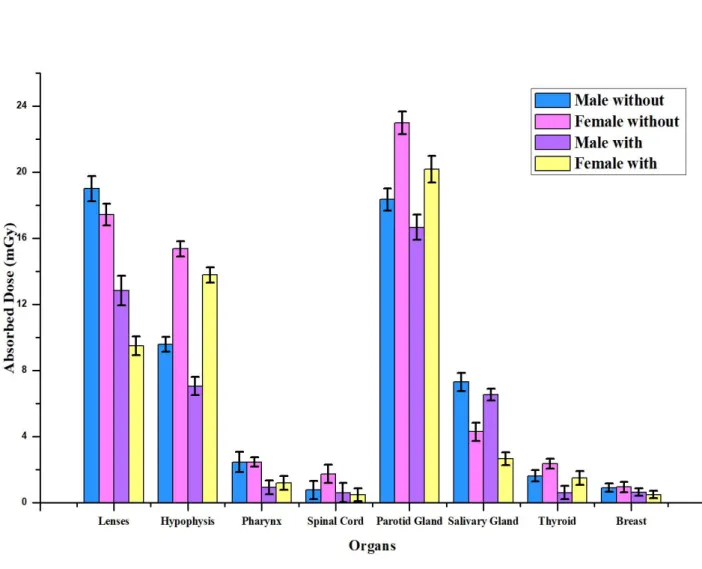
Lenses 19.01 ± 0.77a 17.44 ± 0.66 12.83 ± 0.90 9.49 ± 0.57 Hypophysis 9.59 ± 0.45 15.36 ± 0.47 7.06 ± 0.54 13.79 ± 0.46 Pharynx 2.46 ± 0.62 2.46 ± 0.28 0.93 ± 0.41 1.20 ± 0.41 Spinal Cord 0.76 ± 0.56 1.74 ± 0.56 0.61 ± 0.58 0.48 ± 0.38 Parotid Gland 18.35 ± 0.67 22.98 ± 0.69 16.67 ± 0.76 20.18 ± 0.81 Salivary Gland 7.30 ± 0.55 4.29 ± 0.55 6.55 ± 0.36 2.65 ± 0.38 Thyroid 1.62 ± 0.35 2.35 ± 0.30 0.61 ± 0.41 1.49 ± 0.41 Breast 0.91 ± 0.26 0.95 ± 0.31 0.64 ± 0.22 0.49 ± 0.22 aStandard deviation
absorbed in the parotid glands with 22.98 mGy without the use of bismuth shielding. It is noteworthy that the female phantom has smaller dimensions and weight than the male. Also, the parotid glands obtained 12.2% reduction in dose with the use of bismuth shielding. In the male phantom the parotid glands had a dose of 18.35 mGy without the use of bismuth shielding, having a small reduction of only 9.1% with the use of the bismuth shielding, compared to its non-use.
In the male phantom the highest absorbed dose value occurred in the lenses of 19.01 mGy without the use of bismuth shielding. The dose reduction was 32.5% with the use of the bismuth shielding. In female phantom the absorbed dose of the lenses was 17.44 mGy without the use of the bismuth shielding and had a reduction of the absorbed dose of 45.5% with the use of bismuth shielding.
In organs not directly irradiated such as: salivary glands, pharynx, spinal cord, thyroid and breasts there were small deviations in dose values with the use of reduction techniques. However, dose values in these organs are relatively lower compared to directly irradiated organs.
It is expected that eye shielding would degrade image quality and would increase the image noise, however the results of this work suggests that it might be an acceptable procedure to be used for dose reduction mainly during CT examinations that would provide high doses to radiosensitive organs. The analysis of noise in the image of the head central slice presented acceptable values for soft tissues, less than 1%. Using the graphic in the Figure 4, it is possible to verify that the CT scan with the use of bismuth shielding promotes a considerable reduction in absorbed dose in eye lens, hypophysis, parotid and salivary gland. In thyroid, breasts, pharynx and spinal cord as the use of bismuth shielding promote a small decrease in the absorbed dose.
Figure 4: Influence of bismuth shielding on absorbed doses for some organ positions of the
male and female phantom.
4. CONCLUSION
The absorbed doses were determined during head CT scans with and without eye bismuth shielding using Alderson Rando male and female anthropomorphic phantom. The use of eye bismuth shielding promotes a reduction in the organ absorbed dose in both phantoms. Dose values were significantly reduced and they suggested that the use of bismuth shielding would be a proper procedure for protection during a head CT scan.
This study was financed in part by the Coordenação de Aperfeiçoamento de Pessoal de Nível Superior - Brasil (CAPES) - Finance Code 001. Also, this work was supported by the FAPEMIG. The Technology Center in Nuclear Medicine of UFMG is acknowledged for producing the images and CDTN/CNEN for the use of the phantom.
REFERENCES
[1] ALONSO, T. C; MOURÃO, A. P; SANTANA, P. C. Assessment of breast absorbed doses dur-ing thoracic computed tomography scan to evaluate the effectiveness of bismuth shielddur-ing.
Ap-plied Radiation and Isotopes, v. 117, p. 55-57, 2016.
[2] GOO, H. W. CT radiation dose optimization and estimation: an update for Radiologists.
Kore-an J Radiol, v. 13, p. 1-11, 2012.
[3] ALEME, C; LYRA, M. A; MOURÃO, A. P. Evaluation in the use of Bismuth Shielding on cervical spine CT scan using a Male Phantom, In: INTERNATIONAL SYMPOSIUM ON
SOLID STATE DOSIMETRY, 2014, Cusco., Peru, p. 664-669.
[4] LIAO, Y. L., LAI, N. K., TYAN, Y. S., & TSAI, H. Y. Bismuth shield affecting CT image quality and radiation dose in adjacent and distant zones relative to shielding surface: a phantom study. Biomedical Journal, v. 42, p. 343-351, 2019.
[5] GBELCOVÁ, L; NIKODEMOVA, D; HORVÁTHOVÁ, M. Dose reduction using Bismuth shielding during paediatric CT examinations in Slovakia. Radiation Protection Dosimetry, v. 147, p. 160-163, 2011.
[6] COLLETTI, P. M; MICHELI, O. A; LEE, K. H. To shield or not to shield: application of bis-muth breast shields. AJR, v. 200, p. 503-507, 2013.
[7] GIADDUI, T; CUI, Y. J; GALVIN, W; CHEN, Y; YU, Y; XIAO, Y. Characteristics of Gaf-chromic XRQA2 films for kV image dose measurement. Medical Physics, v. 39, p. 842-850, 2012.
[8] MOURÃO, A. P; ALONSO, T. C; SILVA, T. A. Dose profile variation with voltage in head CT scans using radiochromic films. Radiation Physics and Chemistry, v. 95, p. 254-257, 2014. [9] COSTA, K. C; GOMEZ, A. M. L; ALONSO, T. C; MOURÃO, A. P. Radiochromic film
cali-bration for the RQT9 quality beam. Radiantion Physics and Chemistry, v. 140, p. 370-372, 2017.
[10] CEMBER, H. Introduction to health physics, 3rd ed. New York: MCGraw-Hill, 1996.
[11] National Institute of Standards and Technology. NIST. http:www.nist.gov. Access Feb 17th