The Brazilian Journal of
INFECTIOUS DISEASES
w w w . e l s e v i e r . c o m / l o c a t e / b j i d
Original article
Validity of the polymerase chain reaction in the diagnosis of
clinically suspected cases of American visceral
leishmaniasis
Celia Maria Silva Pedrosa
a,∗, Ricardo Arraes de Alencar Ximenes
b,
Wendell Alexandre Pinheiro de Almeida
a, Eliana Maria Mauricio da Rocha
aaSchool of Medicine, Universidade Federal de Alagoas (UFAL), Maceió, AL, Brazil
bSchool of Medicine, Universidade Federal de Pernambuco (UFPE), Recife, PE, Brazil
a r t i c l e
i n f o
Article history:
Received 22 April 2012 Accepted 13 October 2012 Available online 15 May 2013
Keywords:
American visceral leishmaniasis Diagnosis
Polymerase chain reaction Peripheral blood
Bone marrow blood
a b s t r a c t
To test the validity of the polymerase chain reaction for diagnosing American visceral leish-maniasis, 88 suspected cases were studied. Diagnosis was confirmed in 47 (53.5%) and ruled out in 41 (46.5%) patients. Samples of bone marrow and peripheral blood were processed by polymerase chain reaction to evaluate the sensitivity and specificity of the test and its agreement beyond chance with microscopy examination. The polymerase chain reaction was positive in bone marrow of 100% of the patients with amastigotes seen with microscopy examination, and in 59.5% in those where no parasite were seen. Agreement beyond chance between visualization of the parasite in bone marrow aspirates and polymerase chain reac-tion was considered weak (Kappa = 0.41). Concordance between polymerase chain reacreac-tion of bone marrow aspirates and of peripheral blood was considered excellent (Kappa = 0.88). The test turned out positive in all bone marrow aspirates of those with the disease and whereas the positivity rate was 58.5% among those without the disease, with specificity rate of 41.5%.
© 2013 Elsevier Editora Ltda. All rights reserved.
Introduction
Clinicians who work in areas where American visceral leish-maniasis (AVL) is endemic are not always able to diagnose the cases due to regional concurrence of other diseases with similar manifestations. Definitive diagnosis is made by the identification of the parasites in tissue aspirates, either spleen or bone marrow. Spleen aspirate yield higher percentage of positivity but, due to bleeding risks, the bone marrow aspirate
∗ Corresponding author at: Rua Papa João Paulo I, 49, Maceió, AL 57052-635, Brazil.
E-mail address:[email protected](C.M.S. Pedrosa).
is more frequently used, despite having lower sensitivity.1
Parasite identification tests are invasive and uncomfortable, require trained personnel and a negative result do not rule out AVL diagnosis. Studies have shown polymerase chain reaction (PCR) to be both highly specific and sensitive for the diagnosis of visceral leishmaniasis.2–7 These studies compared people
with confirmed AVL diagnosis to healthy persons, fitting Type II Study Classification of Sacket and Haynes.8The purpose of
the present study was to test the validity of PCR in a group of patients with clinical suspicion of AVL. The PCR protocol
1413-8670/$ – see front matter © 2013 Elsevier Editora Ltda. All rights reserved.
described by Smyth et al.9was used in this study because, at
the time, it was the standard method for DNA sequencing of regionalLeishmania chagasi.
Materials and methods
The type of study carried out was the “diagnostic test vali-dation”, corresponding to phase III of Sacket and Haynes8
classification. The study included patients with diagnostic hypothesis of AVL admitted to “Hospital Escola Dr. Hélvio Auto” (HEHA), Maceió, Alagoas, between April 2003 and March 2005.
Study population
The “cases” consisted of persons originating from endemic regions, with fever for 14 or more days, pallor, hep-atosplenomegaly, and with or without amastigotes of
Leismania in bone marrow aspirate smears. For those with negative bone marrow aspirate, the inclusion criteria was the hematologic tests (pancytopenia) and favourable response to treatment.10 Patients diagnosed as having AVL were treated
with meglumine antimoniate, except for those with intoler-ance to the first dose of the medication, who were switched to amphotericin-B. Cure was defined according to the following criteria established by the Brazilian Minister of Health (2003)10:
resolution of fever, reduction of visceral size, hemoglobin and leukocyte count elevation, weight gain. The “non-cases”, although with some of the above-mentioned manifestations of the disease, did not fulfill all diagnostic criteria. The variables listed in the protocol were: sex, age, size of liver and spleen, PCR result, and visualization of amastigotes in bone marrow aspirate by microscopy.11Informed consent was
obtained from patients or their representatives. Laboratory tests were performed according to the protocol established by the World Health Organization11and adopted by the Brazilian
Ministry of Health.10
Laboratory examination
Samples of peripheral blood and bone marrow were collected in tubes with EDTA, kept at−20◦C for future processing by
PCR amplification. DNA was extracted from these samples using the GFX genomic blood DNA purification kit (Amersham Pharmacia Biotech) according to manufacture’s instructions. The used primers: AJS3 (5′-GGGGGTGGTAAATAGG-3′) and DB8
(5′-CCAGTTTCCCGCCCCG-3′), amplifies a∼800-base pairs (bp)
sequence of a specific kinetoplast DNA minicircle ofLeishmania
described elsewhere.9
PCR reactions were carried out in a total volume of 50L
of reaction mixture containing 4L of extracted DNA, 0.3M
of the primers AJS3 and DB8 (Invitrogen, USA), 1.25M of
each dNTP (Ludwig Biotecnologia Ltda), 3.0 mM MgCl2 (Lud-wig Biotecnologia Ltda), 2.5 U/L Taq DNA Polymerase (Ludwig
Biotecnologia Ltda), and water free of nuclease (Promega, Madison, WI, USA). The PCR conditions consisted of 3 min at 95◦C, followed by 35 cycles of denaturing at 95◦C for 30 s,
annealing at 60◦C for 30 s, extension at 72◦C for 30 s and a final
extension for 6 min at 72◦ C. PCR products were submitted
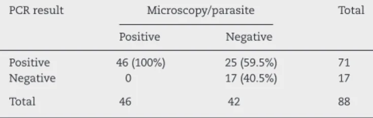
Table 1 – Comparison of PCR and microscopy for detection ofLeishmaniain bone marrow (Kappa= 0.41).
PCR result Microscopy/parasite Total
Positive Negative
Positive 46 (100%) 25 (59.5%) 71
Negative 0 17 (40.5%) 17
Total 46 42 88
PCR, polymerase chain reaction.
to electrophoresis in a 1.2% (w/v) agarose gel stained with ethidium bromide (4.0 mg/L) and identified by visualizing the
bands of expected size under UV transilluminator. A negative extraction control was used to assure absence of contam-ination during DNA extraction process. A negative control containing distilled DNA free water was also used.
Data analysis
Sensitivity and specificity with the respective confidence intervals were estimated for PCR results using bone marrow aspirate and peripheral blood in relation to tissue microscopy. Concordance between PCR results in bone marrow and periph-eral blood were assessed by Kappa statistic.12
Results
The study population consisted of 88 subjects, predominantly males (60.2%), below the age of 15 (55.7%), and of rural origin (64.8%). Of the total, 47 (53.4) were considered “cases” (46 pos-itive for parasites in bone marrow and one negative), and 41 (46.6%) “non-cases”. The classification of subjects into “cases” and “non-cases” was made according to the diagnostic criteria already defined.
All blood samples were subjected to PCR. Tests were positive in 71 subjects. Concordance between amastigote demonstration by microscopy and PCR was considered weak (Kappa0.21–0.40) (Table 1). Compared to bone marrow aspirate PCR showed high sensitivity, but low specificity for disease diagnosis.
PCR in bone marrow aspirates tested positive in all AVL patients (cases) and in 24 (58.5%) of those who did not have active disease, indicating low specificity, as observed in
Table 2.
Table 2 – PCR results in bone marrow aspirates of patients with and without American visceral leishmaniasis prior to treatment.
PCR result American visceral leishmaniasis 95% CI
Cases Non-cases
Positive 47 (100%) 24 (58.5%) 90.5–100.0
Negative – 17 (41.5%) 26.7–57.8
Total 47 (100%) 41 (100%)
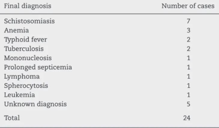
Table 3 – Diagnostic distribution of the false-positive cases.
Final diagnosis Number of cases
Schistosomiasis 7
Anemia 3
Typhoid fever 2
Tuberculosis 2
Mononucleosis 1
Prolonged septicemia 1
Lymphoma 1
Spherocytosis 1
Leukemia 1
Unknown diagnosis 5
Total 24
False positive cases, after the diagnosis of AVL was ruled out, were found to have a variety of other illnesses, as shown inTable 3.
Bone marrow aspirates and peripheral blood samples obtained prior to treatment were evaluated by PCR. The agree-ment beyond chance was 0.88, a result considered excellent (Kappa= 0.88–0.99) (Table 4). This indicates that peripheral blood examination could replace bone marrow aspirates due to better acceptance and facility of collection.
Discussion
In this study patients with suspected diagnosis of AVL were young, 55.7% of them were under 15 years old. The same per-centage of this age group was observed among those with confirmed AVL diagnosis. This result is comparable with those reported by Alves et al.13for AVL cases in Brazil from 2000 to
2004.
Direct microscopic examination and/or culture of spleen aspirate are considered the gold-standard to confirm AVL diagnosis.14 Nevertheless, they are not recommended in
endemic areas due to the potential of serious complications. The demonstration ofLeishmaniaamastigotes in smears from lymph node and bone marrow aspirates, although less sensi-tive, is used as reference by health services in endemic areas.14
Several studies have demonstrated that PCR is highly sensi-tive and specific for leishmaniasis diagnosis.8,15,16 However,
the majority of the authors compared PCR results between patients with AVL and healthy subjects, with the latter group coming from areas different from the former group. In this
Table 4 – Results of PCR in bone marrow and peripheral blood of patients with and without American visceral leishmaniasis (Kappa= 0.88).
Peripheral blood Bone marrow aspirate
Positive Negative
Positive 66 (97.1%) 01 (6.7%)
Negative 02 (2.9%) 14 (93.3%)
Total 68 15
PCR, polymerase chain reaction.
study the evaluation was made adjusting to phase III of Sacket and Haynes8classification, which allows for a better judgment
of the possible usefulness of the test for diagnostic purposes. PCR results in bone marrow aspirates showed poor con-cordance assessed by theKappa statistic with the detection of amastigotes by microscopy, despite its high sensitivity. The results observed in true positive patients on both diagnostic methods are in line with those obtained by Osman et al.5and
Salotra et al.7These authors also reported elevated sensitivity
of PCR in marrow blood aspirates, and concluded that PCR is as good as the search for parasites in lymph nodes or bone marrow in confirmed AVL cases. The low specificity observed diverges from the results of other authors like Piarroux et al.,6
Lachaud et al.3and Disch et al.,16who found 100% specificity.
In their studies cases and controls were not from the same area.
Of the 25 subjects suspected of AVL with only positive PCR, 24 were diagnosed as not having leishmaniasis. The remaining patient had the diagnosis of AVL despite negative parasito-logical examination, on the grounds of epidemioparasito-logical data, clinical presentation, hematological findings showing pancy-topenia, and satisfactory treatment response.
Medical literature reports the existence of asymptomatic cases in endemic areas. In northeast Italy Pampiglione et al.17
demonstrated antibodies against Leishmania in 3.7% of the analyzed population. Six asymptomatic persons underwent liver biopsy, and in one of them amastigotes were found in liver tissue. In Brazil, Evans et al.18 evaluating children
less than 11 years old, found 4.6% with antibodies against
Leishmania. Caldas et al.19 in the same country found
anti-Leishmania antibodies in 34.4% of children (ages 0–5), and Costa et al.20 foundLeishmaniakDNA in asymptomatic
per-sons living in same households of AVL patients. More recently, Adini et al.21identified 2.9% positive sera for anti-Leishmania
antibodies in asymptomatic subjects residents of Israel. These studies suggest that AVL behaves like tuberculosis and Hansen’s disease in regard to the number of infected sub-jects being much larger than the number of those having the disease. In the present study, cases and controls were from endemic regions, thus specificity findings are consistent with those by Fichoux et al.,22Sharma et al.,23and Lambson et al.24
The latter suggests that PCR is a convenient tool to detect asymptomatic carriers of the parasite in endemic areas.
The 17 individuals suspected of having AVL but with neg-ative results on both tests were given other diagnosis. These subjects, if really exposed to the disease, reacted differently to the presence of the parasite. The outcome of infection seems to depend on a complex interaction of virulence fac-tors of the parasite and host immunological mechanisms.25,26
As reviewed by Saha et al.,26 cellular immunity is crucial
for susceptibility or resistance to leishmaniasis, but humoral response also seems to play a role. Parasite antigens do not induce delayed-type hypersensitivity with absence of lym-phocyte proliferation, and inadequate production or secretion of interferon-gamma (IFN-gamma) and interleukin 2 (IL-2), citokynes related to the immune response.27 In the early
stage of infection the IFN-gamma plays a role to control the infection.28 According to Jeronimo et al.29cellular-mediated
providing evidence that genetic risk factors contributes to the disease outcome.30It is possible that the 17 subjects without
infection had been exposed to the parasite but were immuno-logically competent, resisting effectively the development of the disease.
The false-negative PCR in the peripheral blood in the patient with parasites in the bone marrow microscopic exam-ination could be due to: loss of the specimen at the time of the PCR test, time of the test, duration and conditions of specimen storage, presence PCR inhibitors in the blood, or periodicity of the circulating parasites.
Cutaneous tests, such as Montenegro reaction and search for antibodies, revealed that the infectious agent stimulates not only the cellular immunity but also the humoral, without flagging the presence of parasites in the patient. In contrast, a positive PCR, translate detection of specific DNA, meaning that the person harborsLeishmania amastigotes. This refers to AVL found in India and Sudan, where the reservoir and source of infection for the vector is the patient. Working in endemic regions of Brazil studies proved that AVL patients are infective to vectors.20,31Although not yet proved, it is possible
that asymptomatic carriers may serve as source of parasites to sand flies.
A limitation of this study was the fact that PCR was not done in all identified patients during the study period, which could lead to selection bias. To evaluate if this could compromise the validity of the study, comparison was made taking in consideration age, sex, average size of the liver and spleen: patients with AVL who had blood tested by PCR versus those whose blood was not tested, and patients without AVL but with PCR positive versus those with PCR negative. This analysis did not show significant differences (data not presented). It is believed that the inclu-sion of these subjects would not substantially change the results.
Conclusion
The present study used PCR to evaluate persons suspected of having AVL, answering questions formulated in phase III by Sacket and Haynes8 classification. The positivity rate
found was higher in bone marrow aspirates and periph-eral blood of patients diagnosed by microscopic examination. The test was also positive in patients without the dis-ease but living in endemic regions (false-positive). Therefore, PCR should not be valued when used alone for diagno-sis. However, it can be used as an ancillary diagnostic test in persons with strong indication of having the dis-ease but with negative bone marrow aspirate, having the advantage to be performed in peripheral blood samples. It is also possible to use PCR to assess the prevalence of infection in specific population groups, and to evaluate the animal reservoir and vectors involved in the epidemiological chain.
Conflict of interest
All authors declare to have no conflict of interest.
Acknowledgements
The authors wish to thank all the patients for their willingness to participate in this study, and express gratitude to Maria de Fátima Thomas, MD, MPH, for assisting with translations and expert advice regarding research publication.
r e f e r e n c e s
1. Reithinger R, Dujardin JC. Molecular diagnosis of
leishmaniasis: current status and future applications. J Clin Microbiol. 2007;45:21–5.
2. Disch J, Rabello A. A reac¸ão em cadeia da polimerase em sangue periférico para diagnóstico e avaliac¸ão de cura da leishamniose visceral. Rev Soc Bras Med Trop. 2001;34:183. 3. Lachaud L, Dereure J, Chabbert E, et al. Optimized PCR using
patient blood samples for diagnosis and follow-up of visceral leishmaniasis, with special reference to AIDS patients. J Clin Microbiol. 2000;38:236–40.
4. Lachaud L, Marchergui-Hammami S, Chabbert E, et al. Comparison of six PCR methods using peripheral blood for detection of canine visceral leishmaniasis. J Clin Microbiol. 2002;40:210–5.
5. Osman F, Oskan L, Zijlstra EE, et al. Evaluation of PCR for diagnosis of visceral leishmaniasis. J Clin Microbiol. 1997;35:2454–7.
6. Piarroux R, Gambarelli F, Dumon H, et al. Comparison of PCR with direct examination of bone marrow aspiration, myeloculture, and serology for diagnosis of visceral leishmaniasis in immunocompromised patients. J Clin Microbiol. 1994;32:746–9.
7. Salotra P, Sreenivas G, Pogue GP, et al. Development of a species-specific PCR assay for detection ofLeishmania donovaniin clinical samples from patients with kala-azar dermal leishmaniasis. J Clin Microbiol. 2001;39:849–54. 8. Sackett DL, Haynes RB. Evidence base of clinical diagnosis. Br
Med J. 2002;321:539–41.
9. Smyth AJ, Ghosh A, Hassan MQ, et al. Rapid and sensitive detection ofLeishmaniakinetoplast DNA from spleen and blood samples of kala-azar patients. Parasitology. 1992;105:183–92.
10. MS – Ministério da Saúde.Manual de vigilância e controle da leishmaniose visceral. Brasília; 2003.
11. WHO. World Health Organization – the leishmaniases and
Leishmania/HIV co-infections. Fact sheet no. 116; May 1996. 12. OPAS – Organización Pan Americana de la Salud.Métodos de
Investigac¸ão Epidemiológica em Doenc¸as Transmissíveis; 1997. 13. Alves W, Maia NA, Oliveira GM, Sousa W, Bonfim R.
Leishmaniose visceral no Brasil: perfil dos casos no período de 2002 a 2004. Rev Soc Bras Med Trop. 2005;38:489.
14. Boelaert M, Rijal S, Regmi S, et al. A comparative study of the effectiveness of diagnostic tests for visceral leishmaniasis. Am J Trop Med Hyg. 2004;70:72–7.
15. Lambson B, Smyth A, Barker D. Sequence homology within a minicircle class of theLeishmania donovanicomplex. Mol Biochem Parasitol. 1999;101:229–32.
16. Disch J, Maciel FC, Oliveira MC, Orsini M, Rabello A. Detection of circulatingLeishmania chagasiDNA for the non-invasive diagnosis of human infection. Trans R Soc Trop Med Hyg. 2003;97:391–5.
18. Evans TG, Teixeira MJ, Mcauliffe IT, et al. Epidemiology of visceral leishmaniasis in Northeast Brazil. J Infect Dis. 1992;166:1124–32.
19. Caldas AJM, Silva DRC, Pereira CCR, et al. Infecc¸ão por
Leishmania(Leishmania) chagasiem crianc¸as de uma área endêmica de leishmaniose visceral americana na ilha de São Luis-MA, Brasil. Rev Soc Bras Med Trop. 2001;34:445–51. 20. Costa CH, Stewart JM, Gomes RB, et al. Asymptomatic human
carriers ofLeishmania chagasi. Am J Trop Med Hyg. 2002;66:334–7.
21. Adini I, Ephros M, Chen J, Jaffe CL. Asymptomatic visceral leishmaniasis. Northern Israel Emerg Infect Dis. 2003;9:397–8. 22. Fichoux YL, Quaranta JF, Aufeuvre JP, et al. Occurrence of
Leishmania infantumparasitemia in asymptomatic blood donors living in an area of endemicity in Southern France. J Clin Microbiol. 1999;37:1953–7.
23. Sharma MC, Gupta AK, Das VNR, et al.Leishmania donovaniin blood smears of asymptomatic persons. Acta Trop.
2000;76:195–6.
24. Lambson B, Smyth A, Barker DC.Leishmania donovani: development and characterisation of a kinetoplast DNA probe and its use in the detection of parasites. Exp Parasitol. 2000;94:15–22.
25. Pearson RD, Wheeler DA, Harrison LH, Kay HD. The immunobiology of leishmaniasis. Rev Infect Dis. 1983;5:907–27.
26. Saha S, Mondal S, Banerjee A, et al. Immune responses in kala-azar. Indian J Med Res. 2006;123:245–66.
27. Zwingenberger K, Harms G, Pedrosa C, et al. Determinants of the immune response in visceral leishmaniasis: evidence for predominance of endogenous interleukin 4 over interferon-␥
production. Clin Immunol Immunopathol. 1990;57:242–9. 28. Carvalho EM, Bacellar O, Brownell C, et al. Restoration of
IFN-␥production and lymphocyte proliferation in visceral
leishmaniasis. J Immunol. 1994;152:5949–56.
29. Jeronimo S, Pearson RD, Sousa AQ.Leishmaniaspecies: visceral (kala-azar), cutaneous, and mucosal leishmaniasis. In: Mandell GL, Douglas Jr RG, Bennett JE, editors. Principles and practice of infectious diseases. 6th ed. Churchill Livingstone; 2005. p. 3145–64.
30. Blackwell JM, Fakiola M, Ibrahim ME, et al. Genetics and visceral leishmaniasis: of mice and man. Parasite Immunol. 2009;31:254–66.