C L I N I C A L R E S E A R C H A R T I C L E
Orofacial muscles may be affected in early stages of Becker
muscular dystrophy: A preliminary study
Marloes L.J. Lagarde MSc
1|
Nens van Alfen MD, PhD
2|
Alexander C.H. Geurts MD, PhD
1|
Imelda J.M. de Groot MD, PhD
1|
Lenie van den Engel-Hoek PhD
11
Radboud University Medical Center, Department of Rehabilitation, Donders Institute for Brain, Cognition and Behaviour, Nijmegen, The Netherlands
2
Radboud University Medical Center, Department of Neurology, Donders Institute for Brain, Cognition and Behaviour, Nijmegen, The Netherlands
Correspondence
Marloes L.J. Lagarde, Radboud University Medical Center, Geert Grooteplein 10, 6500 HB Nijmegen, The Netherlands.
Email: [email protected]
Abstract
Background: Dysphagia is reported in patients with Duchenne or Becker muscular
dystrophy. Our clinical experience suggests that, compared with Duchenne patients,
impaired mastication and swallowing occur early in Becker patients relative to their
skeletal muscle involvement. The aim of this study was to assess dysphagia in
Duchenne and Becker patients in relation to ambulatory capacity.
Methods: In patients in the early ambulatory stage, clinical symptoms, quantitative
muscle ultrasound of the orofacial muscles, and maximum bite force were assessed.
The 6-Minute Walk Test (6MWT) was used to measure ambulatory capacity.
Results: Eleven Duchenne and 11 Becker patients were included. Although Becker
patients had a greater 6MWT distance than Duchenne patients, the occurrence of
mastication and swallowing difficulties was similar. The temporalis muscle was
signifi-cantly thicker in Becker patients.
Conclusions: Clinicians should be aware of dysphagia in both groups, even when
ambulation is still well preserved.
K E Y W O R D S
Becker muscular dystrophy, Duchenne muscular dystrophy, dysphagia, mastication, orofacial muscles, ultrasound
1
|
I N T R O D U C T I O N
Duchenne muscular dystrophy (DMD) and Becker muscular dystro-phy (BMD) are neuromuscular disorders caused by an X-linked muta-tion in the dystrophin gene. This mutamuta-tion results in a decreased level (BMD) or absence (DMD) of functional dystrophin.1 Mastica-tion (ie, chewing) and swallowing problems due to weakness of
orofacial muscles are observed in patients with either DMD or BMD. In a previous study of DMD patients in the early and late ambulatory stages, 60% had mastication difficulties.2In another study of DMD
patients (9–26 years old), 90% had swallowing problems when assessed with videofluoroscopy.3There is more literature on DMD
than on BMD in this respect, but in both populations orofacial mus-cle weakness may lead to mastication and swallowing difficulties.2-7
Using quantitative muscle ultrasound (QMUS) in patients with DMD, hypertrophy and pseudohypertrophy of the orofacial muscles (tem-poralis muscle and tongue) have been reported.2In addition, previ-ous research has shown a significantly reduced maximum bite force
Abbreviations: 6MWT, 6-Minute Walk Test; BMD, Becker muscular dystrophy; DMD, Duchenne muscular dystrophy; MBF, maximum bite force; N, Newton; QMUS, quantitative muscle ultrasound; SLT, speech language therapist.
This is an open access article under the terms of the Creative Commons Attribution-NonCommercial License, which permits use, distribution and reproduction in any medium, provided the original work is properly cited and is not used for commercial purposes.
© 2019 The Authors. Muscle & Nerve published by Wiley Periodicals, Inc.
(MBF) in all ambulatory stages of DMD, leading to mastication diffi-culties.2,8Of interest, in BMD patients, a similar level of swallowing
problems has been found as in patients with DMD when groups were matched according to their functional capacities.5
QMUS of the orofacial muscles is useful to explain underlying mechanisms of mastication and swallowing problems.9-11QMUS has
previously been used in patients with DMD to assess the muscle abnormality underlying dysphagia,2,6 but no data on thickness or
echogenicity of orofacial muscles are available for patients with BMD.
Although BMD patients have a milder disease progression in terms of ambulatory capacity than those with DMD, our clinical expe-rience suggests that impaired mastication and swallowing occur early in BMD relative to their limb muscle involvement. This notion has led to the hypothesis that the orofacial muscles of BMD patients may show severe dystrophic changes compared with DMD patients, rela-tive to ambulatory capacity. Confirmation of this hypothesis is impor-tant to gain insight into the clinical course of BMD and to evaluate whether early assessment of mastication and swallowing difficulties in BMD is warranted. Hence, the aim of this study was to assess differ-ences in dystrophic changes of orofacial muscles and mastication and swallowing difficulties between DMD and BMD patients in relation to their ambulatory capacity.
2
|
M E T H O D S
2.1
|
Participants
The charts of patients with a genetically confirmed diagnosis of DMD or BMD (in-frame vs out-of-frame exon deletions) who visited the interdisciplinary outpatient clinic of the pediatric and adult cen-ter for neuromuscular disorders of the Radboud University Medical Center between December 2015 and December 2016 were reviewed. All patients who were still in the early ambulatory stage were included for analysis.12 Data had been collected during the
annual visits as part of their regular care. The research was con-ducted in accordance with national and international ethics stan-dards. The study was approved by the regional medical-ethical committee, and informed consent was waived because of the retro-spective nature of the study.
2.2
|
Assessment
At each visit, all patients were seen by a speech language therapist (SLT) and a physiotherapist. Only data from standardized parts of the SLT assessment (questionnaire, MBF measurements, and QMUS) were included for analysis. Age, height, weight, and corticosteroid treatment of the patients were noted. The 6-Minute Walk Test (6MWT) was performed by the physiotherapist. The number of meters walked was used to assess patients’ walking capacity. This number was corrected for age and described as a z-score.13
2.2.1
|
Questionnaire
Mastication and swallowing symptoms were determined using a semi-structured questionnaire about feeding difficulties.6Items pertaining to
mastication difficulties, symptoms of choking (thin liquid and solid food), and food sticking in the throat were scored on a 4-point scale (1, no prob-lems; 2, once a week; 3, once a day; 4, several times a day). These scores were used to determine whether patients had mastication and/or swallowing difficulties. The scores on the questionnaire were dichotomized (1 indicating no difficulties and 2–4 indicating incremental difficulties).
2.2.2
|
MBF
The MBF was measured using the Bite Force Gauge of the VU univer-sity (VU-BFG, Amsterdam, the Netherlands), a device to measure maximum voluntary bite force in kilograms.14The VU-BFG was placed between the incisors of the patients. The patients were asked to bite three times as firmly as possible. The maximum value in kilograms was converted to Newtons to make it comparable to the literature.2The
MBF was corrected for height and converted to a z-score (based on our own laboratory values and regression analysis of 68 healthy par-ticipants).2z-Scores were calculated using the following equation:
z =
MBF in N
9:807 − −21:5 + 0:266*heightð ð ÞÞ
7:2
2.2.3
|
QMUS
For all examinations the z.one ultra Convertible Ultrasound system (Zonare medical systems, Mountain View, CA) was used. QMUS was performed to determine muscle thickness and echogenicity of the masticatory muscles (masseter muscle and temporalis muscle), submental muscles (digastric muscles and geniohyoid muscles), and the muscles of the tongue (superior longitudinal muscle and transverse muscle of the tongue). Muscle thickness shows the presence of atrophy or, in case of dystrophinopathies, hypertro-phy of affected muscles. Hypertrohypertro-phy is an increase in the size of muscle fibers without an increase in connective tissue, while pseudohypertrophy is an increase in the size of the muscle as a consequence of infiltration by fat and fibrosis.15The mean echogenicity of selected regions of interest was calculated using the histogram function in a custom software program for quantitative muscle image analysis (QUMIA) developed at our center, resulting in a number between 0 and 255.16We previously reported our
reference values for healthy subjects.9-11With these data, the muscle thickness and echogenicity of the orofacial muscles were described as z-scores. z-Scores above 2 were considered to be abnormal.10,17
2.3
|
Statistical analysis
All statistical analyses were performed using IBM SPSS, version 22.0 (IBM Corp, Chicago, IL). Descriptive statistics were used to report the patient
characteristics and the z-scores of the 6MWT, MBF, and QMUS results. For comparing the two groups (DMD and BMD) with respect to age, weight, height, and z-scores of the 6MWT, MBF and QMUS results, two-sided independent-samples Mann–Whitney U tests with a significance level of .05 were used. A nonparametric test was chosen because of the small sample size.
3
|
R E S U L T S
3.1
|
Subjects
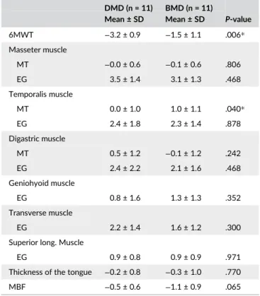
The charts of 81 patients with DMD and 11 patients with BMD were reviewed for this study. Eleven of the 81 boys with DMD (14%) and all boys with BMD were in the early ambulatory stage and were included for further analysis. Table 1 shows the age, height, and weight of the patients, the proportion with mastication and swallowing difficulties, and the percentage receiving treatment with corticosteroids. Age was significantly higher for the BMD patients, as were height and weight.
3.2
|
6MWT
The mean distance on the 6MWT in the boys with BMD was 512.0 m (range, 420–615 m). DMD patients walked a mean of 359.6 m (range, 288–435 m). Boys with DMD had a significantly lower z-score on the 6MWT than boys with BMD, indicating a lower walking capacity in the former (Table 2). An abnormal z-score (<−2) was observed in nine DMD and three BMD patients.
3.3
|
Thickness and echogenicity of orofacial
muscles
Table 2 shows the means and standard deviations of the z-scores for muscle thickness and echogenicity of the orofacial muscles. There were no significant differences in these measures between patients with BMD and DMD, except for the thickness of the temporalis
muscle, which was significantly greater in patients with BMD. The thickness of the temporalis muscle was abnormal (z-score > 2) in two of the BMD patients and none of the DMD patients. The echogenicity of the temporalis muscle was also abnormal in these two BMD patients. The echogenicity of the masseter muscle was abnormal in eight of the BMD and 10 of the DMD patients. The thickness of the masseter muscle was normal in all patients. Hypertrophy of the tongue did not occur in either DMD or BMD patients.
3.4
|
MBF
The mean MBF of DMD patients was 74.8 N with a standard devia-tion of 23.5 N. BMD patients scored a mean MBF of 87.6 N with a standard deviation of 49.9 N. When corrected for height, MBF was lower in patients with BMD than in patients with DMD. This differ-ence, however, did not reach significance (Table 2).
4
|
D I S C U S S I O N
Our results show that, despite the better ambulatory capacity of BMD patients, the orofacial muscles are at least as severely affected in T A B L E 1 Characteristics of the included patients in the early
ambulatory stage (n = 22) DMD (N = 11) Mean ± SD/N (%) BMD (N = 11) Mean ± SD/N (%) P-value Age (years) 7.7 ± 1.7 10.7 ± 2.4 .004 Height (cm) 122.5 ± 12.2 144.3 ± 15.2 .002 Weight (kg) 24.8 ± 5.8 38.3 ± 10.2 .002 Mastication difficulties 3 (27%) 2 (18%) Swallowing difficulties 1 (9%) 1 (9%) Corticosteroids treatment 9 (82%) 1 (9%)
T A B L E 2 Assessments of the included patients in the early ambulatory stage (n = 22) DMD (n = 11) BMD (n = 11) P-value Mean ± SD Mean ± SD 6MWT −3.2 ± 0.9 −1.5 ± 1.1 .006* Masseter muscle MT −0.0 ± 0.6 −0.1 ± 0.6 .806 EG 3.5 ± 1.4 3.1 ± 1.3 .468 Temporalis muscle MT 0.0 ± 1.0 1.0 ± 1.1 .040* EG 2.4 ± 1.8 2.3 ± 1.4 .878 Digastric muscle MT 0.5 ± 1.2 −0.1 ± 1.2 .242 EG 2.4 ± 2.2 2.1 ± 1.6 .468 Geniohyoid muscle EG 0.8 ± 1.6 1.3 ± 1.3 .352 Transverse muscle EG 2.2 ± 1.4 1.6 ± 1.2 .300
Superior long. Muscle
EG 0.9 ± 0.8 0.9 ± 0.9 .971
Thickness of the tongue −0.2 ± 0.8 −0.3 ± 1.0 .770
MBF −0.5 ± 0.6 −1.1 ± 0.9 .065
MT, muscle thickness; EG, echogenicity.
6MWT, MT, EG, thickness of the tongue and MBF were described in z-scores.
*Statistically significant differences in z-scores between DMD and BMD patients, determined with independent samples Mann–Whitney U tests.
BMD as in DMD patients, which confirms our hypothesis. Indeed, the proportion of patients in our cohort with mastication and swallowing difficulties in the early ambulatory stage was equal for DMD and BMD patients. The prevalence of these difficulties in the DMD group was comparable to a previous study reporting a different cohort of DMD patients.2 In the same study, MBF was measured in DMD
patients in the early ambulatory stage and these results were similar to the MBF results in the present study.2Despite their better
ambula-tory capacity, BMD patients did not differ from DMD patients with regard to MBF.
In addition, in the present study, the temporalis muscle was signifi-cantly thicker in patients with BMD with an abnormal z-score for echogenicity, showing a relatively early manifestation of pseudo-hypertrophy of this muscle in BMD patients. Pseudopseudo-hypertrophy of the temporalis muscle has previously been observed on MRI in DMD patients who were 8–16 years old.18In our cohort, pseudohypertrophy
of the temporalis muscle (z-score muscle thickness > 2) was present in two BMD patients of 5–14 years old, but in none of the DMD patients. Straathof et al. stated that it remains unclear whether the increased mus-cle mass of the temporalis musmus-cle results in an increased or a decreased bite force,18but it seems likely that the limited MBF in early stages of DMD and BMD is probably due to the early dystrophic changes of the masseter muscle and the pseudohypertrophy of the temporalis muscle.
An interesting question is why BMD patients would have early involvement of masticatory muscles. One explanation may be that only one patient in the BMD group used corticosteroids. Corticoste-roid treatment in DMD patients usually starts as soon as motor devel-opment reaches a plateau phase.12In BMD patients, however, there
is no guideline for starting corticosteroid treatment. It is unknown whether corticosteroid treatment may have a protective effect on the orofacial muscles. Previous studies showed early cardiomyopathy in BMD patients. In a study of Bredman et al. a cardiac-specific myosin was found in the masticatory muscles, but not in other skeletal muscles,19 which points at another possible explanation for early
involvement of the masticatory muscles in BMD. Further investigation is warranted to gain insight into why early dystrophic changes would occur in the cardiac and masticatory muscles in BMD patients.
Our results showed no differences in dystrophic changes between DMD and BMD patients in the early ambulatory stage regarding the muscles that are responsible for laryngeal elevation (digastric and geniohyoid muscles). These findings confirm the results of previous research, showing that dystrophic changes of the sub-mental muscles occur in a relatively late stage of DMD.20Changes in the submental muscles at a later stage may still lead to differences in laryngeal elevation between older DMD and BMD patients.8 Our results complement the findings of Yamada et al. 20175who used
videofluoroscopy and found that swallowing difficulties in BMD patients are similar to the difficulties in DMD patients, when patients are matched according to functional ability. We found that BMD patients show dystrophic changes of masticatory muscles earlier than DMD patients, relatively to their gait capacity.
A limitation of the current study is the small number of patients. This could have led to an equal number of patients with swallowing
and mastication difficulties in both groups. A strength is that the find-ings were confirmed using different assessment tools (questionnaire, QMUS, and MBF). Analyses of larger groups of patients and a longitu-dinal follow up through the different ambulatory stages are needed to determine more specific differences in dystrophic changes of both limb and orofacial muscles between DMD and BMD patients. We believe that the results of our study warrant awareness of dysphagia and mastication and swallowing difficulties in BMD patients, even when ambulation is still well preserved. When there is a clinical suspi-cion of dysphagia, instrumental assessment is recommended.
E T H I C A L P U B L I C A T I O N S T A T E M E N T
We confirm that we have read the Journal's position on issues involved in ethical publication and affirm that this report is consistent with those guidelines.
None of the authors has any conflict of interest to disclose.
R E F E R E N C E S
1. Koenig M, Beggs A, Moyer M, et al. The molecular basis for Duchenne versus Becker muscular dystrophy: correlation of severity with type of deletion. Am J Hum Genet. 1989;45:498.
2. van den Engel-Hoek L, de Groot IJ, Sie LT, et al. Dystrophic changes in masticatory muscles related chewing problems and malocclusions in Duchenne muscular dystrophy. Neuromuscul Disord. 2016;26: 354-360.
3. Hanayama K, Liu M, Higuchi Y, et al. Dysphagia in patients with Duchenne muscular dystrophy evaluated with a questionnaire and videofluorography. Disabil Rehabil. 2008;30:517-522.
4. Willig T, Paulus J, Saint JL, Beon C, Navarro J. Swallowing problems in neuromuscular disorders. Arch Phys Med Rehabil. 1994;75:1175-1181.
5. Yamada Y, Kawakami M, Wada A, Otsuka T, Muraoka K, Liu M. A com-parison of swallowing dysfunction in Becker muscular dystrophy and Duchenne muscular dystrophy. Disabil Rehabil. 2018;40:1421-1425. 6. van den Engel-Hoek L, Erasmus CE, Hendriks JC, et al. Oral muscles
are progressively affected in Duchenne muscular dystrophy: implica-tions for dysphagia treatment. J Neurol. 2013;260:1295-1303. 7. Pane M, Vasta I, Messina S, et al. Feeding problems and weight gain
in Duchenne muscular dystrophy. Eur J Paediatr Neurol. 2006;10: 231-236.
8. Ueki K, Nakagawa K, Yamamoto E. Bite force and maxillofacial mor-phology in patients with Duchenne-type muscular dystrophy. J Oral Maxillofac Surg. 2007;65:34-39.
9. van den Engel-Hoek L, Lagarde M, Van Alfen N. Ultrasound of oral and masticatory muscles: why every neuromuscular swallow team should have an ultrasound machine. Clin Anat. 2017;30:183-193. 10. van den Engel-Hoek L, van Alfen N, de Swart BJ, de Groot IJ, Pillen S.
Quantitative ultrasound of the tongue and submental muscles in chil-dren and young adults. Muscle Nerve. 2012;46:31-37.
11. Lagarde M, van den Engel-Hoek L. Quantitative ultrasound of orofacial muscles in infants from 6 months to 5 years: collecting nor-mal values. Curr Med Imaging Rev. 2017;13:332-338.
12. Bushby K, Finkel R, Birnkrant DJ, et al. Diagnosis and management of Duchenne muscular dystrophy, part 1: diagnosis, and pharmacological and psychosocial management. Lancet Neurol. 2010;9:77-93. 13. Goemans N, Klingels K, Van den Hauwe M, et al. Six-minute walk
test: reference values and prediction equation in healthy boys aged 5 to12 years. PLoS One. 2013;8:e84120.
14. Weijenberg RAF, Lobbezoo F, Knol DL, Tomassen J, Scherder EJA. Increased masticatory activity and quality of life in elderly persons with dementia-a longitudinal matched cluster
randomized single-blind multicenter intervention study. BMC Neu-rol. 2013;13:26.
15. Cros D, Harnden P, Pellissier J, Serratrice G. Muscle hypertrophy in Duchenne muscular dystrophy. J Neurol. 1989;236:43-47.
16. Pillen S, van Keimpema M, Nievelstein RA, Verrips A, van Kruijsbergen-Raijmann W, Zwarts MJ. Skeletal muscle ultrasonogra-phy: visual versus quantitative evaluation. Ultrasound Med Biol. 2006; 32:1315-1321.
17. Pillen S, Tak RO, Zwarts MJ, et al. Skeletal muscle ultrasound: correla-tion between fibrous tissue and echo intensity. Ultrasound Med Biol. 2009;35:443-446.
18. Straathof C, Doorenweerd N, Wokke B, et al. Temporalis muscle hypertrophy and reduced skull eccentricity in Duchenne muscular dystrophy. J Child Neurol. 2014;29:1344-1348.
19. Bredman J, Wessels A, Weijs W, Korfage J, Soffers C, Moorman A. Demonstration of ‘cardiac-specific’ myosin heavy
chain in masticatory muscles of human and rabbit. Histochem J. 1991;23:160-170.
20. Lagarde M, Knuijt S, Groothuis J, de Groot I, van den Engel-Hoek L. Lon-gitudinal changes in oral and masticatory muscles in Duchenne muscular dystrophy: a disturbed balance. Neuromuscul Disord. 2016;26:S120.
How to cite this article: Lagarde MLJ, van Alfen N, Geurts ACH, de Groot IJM, van den Engel-Hoek L. Orofacial muscles may be affected in early stages of Becker muscular dystrophy: A preliminary study. Muscle Nerve. 2020;61: 213–217.https://doi.org/10.1002/mus.26771