Braz. J. of Develop.,Curitiba, v. 6, n. 10, p. 79252-79266, oct. 2020. ISSN 2525-8761
Analysis of self-etch adhesives behavior in class I composite restorations with
different treatment of enamel margins and light-activation sources
Análise do comportamento de adesivos autocondicionantes em restaurações de
resina composta classe I com diferentes tratamentos das margens de esmalte e
fontes de ativação de luz
DOI:10.34117/bjdv6n10-380Recebimento dos originais: 08/09/2020 Aceitação para publicação:19/10/2020
Maria Malerba Colombi Humel
PhD, Department of Restorative Dentistry, Piracicaba Dental School, University of Campinas Address: Av. Limeira, 901 - Areião, Piracicaba - SP, 13414-903
E-mail: mmchumel@gmail.com
Lucia Trazzi Prieto
PhD, Department of Restorative Dentistry, Piracicaba Dental School, University of Campinas Address: Av. Limeira, 901 - Areião, Piracicaba - SP, 13414-903
E-mail: lucinhatrazzi@hotmail.com
Josué Junior Araujo Pierote
PhD, Department of Restorative Dentistry, Piracicaba Dental School, University of Campinas Address: Av. Limeira, 901 - Areião, Piracicaba - SP, 13414-903
E-mail: josuepierote@hotmail.com
Cintia Tereza Pimenta de Araujo
Adjunct Professor, Department of Dentistry, Faculty of Sciences of Health, Federal University of Jequitinhonha and Mucuri Valley
Address: Rua da Glória, nº 187, Centro, Diamantina, MG, CEP 39100-000 E-mail: ctpimenta@gmail.com
João Victor Frazão Câmara
MSc student, Department of Biological Sciences, Bauru Dental School, University of São Paulo Adress: Alameda Dr. Octávio Pinheiro Brisolla, 9-75 - Jardim Brasil, Bauru - SP, 17012-901
E-mail: jvfrazao92@hotmail.com
Isabel Ferreira Barbosa
PhD, Department of Restorative Dentistry, Piracicaba Dental School, University of Campinas Address: Av. Limeira, 901 - Areião, Piracicaba - SP, 13414-903
E-mail: barbosa.isabelferreira@gmail.com
Guereth Alexsanderson Oliveira Carvalho
MSc student, Department of Dental Clinic, Federal University of Piauí
Address: Campus Universitário Ministro Petrônio Portella - Ininga, Teresina - PI, 64049-550 E-mail: guerethcarvalho@gmail.com
Braz. J. of Develop.,Curitiba, v. 6, n. 10, p. 79252-79266, oct. 2020. ISSN 2525-8761
Luis Alexandre Maffei Sartini Paulillo
Full Professor, Department of Restorative Dentistry, Piracicaba Dental School, University of Campinas
Address: Av. Limeira, 901 - Areião, Piracicaba - SP, 13414-903 E-mail: paulillo@fop.unicamp.br
ABSTRACT
Objective: To evaluate the microtensile bond strength (MTBS), knoop microhardness (KHN) and gap formation of class I restorations restored with self-etching adhesives and resin composites light-activated by either halogen or LED light curing units. Materials and Methods: Class I cavities were prepared in one hundred and forty-four human third molars. Three self-etching adhesives (Clearfil S3 Bond - S3, Clearfil Protect Bond – ProtectB and One-Up Bond F Plus - OneUp) were applied to the cavities, which had the enamel margins either etched with 35% phosphoric acid or left unecthed. The cavities were incrementally restored with TPH3 restorative composite, which was light-activated using Light Emitted by Diode (Hadii-Cal) or Halogen Lamp (Optilux 501). Epoxy resin replicas were obtained from the restored teeth, which were then submitted to thermal cycling. Afterwards, new replicas were obtained and the gaps at the resin composite/enamel margin interface were analyzed by Scanning Electronic Microscopy. Half sample was randomly tested for microtensile bond strength test (n=6) while the other half had the composite tested for KHN (n=6). Results: The etched enamel contributed to avoid gap formation only when OneUp adhesive system was used. No significant difference in MTBS values was found among groups. For KHN analysis, all restorations light-activated with LED showed higher KHN values than those light-activated with halogen lamps. In addition, the resin composites used to restore cavities with acid etched enamel margins showed higher KHN means than those used in cavities having unteched enamel margins. Conclusion: The resin composite bonded to cavities with S3 showed the lowest KHN values at the intermediate and bottom. ProtectB showed no significant differences for the different surface depths.
Keywords: Dentin-Bonding Agents, Composite Resins, Dental Enamel. RESUMO
Objetivo: Avaliar a resistência à microtração (MTBS), a microdureza knoop (KHN) e a formação de fendas de restaurações de classe I restauradas com adesivos autocondicionantes e compósitos de resina ativados por luz de halogênio ou unidades de fotopolimerização LED. Materiais e Métodos: Cavidades de classe I foram preparadas em cento e quarenta e quatro terceiros molares humanos. Três adesivos autocondicionantes (Clearfil S3 Bond - S3, Clearfil Protect Bond - ProtectB e One-Up Bond F Plus - OneUp) foram aplicados nas cavidades, que tiveram as margens do esmalte atacadas com ácido fosfórico 35% ou deixadas unectadas. As cavidades foram restauradas de forma incremental com compósito restaurador TPH3, que foi fotoativado usando luz emitida por diodo (Hadii-Cal) ou lâmpada de halogênio (Optilux 501). Réplicas de resina epóxi foram obtidas dos dentes restaurados, os quais foram submetidos à ciclagem térmica. Posteriormente, novas réplicas foram obtidas e as lacunas na interface resina composta/margem do esmalte foram analisadas por Microscopia Eletrônica de Varredura. Metade da amostra foi testada aleatoriamente para teste de microtração de resistência de união (n = 6), enquanto a outra metade teve o composto testado para KHN (n = 6). Resultados: O esmalte acondicionado contribuiu para evitar a formação de gap apenas quando o sistema adesivo OneUp foi utilizado. Nenhuma diferença significativa nos valores de MTBS foi observada entre os grupos. Para a análise KHN, todas as restaurações ativadas por luz com LED apresentaram valores de KHN mais elevados do que aquelas ativadas por luz com
Braz. J. of Develop.,Curitiba, v. 6, n. 10, p. 79252-79266, oct. 2020. ISSN 2525-8761
lâmpadas de halogênio. Além disso, as resinas compostas usadas para restaurar cavidades com margens de esmalte condicionadas com ácido apresentaram médias de KHN mais altas do que aquelas usadas em cavidades com margens de esmalte não atacadas. Conclusão: A resina composta colada em cavidades com S3 apresentou os menores valores de KHN no intermediário e no fundo. ProtectB não mostrou diferenças significativas para as diferentes profundidades de superfície.
Palavras-chave: Adesivos Dentinários, Resinas Compostas, Esmalte Dentário.
1 INTRODUCTION
The adhesive system may involve two bonding interaction strategies with the hard tissues: total or self-etching technique, in which enamel and/or dentin demineralization is done simultaneously with monomer infiltration, resulting in hybrid layer formation with minerals and resin.1 Among the self-etching adhesive systems, two-bottle and one-bottle are currently available.2
The bond strength values depend on the restorative material used in direct restorations.3,4 The stress generated at the bonding interface due to polymerization shrinkage may result in bonding failure mainly when the adhesive does not resist the contraction stress.5 Effectiveness of the self-etching adhesive systems in sealing the enamel from the cavity margins is controversial.6 The weak penetration of the acidic monomers into enamel seems to create a bonding interface incapable of resisting the stress generated in the restoration by polymerization shrinkage and temperature ranges in the oral environment.7 For this reason, previous phosphoric acid etching of the enamel margins may increase the bond strength values8, although conflicting results are found in the literature.9
However, some studies evaluating bond strength of resin composites to the dental structure do not take into consideration the stress generated from polymerization shrinkage of those restorative materials since the restorations performed in such studies are placed on flat surfaces, which do not reproduce the clinical situation.8,9 The contraction stress is present at different
intensities, particularly when cavities, such as Class I cavities, to be restored have high C Factor. 10-The contraction stress resulting from the competition between the polymerization shrinkage in the resin composite bonded to the dental structure falls upon the adhesive system13 and such stress
on the cavity walls during polymerization is one of the causes of restoration failure, gap and microleakage at the margins.14 The relationship between the bond strength values and marginal
integrity is still controversial in the literature.15 However, studies assessing the durability of the bonding interface showed a strong correlation between the bond strength values and nanoleakage patterns over time.16,17
Another important aspect to be considered in the cavity restoration is the light source used to activate the adhesive system and resin composite polymerization. Halogen (QTC) light is
Braz. J. of Develop.,Curitiba, v. 6, n. 10, p. 79252-79266, oct. 2020. ISSN 2525-8761
routinely used to activate the polymerization. Many of these curing units do not provide the minimum light intensity recommended to ensure proper resin polymerization18 due to wear of the bulb and filter, contamination of guide light, damages to the optic fiber and overheating. Therefore, poorly polymerized resin composite is expected and a successful restorative treatment is compromised as a consequence.18
In an attempt to solve such problems, light curing units equipped with blue light emitting diodes (LED) were developed.19 In spite of their favorable features, such as long useful life and absence of the use of filters18, the results of using LED units to light-activate composites are controversial and dependent on the of resin composition.20
This study aimed to evaluate the microtensile bond strength (MTBS), cross sectioned Knoop microhardness in resin composite and gap formation in Class I resin composite restorations under the following testing conditions: the use of either one or two-step selfetching adhesive systems, which were applied into cavities having enamel margins either acid etched or not; the light-activation of all adhesive systems and resin composite layers with either LED or Halogen curing units.
2 MATERIALS AND METHODS
One hundred and forty-four healthy human third molars were selected. The teeth were cleaned, included in polystyrene resin and their occlusal surfaces were wet polished with 320-grit SiC paper under running water (Politriz, AROTEC – São Paulo, SP) to expose a flat enamel surface without exposing dentin.
Class I cavities were made using #56L diamond burs (KG Sorensen, Barueri, SP, Brazil) at high-speed, under air–water cooling. A custom-made preparation device allowed the standardization of the cavity dimensions to 5 mm mesio-distal length, 4 mm vestibularlingual width and depth of 3 mm, leaving all cavity margins on enamel. The bur was always replaced after the preparation of five cavities.
Three dental adhesive systems were used to restore the cavities: Clearfil Tri-S Bond (pH= 2.7; Kuraray Medical Inc., Okayama Jp), Clearfil Protect Bond (pH = 2; Kuraray Medical Inc. and One-Up Bond F Plus (pH = 1.2; Tokuyama Dental Corp., Tokyo, Jp). The application techniques of each system are presented in Table 1.
Braz. J. of Develop.,Curitiba, v. 6, n. 10, p. 79252-79266, oct. 2020. ISSN 2525-8761
Table 1: Manufacturers` instructions for the application of each adhesive system.
Two light curing units were used to photoactivate the adhesive systems and composite: a halogen light curing unit, Optilux 501 (Kerr Corporation, Orange, USA), or LED curing unit, Radii-Cal (SDI Limited, Victoria, AUS). Prior to the restorative procedure, light intensity (mW/cm2) of both curing units was measured using Ophir 10A-V2-SH power meter (Ophir Optronics, Har – Hotzvim, P.O.B. 45021, Jerusalem 91450, Israel) coupled to a NOVA microprocessor (Ophir Optronics, Har – Hotzvim, P.O.B. 45021, Jerusalem 91450, Israel).21 Thus, the curing units had the time of light exposure changed to provide similar energy density. Therefore, since Radii-Cal mandatorily operates in Soft-Start mode, resin composite layers were only exposed to light 15 seconds after the curing unit was turned on to ensure that both halogen and LED curing units provided similar power density (1.200 mW/cm2). Each resin increment was exposed to light for 40 seconds (1.2 J/s).
After cavity preparation, teeth were assigned to the following experimental groups (n=12): groups 1 and 2: 35% phosphoric acid etching on the enamel margins + Clearfil S3 Bond; groups 3 and 4: Clearfil S3 Bond; groups 5 and 6: 35% phosphoric acid etching on the enamel margins + Clearfil Protect Bond; groups 7 and 8: Clearfil Protect Bond; group 9 and 10: 35% phosphoric acid etching on the enamel margins + One-Up Bond F Plus; groups 11 and 12: One-Up Bond F Plus. Groups 1,3,5,7,9 and 11 were restored using Radii-Cal light curing unit, while groups 2,4,6,8,10 and 12 were light-activated using Optilux 501.
All groups were restored with A3-shade TPH3 Micro Matrix Restorative (Dentsply/Caulk Int., York, USA) resin composite using incremental oblique technique and the last layer at horizontal technique. Each cavity was filled with six 2-mm thick increments, to ensure total irradiance provided to the resin composite. The finishing and polishing procedures were performed with medium-, fine-, and extra fine- grit aluminum oxide disks (SoftLex – 3M/ESPE)fine-, respectively. After polishingfine-, the specimens were stored in distilled water at 37ºC for 24 h. Molds of the occlusal surface were
Clearfil S3 Bond Clearfil Protect Bond One-Up Bond F Plus
Gently dry the dentinal suface Gently dry the dentinal surface Gently dry the dentinal surface Apply Clearfil 3 Bond
on dentin and enamel
Apply primer starting by enamel until complete filling
dentin
Apply the system on the dentin and enamel
Wait 20 seconds
Friction brush for 15 seconds on entire cavity and dry with strong air jet
Friction brush for 15 seconds on entire cavity
Dry lightly Apply adhesive starting by
dentin and then enamel Dry lightly
Light cure for 10 seconds
Dry with light air jet and Light cure for 10 seconds
Apply the system again
(without friction), dry and Light cure for 10 seconds
Braz. J. of Develop.,Curitiba, v. 6, n. 10, p. 79252-79266, oct. 2020. ISSN 2525-8761
obtained from each specimen using addition silicon (Aquasil TM, Dentsply/Caulk-Milford, DE, USA). The molds were filled with epoxy resin (Resina Epóxica Buehler, IL 60044-1699, USA) to created epoxy resin replicas, which were sputter coated with a thin layer of gold (Denton Vacuum Desk II; Denton Vacuum LLC, Moorestown, NJ), and were analyzed using Scanning Electronic Microscopy (SEM; JSM 5900 LV; JEOL,Peabody, MA), to evaluate the restoration margins. Three distinct regions were evaluated on each of the four margins corresponding to buccal, lingual, mesial, and distal adhesive interfaces. The photomicrographs of all the occlusal surfaces had 300X magnification (n=6) and were evaluated using a software with a μm scale specific for such purpose (Image Tools 1.4.1, Jim MacGowan, JP, 2006).
The specimens were then submitted to thermal cycling consisting of 2000 cycles at temperatures ranging from 5 to 55°C ±1°C with dwell time of 60 s in each bath and with a transfer time of 7 s (MCT2, AMM2, São Paulo, Brazil).22 After thermal cycling, new epoxy resin replicas
were obtained from the occlusal surfaces as described above, and another SEM analysis was performed on the restoration margins.
Mechanical tests
After thermal cycling and storage in relative humidity for 24 h at 37 ± 1°C, the specimens were prepared MTBS test and Knoop microhardness (KHN) cross section analysis. Half restored teeth were used for the MTBS test (n=6) while the other half was used for KHN test (n=6).
Microtensile bond strength analysis
The teeth assigned to MTBS test were serially sectioned in the long axis as well as perpendicular to the long axis in buccal-lingual directions using a slow-speed diamond saw (Isomet, Buehler Ltd, Lake Bluff, IL, USA) to obtain sticks having a cross sectional area of approximately 0.9 mm2. Therefore, the bonding interface to be tested was that obtained at the lingual dentin wall of the cavity. The specimens were fixed with cyanoacrylate glue (Super Bond gel, loctite, Henkel, Brazil) to the grips attached to a universal testing machine (Model 4411, Instron Corp, Canton, MA, USA), and were tested in tension at a speed of 0.5 mm/min until failure occurred. Afterwards, the samples were removed from the grips and the fractured area was measured using a digital caliper (Mitutoyo, Japan). Data were obtained in kgf and were converted in Mega Pascal (MPa).
Cross-section Microhardness analysis
Each restored tooth was sectioned in the middle using a slow-speed diamond saw (Isomet, Buehler Ltd, Lake Bluff, IL, USA), parallel to the long axis to originate two halves, which were
Braz. J. of Develop.,Curitiba, v. 6, n. 10, p. 79252-79266, oct. 2020. ISSN 2525-8761
embedded in polystyrene resin to facilitate polishing. The sectioned surface was wet ground with 600, 1200 and 2000 SiC papers and was polished with diamond paste (Arotec Ind. Com., São Paulo, Brazil) in a polishing machine under water cooling.
Three indentations were made on the polished resin composite surface 100 μm, 1500 μm, and 2900 μm below the top of the composite restoration, respectively, and always 100 μm far from the resin composite/dentin bonding interface, under a 25g-load for 20s (HMV- 2000, Shimadzu, Japan). Data was statistically analized by ANOVA and Tukey’s post hoc test at a pre-set alpha of 0.05.
3 RESULTS
Analysis of gap at the restoration margins
The results related to gap formation at the restoration margins are shown in Table 2. Most self-etching adhesives presented significant difference in gap formation prior to and after thermal cycling, regardless of the treatment of enamel margins. The only exception was observed for One Up when the enamel margin was acid etched, where no significant difference in gap formation was observed between values obtained before and after thermal cycling.
Table 2: Means and standard desviation of Gap analysis (μm) according to enamel margin treatments, before and after thermal cycling.
Adhesives
Before thermocycling After thermocycling
No etched enamel margins Etched enamel margins No etched enamel margins Etched enamel margins
One Up 4.85 (1.19)Aa 4.97 (1.19)Aa 25.07 (2.09)Aa* 12.81 (2.09)Ba
S3 Bond 4.49 (1.19)Aa 2.25 (1.19)Aa 18.76 (2.09)Aa* 16.07 (2.09)Aa*
Protect B 8.35 (1.19)Aa 5.41 (1.19)Aa 15.21 (2.09)Aa* 12.9 (2.09)Aa*
Means followed by the same upper case letter are not significantly different when comparing the effect of enamel margin treatment; Means followed by lower case letters are not significantly different when comparing products; means of products after thermocycling followed by asterisks are significantly different from means before thermocycling.
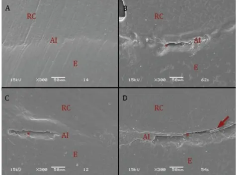
For OneUp, the highest gap formation was observed after thermal cycling when the enamel margin was left unetched. (Figure 1). For S3 and ProtectB, a significant difference was observed between values obtained before and after thermal cycling. There was no significant difference between the group with and that without acid etching (Figures 2 and 3). No significant difference was noted among adhesives systems, regardless of enamel treatment and thermal cycling.
Braz. J. of Develop.,Curitiba, v. 6, n. 10, p. 79252-79266, oct. 2020. ISSN 2525-8761
Figure 1: Scanning Electronic Microscopy of One Up adhesive before (A) and after (B) thermal cycling with etched enamel and before (C) and after (D) thermal cycling without etching. AI - Adhesive Interface; RC – Resin Composite; E – Enamel; * - Gap formation. Vector indicates adhesive fragment debonded from interface.
Figure 2: Scanning Electronic Microscopy of S2 Bond adhesive before (A) and after (B) thermal cycling with etched enamel and before (C) and after (D) thermal cycling without etching. AI - Adhesive Interface; RC – Resin Composite; E – Enamel; * - Gap formation.
Braz. J. of Develop.,Curitiba, v. 6, n. 10, p. 79252-79266, oct. 2020. ISSN 2525-8761
Figure 3: Scanning Electronic Microscopy of Protect B adhesive before (A) and after (B) thermal cycling with etched enamel and before (C) and after (D) thermal cycling without etching. AI - Adhesive Interface; RC – Resin Composite; E – Enamel; * - Gap formation. Vector indicates adhesive fragment debonded from interface.
Microtensile Bond Strength Test
Regarding the MTBS test, the ANOVA showed no significant differences among factors and products (p ≤ 0.05).
Knoop microhardness test
The ANOVA showed a significant difference for the factors: “acid etching of enamel margins”, “light curing units”, and between interaction “adhesive x depth”. Table 3 shows the KHN values of composites light-cured either with LED or with Halogen curing units. The resin composites from cavities restored using LED presented higher values than those from cavities restored using Halogen. Whereas for the acid etched groups, the resin composites in cavities with previous enamel etching exhibited greater KHN results when compared with composite KHN values in cavities without enamel etching.
Table 3: Mean and standard deviation of composite microhardness test for light source units (KHM).
Light source Mean Stand. Error
LED 46.64 0,99 A
Halogen 42.19 0,99 B
Means followed by different letters are significantly different.
For adhesive x depth factors (Table 4), OneUp e S3 adhesives showed higher KHN values
on the top than in the middle and bottom. For ProtectB adhesive, no significant difference in the KHN values was observed among the three depths. When the composite KHN was evaluated within
Braz. J. of Develop.,Curitiba, v. 6, n. 10, p. 79252-79266, oct. 2020. ISSN 2525-8761
depths, the resin composite in cavities restored with ProtectB presented higher KHN values on the top than on middle and bottom. S3 adhesive presented the lowest means with statistically significant difference in middle and bottom depths. At the top, all adhesives presented the similar hardness.
Table 4: Means and standard desviation of composite microhardness test on adhesive and depths compared by Tukey Test (KHM).
Adhesives Base Middle Top
One Up 37.5 (0,72) Bb 36.5 (0,72) Bb 55.7 (3,25) Aa
Protect Bond
61.5 (0,72) Aa 60.5 (0,72) Aa 56.1 (3,25) Aa
S3 Bond 13.5 (0,72) Bc 12.5 (0,72) Bc 65.9 (3,25) Aa
Means followed by different letters (upper case letters: row; lower case letters: column) are significantly different.
4 DISCUSSION
The success of direct restorations with composite resin depends on some factors, such as restoration incremental technique, the light curing units, and the adhesive system. This study analyzed many factors in only one sample, so the interaction among all factors in class I cavities could simulate the clinical situation more precisely. The bonding of selfetching adhesive systems to enamel and dentin surfaces may be susceptible to degradation. The results of present study corroborate with the results of previous studies7,11, as the gap values increased after thermal cycling for all self-etching adhesive systems applied to enamel without previous acid etching. This degradation may be attributed to the hydrolysis of hydrophilic monomers present in the adhesive layer since such layer is considered a permeable membrane. Furthermore, water sorption compromises polymer network formation, impairing mechanical properties of the polymers.23,24
Besides the exposure to water during thermal cycling, the specimens are subjected to temperature ranges capable of generating mechanical stresses due to the differences in the coefficient of thermal expansion (CTE) among substrates, resulting in gap formation as a result of the bond failure at the tooth-restoration interface.25 The CTE of resin composites (28 to 50 ppm/_C) may be at least twice higher than that of tooth structure (9 to 11 ppm/C).26 Therefore, such difference in expansion and contraction of composites in comparison to those of tooth substrate may cause additional stress at the margin of the restoration that contributes to fatigue failure of the bond between the composite and tooth structure.27 Price et al.28 also reported that thermal cycling had a
very significant negative effect on bond strength of resin composites to human dentin when a high C factor testing design was used.
The previous acid etching of enamel margins with 35% phosphoric acid contributed to avoid gap formation after termocycling only when OneUp adhesive system was used. It may be inferred that, for this system, acid etching increased bonding stability. The best result presented by OneUp
Braz. J. of Develop.,Curitiba, v. 6, n. 10, p. 79252-79266, oct. 2020. ISSN 2525-8761
adhesive may be explained by the presence of monomer MAC- 10, which is a hydrophobic monomer and has spacer groups of 10 carbon atoms, resulting in limited dissolution of water and in a polymer hydrolytically more stable.29
On the other hand, the acid etching of enamel margins prior to the application of the Kuraray’s products S3 and ProtectB did not influence the results of gap formation. According to
Yoshida et al.30, the 10-MDP acidic monomer, which is present in both adhesive systems, has the ability to etch and penetrate into the enamel, besides its ability to react chemically with hydroxyapatite.30 Due to previous acid etching of the substrate, the decrease in the amount of hydroxiapatite crystals may have compromised the product bonding effectiveness, which may justify the null effect of etching the enamel margins with phosphoric acid.
Although the results obtained by gap analysis differed according to the treatment of the enamel margins and the thermal cycling, such treatments did not seem to have influenced the MTBS values on dentin. Since the MTBS test was performed in Class I cavities, it is important to take into consideration the stress generated by composite shrinkage and the cavity factor, which may affect the MTBS results for the adhesive systems. Shirai et al., 200512 affirmed that most studies do not take into consideration the stress generated by polymerization shrinkage when investigating bond durability of adhesives. The contraction stress increases when restorations are placed in cavities with high C-factor, such as the Class I cavities used in the current study, allowing less possibility of stress relief after shrinkage. Therefore, the restoration interfaces may become more susceptible to degradation, explaining in part the fast degradation of adhesive interfaces from Class I restorations of in vivo studies when adhesives presenting reliable in vitro results are used.31
When self-etching adhesive systems are tested in conditions closer to clinical situation, different compositions and presentation modes of the bonding agents may not result in higher bond strength values, as demonstrated in the current study. Although the effect of acid etching of enamel margins on the gap values was significant only for OneUp adhesive, a reduction in gap values for S3 and ProtectB adhesives was observed. Burrow et al.32 affirmed that resinous monomers absorb a significant quantity of water. This fact becomes even more critical for the self-etching adhesives because of their more hydrophilic nature, so water ends up penetrating between the interfibrilar spaces, plasticizing the resinous matrix and impairing the mechanical properties of the polymer.33
Analyzing the data obtained from the microhardness test of composites regarding the different light source units, the LED provided higher composite KHN values than Halogen light. These results may be justified by the superposition between the LED emission spectrum and
Braz. J. of Develop.,Curitiba, v. 6, n. 10, p. 79252-79266, oct. 2020. ISSN 2525-8761
absorption spectrum of the photoinitiator used in the resin composite TPH334, since the irradiance was similar in both curing units.
The resin composite in cavities restored with ProtectB showed no significant difference in KHN values among depths. Besides the hydrophilic monomer in its composition, this adhesive system has hydrophobic dimethacrylates, which are applied after the application of a primer composed mainly of hydrophilic monomers. Therefore, after light-activation, such hydrophobic adhesive layer creates a barrier that prevents the contact of the hydrophilic monomers containing water and resin composite.29 As a consequence, the hydrophobic resin layer not only makes the bonding interface less permeable and more stable but also avoids any chemical incompatibility between acidic hydrophilic monomers and resin composite, so proper resin composite polymerization is expected.35-40
On the other hand, the use of OneUp and S3 Bond resulted in lower resin composite KHN
values in the middle and bottom of the cavity. Because both systems have low viscosity, it is possible to expect that once applied to the cavity, the bonding agents tend to flow towards the bottom of the cavity, forming puddles before its photoactivation. Because these adhesive puddles have higher amount of acidic monomers, which can interact with resin composites and compromise composite polymerization, they might have influenced the low hardness values of the middle and base depths. According to Tay et al. 23, bond strength seems to be inversely proportional to acidity of one-step adhesives. Since OneUp adhesive is the most acidic one used in the study (pH=1.2), this low pH seems to have influenced the hardness of the dental composite.
Therefore, further studies evaluating the clinical longevity of self-etching adhesives when exposed to challenges, such as high C-factor, low bond strength values to enamel margins and hydrolytic degradation in the oral environment, should be performed for a better understanding of these systems.
5 CONCLUSION
Based on the results of this study, it is possible to conclude that the type of adhesive and treatment of enamel margins, as well as the type of light source, seem to influence the Knoop microhardness values of dental composite and gap formation on enamel margins. Moreover, the self-etching systems did not show difference in microtensile bond strengths values, regardless of the treatment of enamel margins.
ACKNOWLEDGEMENT
Braz. J. of Develop.,Curitiba, v. 6, n. 10, p. 79252-79266, oct. 2020. ISSN 2525-8761
REFERENCES
1. Perdigão J, Lopes M. Dentin bonding: questions for the new millennium. J Adhes Dent. 1999. 1:191-209.
2. Van Meerbeek B, De Munck J, Yoshida Y, Inoue S, Vargas M, Vijay P, Van Landuyt K, Lambrechts P, Vanherle g. Buonocore memorial lecture. Adhesion to enamel and dentin: current status and future challenges. Oper Dent 2003; 28:215-35.
3. Van Meerbeek B, Van Landuyt K, De Munk J, Hashimoto M, Peumans M et al. Technique-Sensitivity of Contemporary Adhesives. Dent Mater J. 2005; 24(1):1-13.
4. Bolhuis PB, De Gee AJ, Kleverlaan CJ, EL Zohairy AA, Feilzer AJ. Contraction stress and bond strength to dentin for compatible and incompatible combinations of bonding systems and chemical and light-cured core build-up resin composites. Dent Mater. 2006; 22:223-233.
5. De Munk J, Van Landuyt K, Coutinho E, Poitevin A, Peumans M, Lambrechts P et al. Microtensile Bond strength of adhesives bonded to class-I cavity-bottom dentin after thermo-cycling. Dent Mater. 2005; 21(11):999-1007.
6. Perdigão J, Lopes MM & Gomes G. In vitro bonding performance of self-ecth adhesives: II – Ultramorphological evaluation. Oper Dent. 2008 33(5):534-549.
7. Kubo S, Yokota H, Sata Y, Hayashi Y. Microaleakage of Self-etch Primers After Thermal and Flexural Load Cycling. Am J Dent 2001; 14:163-169.
8. Watanabe T, Tsubota K, Takamizawa T, Kurokawa H, Rikuta et al. Effect of prior acid etching on bonding durability of single-step adhesives. Oper Dent. 2008; 33(4):426-433.
9. Pivetta MR, Moura SK, Barroso LP, Lascala AC, Reis A et a. Bond strength and etching pattern of adhesive systems to enamel: effects of conditioning time and enamel preparation. J Esthet Restor Dent. 2008; 20:322-336.
10. Giachetti L, Russo DS, Bambi C, Grandini R. A review of polymerization shrinkage stress: current technique for posterior direct resin restorations. J Contemp Dent Pract. 2006; 7(4): 79-88.
11. De Munk J, Van Landutyt K, Coutinho E, Poitevin A, Peumans M et al. Micro-tensile Bond strength of adhesives bonded to class-I cavity-bottom dentin after thermo-cycling. Dent Mater. 2005; 21:999-1007.
12. Shirai K, De Munk J, Yoshida Y, Inoue S, Lambrechts P et al. Effect of cavity configuration and aging on the bonding effectiveness of six adhesives to dentin. Dent Mater. 2005; 21:110-124.
13. Bowen RL, Nemoto K, Rapson JE. Adhesive bonding of varius materials to hard tooth tissues: forces developing in composite materials during hardening. J Am Dent Assoc 1983: 106(4): 475-7. 14. Ferracane JL, Condon JR, Pham B, Mitchem JC. Relating composite contraction stress to leakage in class V cavities. J Dent Res 1999; 78:482 [Abst. No 3016].
Braz. J. of Develop.,Curitiba, v. 6, n. 10, p. 79252-79266, oct. 2020. ISSN 2525-8761
15. Calheiros FC, Sadek FT, Boara LCC, Braga RR. Polymerization stress related to radiant exposure and its effect on microleakage of composite restorations. J Dent. 2007 (35):946-952.
16. Okuda M, Pereira PN, Nakajima M, Tagami J, Pashley DH. Long-term durability of resin dentin interface: nanoleakage vs. microtensile bond strength. Operat Dent 2002;27:289–96.
17. Carrilho MR, Geraldeli S, Tay F, de Goes MF, Carvalho RM et al. In vivo presentation of the hybrid layer by chlorhexidine. J Dent Res. 2007; 86(6):529-33.
18. Jiménez-Planas A, Martin J, Ábalos C, Llamas R. Developments in polymerization lamps. Quintessence Int. 2008; 39;180.e74-84.
19. Martin FE, A survey of the Efficiency of Visible Light Curing Units. J Dent 1998; 26(3):239-243.
20. Bouschlicher M, Berning K & Qian F. Describling adequacy of cure with maximum hardness ratios and non-linear regression. Oper Dent. 2008;33(3):312-320.
21. Brandt WC, Moraes RR, Correr-Sobrinho L, Sinhoreti MAC, Consani S. Effect of different light-activation methods on push-out force, hardness and cross-link density of resin composite restoration. Dent Mat 2008; 24:846-850.
22. Gale MS, Darvell MW. Thermal cycling procedures for laboratory testing of dental restorations. J Dent. 1999;27:89-99.
23. Tay FR, Pashley DH, Suh BI, Carvalho RM, Itthsgsrun A. Single-step adhesives are permeable membranes. J Dent. 2002; 30:371-382.44
24. Reis A, Grandi V, Carlotto L, Bortoli G, Patzlaff R et al. Effect of smear layer thickness and acidity of self-etching solutions on early long-term bond strength to dentin. J Dent. 2005; 33:549-559.
25. El Araby AM, Talic YF. The effect of thermal cycling on the adhesion of self-etching adhesives on dental enamel and dentin. J Cont D Pract. 2007; 8(2):1-10.
26. Anusavice KJ, Brantley WA. Physical properties of dental materials. In: Anusavice KJ, editor. Phillip’s science of dental materials. 11th edition. Philadelphia: WB Saunders; 2003. p. 41–71.
27. Puckett AD, Fitchic JG, Kirk PC, Gamblin J. Direct composite restorative materials. Dent Clin N Am. 2007;51:659-675.
28. Price RB, Derand T, Andreou P, Murphy D. The effect of two configuration factors, time, and thermal cycling on resin to dentin bond strengths. Biomater. 2003; 24:1013-1021.
29. Van Landuyt KL, Snauwaert J, De Munk J, Peumans M, Yoshida Y, et al. Systematic review of the chemical composition of contemporary dental adhesives. Biomaterials. 2007; 28:3757-3785. 30. Yoshida Y, Nagakane K, Fukuda R, Nakayama Y, Okazaki M, et al. Comparative study on adhesive performance of functional monomers. J Dent Res. 2004;83:454-458.
Braz. J. of Develop.,Curitiba, v. 6, n. 10, p. 79252-79266, oct. 2020. ISSN 2525-8761
31. Hashimoto M, Ohno H, Kaga M, Endo K, sano H et al. In vivo degradation of resin dentin bonds over 1 to 3 years. J Dent Res. 2000;79:1385-91.
32. Burrow MF, Inokoshi S, Tagami J. Water sorption of several bonding resins. Am J Dent. 1999;12:295-298.
33. Santerre JP, Shajii L & Leung BW. Relation of dental composite formulations to their degradation and the release of hydrolyzed polymeric-resin-derived products. C Rev Oral Biol Med. 2001; 12(2):136-151.
34. Stahl F, Asworth SH, Jandt KD, Mills RW. Light emitting diode (LED) polymerization of dental composites: flexural properties and polymerization potential. Biomaterials. 2000; 21(13) 1379-1385.
35. Sanares AME, Itthagarum A, King NM, Tay FR, Pashley DH. Adverse surface interactions between one-bottle light-cured adhesives and chemical-cured composites. Dent Mater. 2001;17:542-556.
36. Damasceno JE, Rodrigues FV, Dias LM, Shibasaki PAN, Lima MJP, Araújo RPC, Foxton RM, Cavalcanti AN. Effect of Dental Erosion and Methods for its Control on the Marginal and Internal Adaptation of Restorations with Different Adhesive Systems.J Health Sci. 2019;21(5esp):437-44.
37. AlHabdan AA. Review of microleakage evaluation tools. J Int Oral Health. 2017;9(4):141-145.
38. Sofan E, Sofan A, Palaia G, Tenore G, Romeo U, Migliau G. Classification review of dental adhesive systems: from the IV generation to the universal type. Ann Stomatol (Roma).2017;8(1):1– 17.
39. Aggarwal V, Singla M, Yadav S, Yadav H. Effect of flowable composite liner and glass ionomer liner on class II gingival marginal adaptation of direct composite restorations with different bonding strategies. J Dent. 2014; 42(5):619-625.
40. Falconí-Borja GM, Molina-Pule CG, Velásquez-Ron BV, Armas-Vega AC. Evaluación del grado de microfiltración en restauraciones de resina compuesta, comparando dos sistemas adhesivos tras diferentes períodos de envejecimiento. Rev Fac Odontol Univ Antioq. 2016;27(2):281-295.