INTRODUCTION
M
icrosurgery is a routine of many surgical specialties, such as Neurosurgery, Reconstructive Plastic Surgery, Vascular Surgery, Cardiac Surgery and Otorhinolaryngology, among others. There are several models described in literature to train microsurgery in experimental environment, employing from synthetic materials, such as plastics and latex, to biological ones - such as corpses1-5.These factors are important in this context of microsurgical models: cost6, time for replication, basic instrumental for reproduction, similarity with structures and environments found in surgical procedures, comparison and evaluation of training results7,8, applicability of developed ability9, use of inert materials, use of guinea pigs and their bioethical issues10, and guidance or supervision during the execution of activities11.
We present a model of easy reproducibility and low cost for microsurgical training, using bovine heart. This model allows to perform microanastomoses and to exercise necessary fine movements with microsurgical instruments. The advantages and disadvantages of this model are also related in the text.
TECHNICAL NOTE
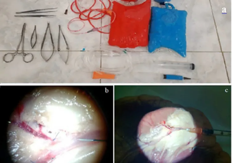
For the model, we used fresh bovine hearts, in which the following procedures were performed: selective catheterization of the coronary arteries, perfusion and removal of clots followed by saline solution infusion containing dyes in a ratio of 4:1. For such method, the traditional quenching ink, which has the advantages of easy acquisition, low cost and immediate dilution without agglutinations in serum, was infused directly into the serum container. As for color, red and blue dyes were used because they present good visual
Ex vivo
model with bovine heart: a proposal for training microscopic
dissection and vascularmicroanastomoses.
Modelo ex-vivo com coração bovino: proposta para treinamento de dissecção
microscópica e de microanastomoses vasculares.
Leonardo desessards oLijnyk1,2; rodoLfo figueiredode CarvaLho1; antonio generoso severino2; krunaL PateL3; geraLdo Pereira jotz4; CarLos eduardoda siLva1,4; MarCo antônio stefani2
Training is a process that requires patience and constant practice. The execution of microscopic procedures is present in the day-to-day of several surgical specialties, but unfortunately experimental models are not easy to access in our environment. We propose a bovine heart model used by residents and young surgeons in the training of microscopic dissection and microanastomoses. It is described the assembly of this model, which can be performed individually and with accessible material to the surgical departments. Our experience in the preparation of the pieces, as well as tips for the process, are described in the text. The bovine myocardial model can be reproduced in any center with benches and surgical instruments. Low cost, fast preparation, and wide availability of the used tissue are among the advantages of this model. We consider the project useful in the training of surgical residents and young surgeons.
Keywords: Microsurgery. Heart. Anastomosis, Surgical.
A B S T R A C T
Olijnyk Ex vivo model with bovine heart: a proposal for training microscopic dissection and vascularmicroanastomoses.
2
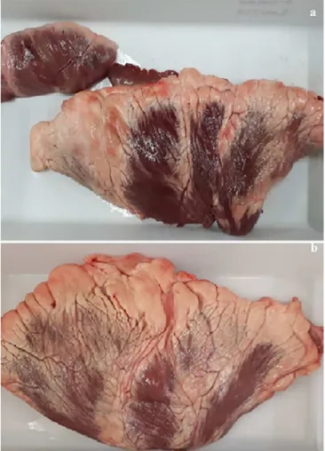
appearance for dissection and have already been used in other works in literature. Figures 1 and 2 show the used instruments, the piece assemblies, and the initial work of the dissection of the vessels. The cardiac muscle was sectioned and extended, which generated more stability to work (Figure 3).
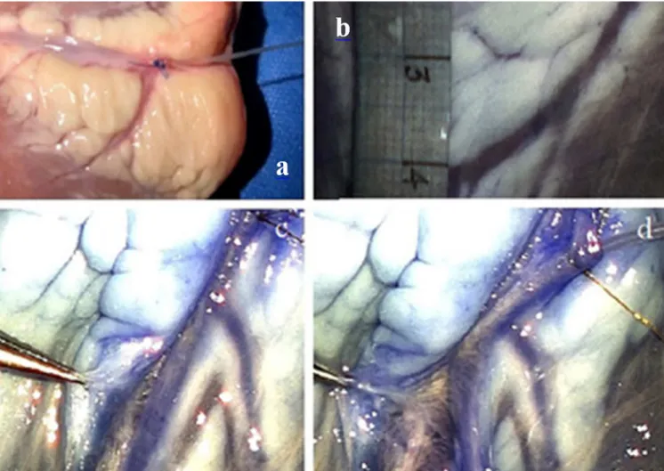
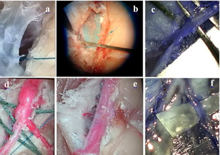
After coronary dissection, extensions from 2cm to 3cm free from perforators in the course of vessels may be found. Larger space to perform microanastomoses could be obtained by ligating perforators. Nylon 9-0 and 10-0 suture wire were used to perform end-to-end, end-to-side, and side-to-side anastomoses, these with the use of sectioned vessels of the piece itself or of other
myocardiums (Figures 4 and 5). The execution is performed with the help of a surgical microscope with magnification ranging from 10x to 14x. In our laboratory there was a Zeiss S88 microscope available for training. In general, from six to ten knots were required for an integral suture, varying with the diameter of the vessel. In more proximal stretches, coronarieswith up to 3mm diameter may be found, while in more distal areas in the piece most vessels are up to 1.5mm in diameter. Then, the surgeon tests the permeability of communication with the diluted dyes. In addition to anastomoses, direct sutures could also be trained in more gibbous parts of the vessels, as shown in figure 4.
The elaboration of the presented model did not require more than 40 minutes to be ready on the lab bench. Not considering suture threads, the cost of non-reusable materials did not exceed U$16. The storage and freezing of the pieces generated inconveniences, such as degradation and loss of handling, thus, we used a single fresh piece. Cleaning the pieces with warm water, leaving the tissue at room temperature (or a little warmer) and using saline for constant humidification are tips that may improve de process.
DISCUSSION
Experimental training models are fundamental in acquiring manual dexterity and familiarization with microscopic environment. Training surgeons, especially ones of the aforementioned specialties, can have their learning curves made easier when they become accustomed to the increase of visual field (up to ten times) and perform movements that require precision on laboratory benches. Likewise, the execution of microanastomosis is always a challenging task and even experienced professionals can benefit from the constant improvement in laboratory practices12.
Olijnyk Ex vivo model with bovine heart: a proposal for training microscopic dissection and vascularmicroanastomoses.
4
The objective of this work is to encourage the microsurgical exercise with a practical, economically advantageous and ex vivo model, so that it does not involve animal sacrifice for the continuous replication of the exercise. A piece can be prepared by a single person and time spent is feasible even for those who have busy routines, such as resident physicians. Reproducibility is simple, since bovine hearts are found in ordinary butcher shops and the other disposable products are accessible. The exception is needle thread, which has a higherprice.
Initially, the young surgeon may find difficulty in preserving perforators, which have very delicate walls and are easily ruptured. At this point, the contrast generated by the dye extravasation significantly helps, under microscope magnification, in the identification of wall lesions of the small vessels. In some surgeries, the dissection of the vessel takes the main time of a procedure, thus the effort in insulating the entire diameter is part of the acquisition process of microsurgical mastery (Figure 4).
Coronary arteries and veins are found in the bovine myocardium. These have a thinner and more malleable wall, what leads to a more laborious dissection process. Also, in general, more tributaries are observed in these vessels. In their turn, coronaries can be more easily isolated, and their firmer wall - typical of arteries, because they have a medium tunica - is more conducive to anastomoses training. The richer anatomy of the heart was crucial to encourage the authors to develop this prototype, in relation to other already proposed ex vivo or inorganic models.
An important aspect in anastomoses is the patency after vessel suturing. The use of dyes after luminal catheterization has been already successfully described in a variety of organs or tissues. Among them, placenta, rat cadavers, and even human parts in anatomical laboratories3,8. In this project, the injection of saline with ink allowed the surgeon good visibility of the flow through the anastomosis and, in case of absence of patency or externalization of the content, the correction could be made instantly, observing the result of new knots or their withdrawal. We mentioned the dilution we usually use, but, if the practitioner desires a more consistent appearance of the solution, the dye/ ink ratio should be increased. More concentrated mixtures leak less through sutures or damaged tributaries.
Figure 3. a) sectioned and extended piece of bovine
The model was not restricted to the use of bovine hearts. Swine pieces were also tested. We preferred the first option, because it presented larger vessels and more availability for acquisition. On the other hand, the porcine heart presents less adipose tissue, what facilitates coronary dissection. In our opinion, some bovine pieces are even inadequate for insulation work and coronary anastomoses due to excess fat in the myocardium (Figure 3). Moreover, if the surgeon wishes to spare the work of coronary dissection, or shorten its duration, he (she) can transpose the small chicken vessels, as described in other articles7, and thus move to the anastomoses phase more quickly.
Numerous authors emphasize the importance of developing microsurgical skills before performing in patients1,6,8,13,14. Surgical services with availability of guinea pigs for microsurgical training also benefit, if young surgeons familiarize themselves with magnification and with microsuture technique before handling animals. Anastomosis practice in vessels of living animals, in laboratory, can be considered a gold standard for this training, since dissection and execution of microanastomoses in anaesthetized guinea pigs offer greater similarity to surgical situations with bleeding and anatomical variability in the different specialties that employ microscopy.
Olijnyk Ex vivo model with bovine heart: a proposal for training microscopic dissection and vascularmicroanastomoses.
6
Training centers with this resource availability should encourage their residents to take full advantage of this opportunity5,15. Works with femoral vein and carotid artery are the most cited in literature6,16.
However, the model of this article presentes other relevant advantages. As already mentioned, its cost was lower when compared to living animal models, for which it is necessary, in addition to the direct acquisition, budget for animal
husbandry, anesthesia material, and postoperative support. Regulation and bioethical considerations are not a problem for the training centers, since the pieces are easily obtained by any citizen and care should always be taken only with proper disposal in the laboratory. Finally, in relation to inorganic (for example, latex13) models, the use of animal tissue offers greater similarity to human surgeries, regardless of the topography or surgical area.
R E S U M O
O treinamento é um processo que exige paciência e constante prática. A execução de procedimentos microscópicos está presente no dia a dia de diversas especialidades cirúrgicas, mas infelizmente modelos experimentais não são de fácil de acesso. Propomos um modelo com coração bovino usado por residentes e jovens cirurgiões no treinamento de dissecção microscópica e microanastomoses. É descrita a montagem deste modelo, que pode ser realizado de maneira individual e com material acessível aos departamentos cirúrgicos. Nossa experiência na elaboração das peças, assim como, dicas para o processo são descritas no texto. O modelo com miocardio bovino pode ser reproduzido em qualquer centro que disponha de bancadas e instrumental cirúrgico. Dentre as vantagens estão o baixo custo, rápido preparo e grande disponibilidade do tecido utilizado. Consideramos o projeto útil no treinamento de residentes cirúrgicos e jovens cirurgiões.
Descritores: Microcirurgia. Coração. Anastomose Cirúrgica.
CONCLUSIONS
Surgeons naturally obtain satisfactory results in the day-to-day routine due to the repetition of the dissection process, identification, and microsuture practice. To reach this point, it takes determination and patience during training period. As coordinators of this project, we aimed to demonstrate an objective and reproducible method to collaborate in the development of
the microsurgical practice. This model serves as a facilitator for supervised learning during the medical residency, or even for the constant exercise of microvascular technique by specialist surgeons.
In Brazil, residents usually lack time or financial conditions to seek a more refined training or even have access to structured laboratories. As described, the use of bovine heart is cost-effective and can help in this preparation stage to work with microstructures and perform vascular surgeries in a valuable way.
REFERENCES
1. Martins PNA, Montero EFS. Treinamento básico em microcirurgia. Comentários e proposta. Acta Cir Bras. 2007;22(1):79-81.
2. Dias IS, Pessoa SGP, Benevides AN, Macêdo JE. Treinamento inicial em microcirurgia. Rev Bras Cir Plást. 2010;25(4):595-9.
3. Aboud E, Al-Mefty O, Yasargil MG. New laboratory model for neurosurgical training that simulates live surgery. J Neurosurg. 2002;97(6):1367-72.
4. Marcondes CA, Pessoa SGP, Pessoa BBGP, Dias IS, Guimarães MGM. Padronização técnica no treinamento em microcirurgia do serviço de cirurgia plástica e microcirurgia reconstrutiva do hospital universitário Walter Cantídio da Universidade Federal do Ceará (HUWC/UFC). Rev Bras Cir Plást. 2014;29(2):283-8.
5. Maluf Junior I, Da Silva ABD, Groth AK, Lopes MAC, Kurogi AS, Freitas RS, et al. Modelo experimental alternativo para treinamento em microcirurgia. Rev Col Bras Cir. 2014;41(1):72-4.
6. Webster R, Ely PB. Treinamento em microcirurgia vascular: é economicamente viável? Acta Cir Bras. 2002;17(3):194-7 .
7. Fann JI, Caffarelli AD, Georgette G, Howard SK, Gaba DM, Youngblood P, et al. Improvement in coronary anastomosis with cardiac surgery simulation. J Thorac Cardiovasc Surg. 2008;136(6):1486-91. 8. De Oliveira MMR, Ferrarez CE, Ramos TM,
Malheiros JA, Nicolato A, Machado CJ, Ferreira MT, et al. Learning brain aneurysm microsurgical skills in a human placenta model: predictive validity. J Neurosurg. 2018;128(3):846-52.
9. Martins PNA. Importância da microcirurgia experimental para transplantes de órgãos. Acta Cir Bras. 2003;18(1):1-6.
10. Kinshoku MR, Rodriguez CAL, Fidalgo RS, Duran CCG, Leme PLS, Duarte IS. Uso racional de modelos animais para pesquisa e ensino de microcirurgia. Rev Col Bras Cir. 2012;39(5):414-7.
Olijnyk Ex vivo model with bovine heart: a proposal for training microscopic dissection and vascularmicroanastomoses.
8
12. Hino A. Training in microvascular surgery using a chicken wing artery. Neurosurgery. 2003;52(6):1495-7; discussion 1497-8.
13. Pessoa BBGP, Pessoa SGP. Treinamento em microanastomoses utilizando tubos de látex. Acta Cir Bras. 2002;17(2):143-6.
14. MacDonald JD. Learning to perform microvascular anastomosis. Skull Base. 2005;13(5):229-40.
15. Martins PNA, Montero EFS. Organização de laboratório de microcirurgia. Acta Cir Bras. 2006;21(3):187-9.
16. Isolan GS, Santis-isolan PMB, Dobrowolski S, Cioato MG, Meyer FS, Antunes ACM, et al. Considerações
técnicas no treinamento de anastomoses microvasculares em laboratório de microcirurgia. J
Bras Neurocirurg. 2010;21(1):8-1.
Received in: 08/29/2018
Accepted for publication: 10/12/2018 Conflict of interest: none.
Source of funding: none.
Mailing address:
Leonardo Desessards Olijnyk E-mail: [email protected]