Cláudia Filipa Martins Afonso
Mestre em Neurociências
Licenciada em Química com minor em Biologia [Habilitações Académicas]
Development of in-cell NMR methodologies
Dissertação para obtenção do Grau de Mestre em Química Bioorgânica
Orientador: Professor Doutor Eurico José da Silva Cabrita, Professor Auxiliar com Agregação, Faculdade de Ciências e Tecnologia, Universidade Nova de Lisboa
Co-orientador: Doutora Filipa Margarida Barradas de Morais Marcelo, Investigador Auxiliar, Faculdade de Ciências e Tecnologia, Universidade Nova de Lisboa
Júri:
Presidente: Professora Doutora Paula Cristina de Sério Branco, Professor Auxiliar, Faculdade de Ciências e Tecnologia, Universidade Nova de Lisboa
Arguente: Professora Doutora Maria dos Anjos Lopez de Macedo, Professor Auxiliar, Faculdade de Ciências e Tecnologia, Universidade Nova de Lisboa
Development of
in-cell
NMR methodologies
Copyright © Cláudia Filipa Martins Afonso, Faculdade de Ciências e Tecnologia da Universidade Nova de Lisboa
When I heard the learn’d astronomer,
When the proofs, the figures, were ranged in columns before me,
When I was shown the charts and diagrams, to add, divide, and measure them,
When I sitting heard the astronomer where he lectured with much applause in the lecture-room, How soon unaccountable I became tired and sick,
Till rising and gliding out I wander’d off by myself,
In the mystical moist night-air, and from time to time,
Look’d up in perfect silence at the stars.
Gostaria de agradecer à Professora Paula Branco pela ajuda prestada como coordenadora durante o meu percurso no mestrado em Química Bioorgânica. Este agradecimento estende-se, de um modo geral, aos restantes Professores deste mestrado.
Queria igualmente agradecer ao meus orientadores, o Professor Eurico Cabrita e a Dra. Filipa Marcelo, por ter me terem aceite como aluna. Ao Professor Eurico Cabrita ainda pela disponibilidade em falar comigo e pelas correções durante a fase de escrita da dissertação.
Um agradecimento especial ao Micael Silva, pela ajuda nas experiências de in-cell e pelo esclarecimento de muitas das dúvidas que lhe coloquei, e ao Doutor Fábio Fernandes, por toda a ajuda durante a aquisição e tratamento de resultados das experiências de FCS.
Gostaria de agradecer aos restantes membros do grupo, sobretudo aos meus colegas de laboratório, o Diogo e a Ana Sofia, pela amizade durante este ano.
Queria agradecer também a ajuda do grupo de cristalografia da FCT-UNL, em particular do Francisco Leisico.
Gostaria também de agradecer aos meus colegas de mestrado, sobretudo à Inês Rosete e ao Mário Almeida, por toda a amizade e ajuda durante estes dois anos. Teria sido mais difícil fazer dois mestrados ao mesmo tempo sem vocês.
O estudo de moléculas num meio biologicamente relevante distingue a espectroscopia de ressonância magnética nuclear (RMN) das restantes técnicas, tais como a cristalografia de raios-X ou a criomicroscopia eletrónica. Devido à sua elevada estabilidade e fraca interação com outros componentes celulares, a proteína GB1 representa a sonda por excelência para estudar os efeitos físico-químicos impostos na estrutura e dinâmica de proteínas pelo aglomerado macromolecular existente nas células, sem comprometer a acquisição de espectros in-cell de RMN.
A presente dissertação teve por objectivo investigar as possíveis interações da GB1 com o lisado e ambiente intracelular de Escherichia coli de modo a inferir sobre os efeitos
físico-químicos impostos por estes dois meios molecularmente densos na estrutura e dinâmica de proteínas. Assim, os parâmetros experimentais críticos para a realização de experiências in-cell
foram inicialmente otimizados. Subsequentemente, pela monitorização dos desvios químicos de protão e azoto correspondentes aos grupos amida do esqueleto proteico e cadeias laterais de lisinas, assim como de protão e carbono das cadeias laterais de resíduos contendo grupos carbonilo, o comportamento da GB1 foi estudado em lisado e E. coli, considerando a proteína
pura em água como referência. Além disso, as interações com o ambiente local foram também avaliadas através da determinação do movimento de translação global da proteína através de
“diffusion-ordered NMR spectroscopy” (DOSY). Os resultados obtidos sugerem que o meio
intracelular é mais viscoso que a correspondente solução composta por 150 mg/ml de lisado e
que os resíduos nas regiões “loop” mais flexíveis e expostas ao solvente, ou próximas destas,
apresentam uma maior preferência pelos componentes celulares em E. coli relativamente ao
lisado. Finalmente, foi feita uma comparação dos coeficientes de difusão obtidos por DOSY e espectroscopia de correlação de fluorescência (FCS), a técnica padrão para o estudo da difusão de proteínas.
PALAVRAS-CHAVE
Abstract
The characterization of molecules within a biologically relevant environment distinguishes nuclear magnetic resonance (NMR) spectroscopy from other molecular-based biophysical techniques, such as X-ray crystallography and cryo-electron microscopy. Due to its exceptional stability and reduced ability to interact in a specific manner with other cellular components, the GB1 protein represents the quintessential probe to investigate the physiochemical effects imposed by the crowded environment on the structure and dynamics of proteins, without simultaneously compromising the ability to obtain in-cell NMR spectra due to binding events.
The general aim of this thesis was to investigate the possible interactions of the GB1 protein with the Escherichia coli lysate and intracellular milieu with the purpose of inferring the
physiochemical effects imposed by these two crowded environments on the structure and dynamics of proteins. Thus, the experimental parameters critical for performing in-cell NMR experiments, including bacterial growth and protein overexpression within E. coli cells, were initially optimized. Subsequently, by monitoring proton and nitrogen chemical shifts of backbone amides and lysines side chains, as well as carbon and proton chemical shifts of side chains containing carbonyl groups, the preferential behaviour of GB1 was analysed in lysate and within cells, considering the pure protein in water as the reference state. Furthermore, interactions with the local environment were further examined by determining the overall translational motion of the protein through diffusion-ordered NMR spectroscopy (DOSY). The results obtained suggest that the intracellular environment is much more viscous than its artificially crowded counterpart and that GB1 exhibits a distinct behaviour in E. coli than in lysate. Specifically, residues at or near the more flexible and solvent-exposed loop regions of the protein display an increased preference for interaction with cellular components within cells compared to lysate. Finally, a comparison of diffusion coefficients obtained with DOSY and fluorescence correlation spectroscopy (FCS), the standard analytical technique for studying protein diffusion, was made.
KEYWORDS
Contents
Agradecimentos ... i
Resumo... iii
Abstract……… v
Contents……… vii
List of Figures ... ix
List of Tables ... xi
Abbreviations ... xiii
1. Introduction ... 1
1.1 In-cell NMR spectroscopy as a new tool for studying proteins in their native environment……….. 2
1.1.1 Overview of methodologies ... 2
1.1.2 Applications, limitations and future perspectives ... 10
1.2 The significance of macromolecular crowding for protein in-cell NMR spectroscopy ... 13
1.2.1 Basic concepts of macromolecular crowding ... 13
1.2.2 The model protein GB1 for probing interactions within biological settings ... 16
1.3 Aims... 18
2. Methods ...21
2.1 Molecular biology ... 21
2.1.1 Production of competent Escherichia coli cells ... 21
2.1.2 Bacterial transformation ... 21
2.2 NMR spectroscopy ... 22
2.2.1 In vitro NMR experiments ... 22
2.2.2 In-cell NMR experiments ... 23
2.2.3 Data acquisition and processing ... 24
2.3.1 Sample preparation ... 28
2.3.2 Data acquisition ... 28
3. Results……….31
3.1 In-cell NMR protocol optimization ... 31
3.2 Study of GB1 backbone amide interactions in lysate and E. coli ... 35
3.3 Study of GB1 side chain interactions in lysate and E. coli ... 41
3.3.1 1H-15N combined chemical shift perturbations of lysine side chains ... 41
3.3.2 1H-13C combined chemical shift perturbations of carbonyl side chains ... 42
3.4 Comparative study between diffusion coefficients obtained by NMR and FCS experiments ... 44
3.4.1 Conjugate reaction between the GB1 protein and the Alexa Fluor 488 succinimidyl ester ... 44
3.4.2 Diffusion studies of the fluorescently labelled 15N-GB1 protein with FCS experiments………. 46
3.4.3 Diffusion studies of the fluorescently labelled 15N-GB1 protein with DOSY experiments ... 48
4. Discussion ... 51
5. Conclusions and Future Perspectives ... 57
6. Bibliography ... 59
7. Appendix ... 71
7.1 1H-15N backbone assignment of GB1 in water, lysate and E. coli cells... 71
7.2 1H-15N lysine side chain assignment of GB1 in water, lysate and E. coli cells ... 74
List of Figures
Figure 1.1 – Overview of possible methodologies for in-cell NMR experiments. ... 6
Figure 1.2 – The use of more complex isotopic labelling schemes allows the study of increasingly larger proteins ... 8
Figure 1.3 – The specific labelling of methyl-group containing amino acids exploits the metabolic pathways involved in their biosynthesis ... 9
Figure 1.4 – Schematic representation of the crowded Escherichia coli cytoplasm ... 14
Figure 1.5 – Schematic cartoon diagram representing the folding and secondary elements of GB1………17
Figure 2.1 – Bacterial transformation with the GB1-encoding plasmid……… 22
Figure 2.2 – Correlation between nuclei in the H2CN pulse sequence………..25
Figure 2.3 – Correlation between nuclei in the HCCO pulse sequence………..25
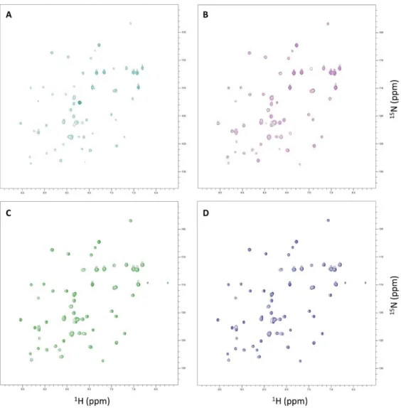
Figure 3.1 – SOFAST1H-15N HMQC spectra from GB1-expressing E. coli acquired at four distinct time-points after induction of protein expression in 15N-labelled minimal medium………31
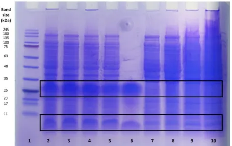
Figure 3.2 – Tricine-SDS-PAGE of samples from in-cell NMR experiments ... 32
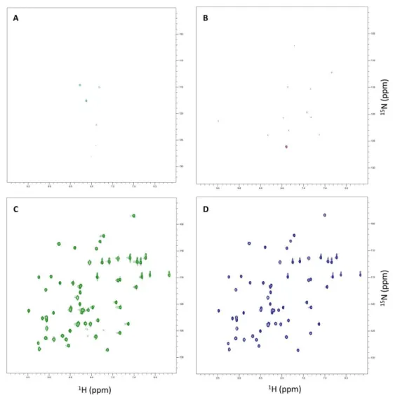
Figure 3.3 – SOFAST 1H-15N HMQC spectra from the supernatant of GB1-expressing E. coli acquired at four distinct time-points after induction of protein expression in 15N-labelled minimal medium. ... 33
Figure 3.4 – Number of GB1-specific 1H-15N cross-peaks of in-cell NMR samples and their respective supernatants acquired at four distinct expression time-periods ... 33
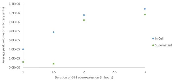
Figure 3.5 – Average peak volume of in-cell NMR samples and their respective supernatants acquired at four distinct expression time-periods ... 34
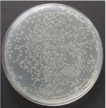
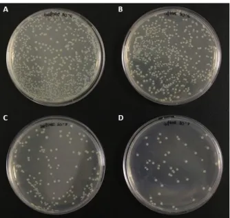
Figure 3.6 – Colony-forming unit assays before and after in-cell NMR experiments………. 35
Figure 3.7 – Combined 1H-15N chemical shift perturbations of the GB1 protein in E. coli lysate………36
Figure 3.8 – Box-plot depicting the combined 1H-15N chemical shift perturbations in E. coli lysate as a function of secondary structural elements in GB1 ... 36
Figure 3.10 – Box-plot depicting the combined 1H-15N chemical shift perturbations in E. coli cells
as a function of secondary structural elements in GB1 ... 37 Figure 3.11 – Proton linewidths of 1H-15H cross-peaks in the spectra of the GB1 protein in water,
lysate and E. coli cells ... 38 Figure 3.12 – Fractional changes in proton linewidths in lysate as a function of secondary structural elements in GB1... 39 Figure 3.13 – Fractional changes in proton linewidths in E. coli cells as a function of secondary structural elements in GB1... 39 Figure 3.14 –1H-15N cross-peak heights in the spectra of the GB1 protein in water, lysate and E.
coli cells. ... 40 Figure 3.15 – Combined 1H-15N chemical shift perturbations of lysine side chains present in the
GB1 protein in E. coli lysate ... 41 Figure 3.16 – Combined 1H-15N chemical shift perturbations of lysine side chains present in the
GB1 protein within E. coli cells ... 42 Figure 3.17 – Combined 1H-13C chemical shift perturbations of carbonyl side chains present in
the GB1 protein in E. coli lysate ... 43 Figure 3.18 – Combined 1H-13C chemical shift perturbations of carbonyl side chains present in
the GB1 protein within E. coli cells... 43 Figure 3.19 – Reaction scheme between the Alexa Fluor 488 succinimidyl ester and the primary amines of the GB1 protein ... 44 Figure 3.20 – Mechanism for the reaction between the Alexa 488 succinimidyl ester and the primary amines of the GB1 protein... 45 Figure 3.21 – Absorption spectra of the free Alexa 488 dye and the fluorescently labelled 15
N-GB1 protein between 250 and 650 nm………46 Figure 3.22 – Autocorrelation curve for the reference dye rhodamine 110 in water at 20oC .... 47
Figure 3.23 – Autocorrelation curve for the 15N-GB1 protein conjugated with the Alexa 488 dye
in water at 20oC ... 47
Figure 3.24 – Autocorrelation curve for the 15N-GB1 protein conjugated with the Alexa 488 dye
in 150 mg/ml of lysate at 20oC ... 48
List of Tables
Table 3.1 – Diffusion coefficients and hydrodynamic radius ratio of the GB1 protein in water and
lysate………40
Table 3.2 – Values obtained in FCS experiments for the 15N-GB1 protein coupled to the Alexa 488
dye in water and lysate. ... 48 Table 3.3 – Values obtained in DOSY experiments for the 15N-GB1 protein coupled to the Alexa
Abbreviations
1D One-dimensional 2D Two-dimensional 3D Three-dimensional 4D Four-dimensional BSA Bovine serum albumin
CI2 Chymotrypsin inhibitor 2 CK2 Casein kinase 2
CPP Cell-penetrating peptide DOL Degree of labelling
DOSY Diffusion-ordered spectroscopy
E. coli Escherichia coli
FCS Fluorescence correlation spectroscopy GB1 B1 domain of protein G
HIV-1 Human immunodeficiency virus-1
HSQC Heteronuclear single quantum coherence IPTG Isopropyl-β-D-thiogalactopyranoside
LB Lysogeny broth
MBP Maltose-binding protein MRI Magnetic resonance imaging MSG Malate synthase G
MW Molecular weight
NMR Nuclear magnetic resonance NOE Nuclear Overhauser effect
PDB Protein Data Bank PEG Polyethylene glycol
pI Isoelectric point PFTs Pore-forming toxins ppm Parts per million
PVP Polyvinylpyrrolidone rpm Revolutions per minute
SLO Streptolysin O
SOD1 Superoxide dismutase 1
1.
Introduction
Biological systems are inherently complex and detailed knowledge on the structure of their basic components, i.e. biomolecules, is required to characterize their intricate behaviour. Information regarding the three-dimensional structures of target biomolecules is particularly relevant in the design of new therapeutic compounds.[1] Since the first structural elucidation of
DNA in 1953 and myoglobin in 1958, the atomic coordinates of more than 132,000 of proteins, nuclei acids and their complexes have been deposited, as of August 2017, in the Protein Data Bank (PDB).[2,3]
Due to the wealth of structural data already collected, the interest of the scientific community is now shifting towards the functional characterization of biomolecules. To fully achieve this, it is essential to validate the structures obtained in vitro within a biologically relevant context.[4] However, most biophysical methods employed in the atomic resolution
study of biomolecules are constrained to artificial conditions, remote from the native cellular environments in which these molecules exist and exert their proper biological functions. For instance, X-ray crystallography, the first and still the leading and most powerful technique today in the field of structural biology, requires pure and regular crystals to generate diffraction patterns following irradiation with X-ray beams.[5] Furthermore, proteins that contain large
mobile or intrinsically disordered regions are usually difficult, if not impossible, to crystallize.[6]
By contrast, nuclear magnetic resonance (NMR) spectroscopy represents the most versatile technique in modern structural biology, due to its unique ability to characterize the behaviour of biomolecules in solution.[7]
NMR spectroscopy exploits the intrinsic property of nuclear spin to provide information on the chemical environment surrounding atoms. In brief, nuclei with nonzero spin quantum number I, termed “NMR-active nuclei”, exhibit a magnetic moment. When placed in a static magnetic field, the individual magnetic moments of each nucleus align themselves in a discrete number of orientations with respect to the field, rotating continuously about its axis in a motion referred to as Larmor precession. Each orientation or spin state is associated with a specific energy. The physical phenomenon responsible for NMR occurs when nuclei, driven by the absorption of electromagnetic radiation arising from a magnetic field oscillating at the Larmor frequency of the spin, change their state. However, since electrons are charged particles that also possess spin, their precession induces local secondary magnetic fields that alter the strength of the magnetic field experienced by nuclei.[8] Therefore, NMR spectroscopy is sensitive to
differences in the chemical environment surrounding nuclei and structural and dynamic information on biomolecules can thus be deduced based on this.[9]
NMR studies have been mostly restricted to dilute and isolated conditions, with buffers being chosen not to mimic the intracellular milieu but instead to optimize parameters such as solubility or signal sensitivity.[10,11] Indeed, a critical distinction between in vitro and intracellular
environments pertains to the high concentration of macromolecules in the latter, which can extend to 400 g/L in the prokaryotic cytoplasm.[12] These high concentrations of macromolecules
lead to excluded volume effects that, in addition to interactions with other molecules and posttranslational modifications, which occur inside living cells and can also influence the structural and dynamic behaviour of biomolecules, are difficult to replicate in vitro. Therefore, an increasingly attractive avenue for acquiring information about biomolecules at atomic resolution in their native physiological environment is provided by in-cell NMR.[13]
1.1
In-cell NMR spectroscopy as a new tool for studying proteins in their
native environment
In-cell NMR is a relatively new application of high-resolution NMR spectroscopy which provides atomic resolution of macromolecular structure and dynamics within a highly crowded, viscous and complex, but biologically relevant environment.[14] For this purpose, the resonance
frequencies of biomolecules are distinguished from the unlabelled cellular environment and made visible in multi-dimensional correlation experiments following isotopic enrichment with NMR-active nuclides, particularly 15N and 13C.[15] This contrasts with the more well-established
technique of in vivo NMR, in which characteristic resonance lines are obtained from one-dimensional (1D) spectra of metabolites containing naturally abundant NMR-active nuclides such as 1H and 31P or, alternatively, after infusion with small molecules labelled with
NMR-sensitive isotopes, mostly 13C, which later diffuse or become actively transported to the interior
of cells in living organisms.[16]
1.1.1 Overview of methodologies
The inherent insensitivity of NMR spectroscopy requires relatively high concentrations of isotopically-enriched biomolecules to be present, in order to distinguish the resonance peaks of interest from the remaining cellular components. To this end, distinct biological systems, delivery methods and labelling techniques can be employed as discussed below, with a special focus on proteins.
Biological systems and delivery methods
The first in-cell NMR proof-of-concept was described in 2001 by Serber and colleagues and performed using an Escherichia coli (E. coli) T7-dependent overexpression system grown in
N-terminal metal-binding domain of mercuric ion reductase (NmerA) and human calmodulin above other cellular components by 1H-15N heteronuclear single quantum coherence (HSQC)
NMR, which allows for the identification of amide bonds in proteins.[17,18] Since then,
recombinant protein expression in E. coli has proven to be a suitable and low-cost strategy for
in-cell NMR studies of proteins that tumble freely in the cytoplasm and do not exceedingly interact with cellular components.
The general protocol consists of a two-step culture, in which transformed bacterial cells are first grown in unlabelled lysogeny broth (LB) medium to an optical density (OD) of 0.6-0.8 and then transferred to isotopically-enriched minimal medium, with the most commonly used strategy employing ammonium chloride (NH4Cl) and glucose as the sole sources of metabolic
precursors. Following induction of gene expression downstream of the T7 promoter, which outperforms endogenous protein synthesis, labelled isotopes become selectively incorporated into the plasmid-encoded polypeptides. This scheme, in which unlabelled LB medium is switched to fresh labelled minimal medium prior to induction, diminishes background resonance peaks and increases spectral quality, because bacterial growth is significantly reduced during overexpression in minimal medium.[18] Accordingly, protein expression, isotopic labelling and
NMR measurements occur in the same cell type and sample preparation is relatively straightforward. However, recombinant expression can be difficult to control and protein concentrations can vary between experiments. Moreover, considerable amounts of recombinant proteins in bacteria can lead to the formation of insoluble intracellular aggregates in the shape of inclusion bodies, which is detrimental to solution-state in-cell NMR.[19]
Nevertheless, if recombinant expression is low and prolonged induction times are necessary to achieve adequate concentrations of intracellular proteins, then diminished cell viability and increased background labelling due to accumulation of metabolites can restrict the applicability of in-cell NMR experiments.
While E. coli remains the best characterized system for in-cell NMR due to its technical simplicity, bacteria may not represent the most adequate framework for studying eukaryotic proteins. Ideally, these proteins should be characterized in a cellular environment which best mirrors the native one. For instance, due to fundamental differences in cell morphology, internal organization and function between prokaryotes such as E. coli and eukaryotes, bacteria may lack the required machinery to fold and affect the post-translational modifications of eukaryotic proteins. Moreover, functional partners will likely be absent and the protein will not be targeted to its proper sub-cellular compartment.[20]
In this context, the first successfully conducted eukaryotic in-cell NMR experiments were reported in Xenopus laevis oocytes by Selenko and colleagues in 2006. Using this model system, the 15N-labelled B1 immunoglobulin-binding domain of streptococcal protein G (GB1, 6.2 kDa)
was recombinantly expressed and purified from E. coli and then delivered to the oocytes by microinjection.[21] This approach provides a higher degree of reproducibility and selectivity than
signals. Xenopus oocytes have long been employed in biomedical research, in part due to their large size (1 mm diameter) and cell volume (1 µL) allowing convenient manipulation and injection of different materials such as nuclei acids and proteins. Considering the small injection volume per cell (50 nL), an in-cell NMR sample with a final volume of 250 µL inside a Shigemi tube containing 200 oocytes requires only 10 µL of labelled protein. The concentration of injected protein required to achieve an adequate signal-to-noise ratio must be determined empirically but is normally in the range of 0.5-3.0 mM.[22]
In other eukaryotic model systems, such as mammalian cells, sample preparation by microinjection is technically unfeasible, because an absurd number of cells would need to be manipulated. Therefore, a distinct delivery mechanism that targets many cells simultaneously while still ensuring high sample deposition and cell viability is required. To this end, three techniques are currently available, namely active transport via cell-penetrating peptide (CPP) tags, passive diffusion through-pore forming toxins and electroporation.
CPPs are short peptides, typically comprising 5 to 30 amino acids, which can translocate across membranes and deliver various cargo molecules, such as proteins, nuclei acids and drugs, with low cellular toxicity.[23,24,25] Although the precise mechanism of entry into cells is still under
debate, it is believed that most CPPs have two or more internalization pathways, depending on experimental conditions such as temperature, concentration and weight of the cargo.[26] The
first CPP was described independently by two groups in 1988, when it was discovered that the trans-activator of transcription (Tat) protein from the human immunodeficiency virus 1 (HIV-1) could enter cells and translocate to the nucleus.[27,28] In 1998, the peptide sequence of Tat
necessary for cellular uptake was identified (47YGRKKRRQRRR57) and the majority of CPPs are
indeed cationic peptides rich in arginine (R) and lysine (K) residues.[29] Based on this knowledge,
Inomata and colleagues established the first method to observe in-cell NMR spectra of proteins delivered by CPPs in 2009.[30] The CPP sequence of Tat was fused to the carboxy terminus human
ubiquitin that was mutated at three sites to prevent binding with ubiquitin-interacting proteins. Following isotopic labelling, the fusion protein was able to translocate through the plasma membrane of cultured HeLa cells pre-incubated with pyrenebutyrate, a negatively-charged counterion with high hydrophobicity that facilitates translocation through lipid bilayers.[30,31]
Furthermore, the authors also demonstrated that the CPP can be covalently conjugated to the protein through a disulphide bond, which is subject to intracellular cleavage by autonomous reduction.[30]
An alternative approach to CPP-mediated delivery for eukaryotic in-cell NMR experiments was also reported in 2009 by means of pore-forming toxins (PFTs). Ogino and colleagues employed the bacterial toxin streptolysin O (SLO) to permeabilize the plasma membrane and introduce the actin-sequestering protein thymosin β4 (Tβ4) into 293F cells.[32] This toxin binds
to cholesterol as a monomer and oligomerizes laterally into ring-shaped transmembrane structures containing 50 to 80 subunits to form pores exceeding 30 nm in diameter, through which proteins up to 150 kDa can diffuse in a concentration-dependent manner.[33,34] At high
At low doses, the plasma membrane can recover by extracellular treatment with Ca2+ ions, which
permeate the cells and trigger the arachidonic acid cascade that promotes resealing.[34,35]
However, incubation of cells with even small amounts of PFTs can induce cellular responses which activate signalling pathways to restore membrane integrity and ion homeostasis. For instance, PFTs stimulate the activity of p38 MAP kinase to re-establish cytosolic ATP and K+ ion
levels while arresting protein synthesis.[36] Therefore, the intracellular environment following
PTF-mediated delivery may not represent the true physiological state of the cell.
More recently, a method based on electroporation to introduce proteins into eukaryotic cells for in-cell NMR studies was described. Since the first successful intracellular delivery of DNA into mammalian cells in 1982, electroporation constitutes an established technique for introducing nucleic acids, proteins and drugs into cells.[37] It relies on the use of short
high-voltage pulses to generate external electric fields that temporarily permeabilize cell membranes. Pore formation at the molecular level is achieved in a timescale of nano- to microseconds by infiltration of water molecules into the bilayer, which leads to reorientation of the adjacent lipids with their hydrophilic head groups pointing towards the water molecules. Once the external electric field is discontinued, pore resealing occurs and it is usually completed only within seconds or minutes after the end of exposure.[38] Using this approach, the group of Selenko
demonstrated in 2016 the feasibility of electroporation to observe isotopically labelled α -synuclein in various mammalian cell lines by NMR spectroscopy.[39,40]
The approaches for eukaryotic in-cell NMR experiments outlined above depend upon the heterologous expression of proteins in bacterial cells. Instinctively, a protein is best expressed in its natural cellular environment, where the entire machinery required for synthesis, folding, maturation and targeting is present. Conceptually different from E. coli expression and protein delivery, a eukaryotic expression system thus represents the ideal choice to support structural and dynamic studies of eukaryote-specific proteins.[41] In this context, Bertrand and colleagues
reported the observation of in-cell NMR spectra of S. cerevisiae and human ubiquitin following overexpression under the control of a methanol-induced AOX1 promoter in the yeast Pichia pastoris.[42] Another strategy for eukaryotic in-cell NMR using the baculovirus expression system
in insect cells was also later described.[43] Furthermore, the research group of Banci exploited
the recently developed mammalian cell-based expression systems, which rely on transient transfection with vectors designed for high-level protein production, to record NMR spectra of in living human cells.[44,45] An overview of the possible approaches discussed so far for in-cell
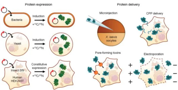
Figure 1.1 – Overview of possible methodologies for in-cell NMR experiments. The expression and isotopic labelling of endogenous proteins can be accomplished in distinct biological systems, such as bacteria, yeast, insect and mammalian cells. The introduction of exogenous proteins can be achieved through microinjection into Xenopus laevis oocytes or to human cells by cell-penetrating peptides (CPPs), pore-forming toxins or electroporation.[4]
Labelling techniques
Isotopic enrichment of biological macromolecules can be achieved by uniform or selective labelling. The specific labelling strategy will depend on the macromolecule under study, as well as the type of cells that are employed.
In the most frequently used scheme, the target protein is overexpressed from E. coli in an isotopically enriched minimal media containing 15NH
4Cl and/or 13C-glucose as the only sources
of nitrogen and carbon, thus becoming uniformly labelled.[46] In addition to being cost-effective,
the production of proteins in prokaryotic expression systems such as E. coli occurs without the post-translational modifications that are usually present in polypeptides derived from eukaryotes. Once delivered into eukaryotic cells, the residue-specificity and kinetics of post-translational modifications can be followed by time-resolved NMR spectroscopy.[47] An
alternative strategy consists of selective isotopic labelling. This is achieved by supplementing the minimal-expression media with labelled amino acids that are located at the end of biosynthetic pathways, such as lysine, arginine and histidine. Due to less extensive labelling, the resulting spectrum naturally exhibits a lower resolution than if it had been obtained for a uniformly enriched target, but is basically devoid of background signals.[46] The possibility of using
auxotrophic E. coli host strains, which require an exogenous source of specific amino acids for
their growth, allows the selective labelling of the remaining amino acids.[48] However,
In theory, uniform 13C [U-13C] enrichment can provide advantages over uniform 15N [U-15N] labelling, largely because the gyromagnetic ratio of 13C is 2.5 times higher than 15N, thus
enhancing the intrinsic sensitivity of the former nucleus to NMR signal detection.[49] However,
in practice, U-15N labelling is mostly favoured due to lower sample preparation costs and natural
abundance. Furthermore, the chemical shifts associated with 13C are confined to a narrower
range than 15N and 1H-15N HSQC spectra are easier to interpret than their 13C counterparts.
Selective 13C-isotopic labelling of individual amino acids, particularly their methyl and methylene
side chain residues, constitutes a more suitable strategy for enhancing sensitivity and minimizing background signals due to unwanted metabolic reactions.[46]
In NMR spectroscopy, the lifetime of the excited state is significantly influenced by the rate of molecular tumbling. As molecules become larger, the overall random tumbling motion begins to slow and the transverse relaxation rate increases. Since the linewidth of NMR signals is inversely proportional to the transverse relaxation rate, the spectra of macromolecules are characterized not only by increased complexity, but also display broader resonances. Moreover, biomolecular NMR experiments frequently rely on coherence transfer through scalar or dipolar couplings between nuclei. Because transfer steps are subjected to transverse relaxation, pulse sequences that use prolonged delays or require multiple transfer steps become less sensitive for larger biomolecules.[50]
The relaxation rate of 1H nuclei in proteins is primarily affected by the substantial number
of dipolar interactions with neighbouring protons. For the 15N and 13C heteronuclei, however,
the predominant relaxation pathway is the direct dipolar interactions with covalently-bound protons.[50] One popular, albeit expensive avenue for attenuating nuclear relaxation that arises
for higher molecular weight proteins involves their overexpression in bacteria grown in deuterated media. Bacteria are sufficiently robust to grow in such media, although at a slower rate and producing lower yields of recombinant proteins.[51] Due to chemical exchange following
H2O-based purification and refolding, protons are re-incorporated at the amide labile sites. Thus,
proteins that are deuterated at aliphatic and aromatic positions but otherwise protonated at the amide sites can be prepared and observed in a 1H-15N heteronuclear correlation spectrum.
By reducing proton density, perdeuteration decreases the transverse relaxation rates of the remaining protons, which results in a narrowing of their 1H signal linewidths. Furthermore,
deuteration of aliphatic sites greatly enhances the lifetime of transverse coherences which, in conjunction with 13C and 15N labelling, extends the size limit of proteins that can be studied by
three-dimensional (3D) or four-dimensional (4D) NMR experiments.[50]
However, deuteration removes many of the protons that are necessary for structural and dynamic characterization by nuclear Overhauser effect (NOE)-based distance restraints and two-dimensional (2D) 1H-13C NMR experiments of larger proteins. One ingenious labelling strategy
chemical shift degeneracy and longer transverse relaxation times.[52] Furthermore, protons in
methyl groups do not chemically exchange with water as amide protons do, which can reduce and even broaden the resonance frequency intensity beyond detection. Therefore, methyl groups constitute powerful probes of structure and dynamics of high-molecular weight proteins, since they regularly occur in the hydrophobic core and can connect distant regions of primary structure.
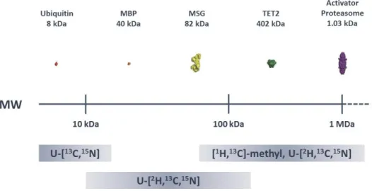
Figure 1.2 – The use of more complex isotopic labelling schemes allows the study of increasingly larger proteins.
Structural representations are plotted as a function of molecular weight (MW) for ubiquitin, maltose-binding protein (MBP), malate synthase G (MSG), TET2 and proteasome activator complex. Adapted from Plevin et al., 2012.[50]
Excluding post-translational modifications, there are six-methyl containing amino acids in proteins. A variety of strategies for the selective labelling of these groups, while retaining the advantages of perdeuteration have been developed since the late nineties, most of which exploit the metabolic pathways involved in the biogenesis of methyl-containing amino acids. The simplest of these involves the overexpression of proteins in bacteria grown in deuterated media using protonated 13C-pyruvate as the only source of carbon. Pyruvate is a direct precursor to
alanine, valine, leucine and isoleucine and its methyl group is incorporated into these amino acids with minimal hydrogen exchange. Using this scheme, it was possible to obtain highly perdeuterated proteins that remain 40-80% protonated at the methyl groups of alanine, valine,
leucine and isoleucine at the γ2-position only.[53] However, due to proton exchange at the methyl
group of 13C-pyruvate with D
2O during protein overexpression, a mixture of proteins containing
all four possible methyl isotopomers (13CH
3, 13CH2D, 13CHD2 and 13CD3) is obtained, which reduces
the sensitivity of NMR spectra. Subsequently, a more selective 1H,13C-labelling strategy was
developed for the production of deuterated proteins with protonation restricted to isoleucine
at the δ1 position and leucine/valine, respectively, by adding [3,3-2H2,U-13C]-α-ketobutyric and
[3,3-2H
2,U-13C]-α-ketoisovaleric acids to otherwise D2O-based minimal medium one hour before
induction of expression.[54,55]More recently, it was demonstrated that α-ketoisovaleric acid in
from the methyl probe to the backbone and thus is more suitable for NMR studies of larger proteins.[56]
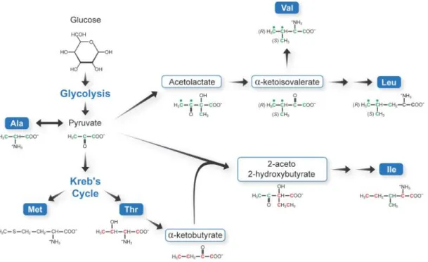
Figure 1.3 – The specific labelling of methyl-group containing amino acids exploits the metabolic pathways involved in their biosynthesis. Pyruvate serves as a precursor to the methyl-containing amino acids alanine, valine, leucine and isoleucine. Alanine is synthesized directly from pyruvate by a transamination reaction. Acetolactate, a precursor to valine and leucine, is obtained from the condensation reaction of two pyruvate molecules. Threonine is converted by a deaminase into α-ketobutyrate, which is then combined with pyruvate to yield isoleucine. The δ1-methyl group
of isoleucine is derived from α-ketobutyrate, while pyruvate originates the methyl group at the γ2-position.[50]
In addition to 15N and 13C, 19F has also proved to be a valuable probe in the study of
proteins. Due to its high gyromagnetic ratio, 83% relative to the proton, and large chemical shift dispersion, which approaches 1000 ppm, this isotope constitutes a very sensitive reporter of structural and conformational changes.[57,58] Moreover, since the ½-spin 19F nucleus is the only
stable isotope of fluorine, with an abundance of 100%, and is not present in naturally occurring biomolecules, the resulting NMR spectra is virtually devoid of background signals. Accordingly, 1D 19F NMR experiments generally require lower concentrations and shorter acquisitions times
than multi-dimensional NMR methods and, therefore, proteins can be more easily studied near physiological conditions.[58] Also, conveniently enough, the introduction of a single fluorine in
exchange for hydrogen at an aromatic side chain site does not lead to meaningful biological or structural changes.[58,59]
A wide variety of methods exist for incorporating fluorine into the protein of interest. Even though chemical synthesis is problematic for any protein of considerable size, several have
been labelled using a “semi-synthetic” approach. In this method, a peptide fragment containing
following protein expression in growth media containing the desired amino acid analogue. For instance, proteins with fluorinated aromatic amino acids can be easily prepared by either using auxotrophic strains or adding glyphosate, an inhibitor of aromatic amino acid biosynthesis.[61,62]
The use of natural translational machinery to introduce fluorinated analogues of amino acids seldom approaches an efficiency of 95% at a specific location. Moreover, this approach modifies every position of one amino acid simultaneously with varying efficiencies, which can produce protein molecules with heterogeneous structural perturbations. An alternative method for site-specific incorporation of 19F-labelled amino acids into proteins with high translational
efficiency and fidelity relies upon the use of an orthogonal tRNA/aminoacyl-tRNA synthetase pair.[63] This technology is based on the degeneracy of three stop codons in mRNA: UAA (ochre),
UGA (opal) and UAG (amber).[64] The amber codon is the less commonly used in E. coli (≈7%) and
does not terminate the essential genes required for bacterial growth and survival. Moreover, some species do not use the amber codon to signal termination of polypeptide synthesis, but instead to introduce an amino acid.[65] This allowed the development of a genetically engineered
translational system in which an exogenous tRNACUA and its cognate aminoacyl-tRNA synthetase
specifically recognise and incorporate an unnatural 19F-labelled amino acid into proteins.[66]
1.1.2 Applications, limitations and future perspectives
Since its inception, in-cell NMR spectroscopy has progressively emerged as a promising methodology to investigate the structure and dynamics of biomolecules, particularly proteins, at atomic resolution in their natural environment.
Traditionally, most of the work in this field has focused on prokaryotic hosts such as E. coli
to achieve recombinant protein expression, because it is easily the most affordable and well-established method. Using the E. coli overexpression in-cell design, interactions with cofactors and other proteins following sequential expression of independent induction systems, as well as pH determination, conformation and three-dimensional structures have been investigated. For instance, Burz and colleagues developed a protocol for mapping structural changes that occur following protein-protein interactions, which was designated by STINT-NMR. Bacterial cells transformed with two plasmids, each containing different inducible promoters to express the target and interactor proteins at separate time points, are first grown overnight on LB media. The culture is then transferred to [U-15N] minimal medium and expression of the target protein,
whose NMR structure is known, is induced with L-arabinose for a specific extent of time. Cells are then washed and placed in unlabelled medium, with the interactor now being produced following induction of expression with isopropyl-β-D-thiogalactopyranoside (IPTG).[67,68] This
approach was used to characterize the interaction between ubiquitin (8.5 kDa) and two protein ligands, AUIM (4 kDa) and STAM2 (50 kDa), with results demonstrating chemical shift differences and line broadening between the in-cell1H-15N HSQC spectra of free and bound ubiquitin.[68] A
small compounds capable of weakening or enhancing interactions between two components within a biomolecular complex.[69]
In addition, the dependence of the chemical shift and linewidth on binding events has also been used to study protein interactions with cofactors in E. coli cells. As an example, Hubbard and colleagues investigated the ion binding preference of the signal transduction protein CheY (15 kDa) under physiological conditions in 2003. By comparing the in vitro spectra of the protein in the presence of different ions with the in-cell spectrum, the authors demonstrated that CheY preferentially binds to Mg2+ ions in the E. coli cytoplasm.[70] In the same year, the research group
of Dötsch illustrated the relevance of in-cell NMR for determining the intracellular pH of E. coli. By determining the intensity ratios of Cε1-H and Cδ2-H cross-peaks in histidine residues, which depend on the protonation and tautomerization state of the imidazole ring, a value of 7.1±0.1 was determined for the cytoplasmic pH in E. coli.[71]
Finally, in-cell NMR in bacteria has also been employed to study the 3D structure of
proteins at concentrations that approach the physiological ones. For instance, the structure of the metal binding protein TTHA1718 at a concentration of 3-4 mM was solved de novo in E. coli
cells by Sakakibara and colleagues in 2009. Because the short in-cell sample lifetime does not allow sufficient structural information to be recorded using typical high-dimensionality NMR experiments, the authors relied on nonlinear sampling schemes and distinct labelling strategies, such as [U-13C,15N] and 13C-methyl selective labelling for backbone and side chain assignment,
respectively, to reduce measurement times. To improve the signal-to-noise ratio and ensure that only the data of viable cells were acquired, each of the six triple-resonance experiments was repeated several times with fresh samples and the data was later combined.[72] Similarly,
the structure of the GB1 protein at a concentration of 250 µM in E. coli cells was also recently described in 2016.[73]
Despite being initially established in prokaryotic systems, the full potential of in-cell NMR as a method for bridging structure and function of biomolecules in their native environment can only be reached when higher eukaryotic cells become routinely employed in such studies. Much progress has been recently made in this area with the development of different approaches for the expression and delivery of proteins into mammalian cells, including human.
In this context, residue-level information on cellular processes associated with protein modification and maturation have been obtained. For instance, Selenko and colleagues used time-resolved NMR to elucidate the stepwise sequence of phosphorylation events catalysed by casein kinase 2 (CK2) in Xenopus oocytes. Experiments were performed using a model substrate,
XT111-132GB1, composed of the regulatory protein sequence of the viral SV40 large T antigen
fused to the GB1 protein, the latter functioning as a solubility-enhancement tag. The authors observed that CK2 phosphorylation of XT111-132GB1 proceeds via a two-step process, with
intermediate release of the mono-phosphorylated substrate.[74] Moreover, the research group
of Banci used NMR experiments to follow the maturation events of superoxide dismutase 1 (SOD1) in human cells.[75] SOD1 is a 32 kDa homodimeric enzyme containing an intramolecular
radicals to hydrogen peroxide and is involved in the onset of the familial form of amyotrophic lateral sclerosis, a fatal neurodegenerative disease.[76] The addition of Zn2+ ions to the culture
medium induced the dimerization of SOD1, while treatment with Cu2+ alone was not sufficient
to produce the mature protein, because copper incorporation in SOD1 is tightly depends on the action of a specific chaperone. The role of this chaperone, the CSS protein, was further studied
in-cell. When Cu2+ ions were added to cells co-expressing both SOD1 and CSS, full maturation
was observed.[75] Together, these examples provide a brief look on the applicability of in-cell
NMR to study cellular events.
In addition to proteins, in-cell NMR has also been extended to include nuclei acids. The study of these biomolecules inside a cellular environment is, however, challenging due to the underlying inability to overexpress and, therefore, label them. Furthermore, nucleic acids are extremely sensitive to the action of hydrolytic enzymes, such as DNase and RNase, that catalyse their cleavage inside living cells. These difficulties were overcome by Hӓnsel and colleagues in 2009 to produce the first in-cell NMR spectra of synthetically prepared [U-13C,15N]-labelled DNA
and RNA sequences, which were chemically modified against enzymatic degradation and then microinjected into Xenopus oocytes.[77]
A favourable outcome of in-cell experiments depends on overcoming some obstacles. The relative insensitivity of NMR spectroscopy requires a substantial amount of the biomolecule of interest to be soluble inside cells. For most proteins, this is likely problematic because exceeding their physiological concentration, which is commonly on the order of nM to lower µM, can also conceivably affect their behaviour. The steady development of high-field magnets and cryogenic probes will increase the sensitivity of the technique and reduce the required concentration for acquiring NMR experiments with adequate spectral quality. Furthermore, the widespread use of selective labelling schemes based on methyl group detection will enable a further increment in sensitivity.[78]
Nevertheless, the most crucial factor that limits the length and type of experiments that can be recorded is sample viability. In a standard experimental setup, sedimentation and lysis with subsequent leakage of cytoplasmic constituents to the extracellular space naturally occurs within a certain time frame. To stabilize cells and minimize their degradation, some authors have suggested their encapsulation in embedding medium, such as low-melting agarose and Ficoll solutions.[79,80] However, prolonged cell viability invariably requires an exchange of nutrients and
oxygen, while simultaneously removing the metabolic waste and maintaining the external pH within a narrow range. For this purpose, bioreactors compatible with conventional NMR instruments have been designed and employed for in-cell NMR in bacteria and human cells.[81,82]
globular domains of the same size. This was demonstrated by the research group of Pielak when
a fusion protein constructed from the globular ubiquitin and the disordered α-synuclein was
visualized within E. coli and subsequently in the cell lysate. The peaks associated with ubiquitin were absent in the in-cellspectrum, while those corresponding to α-synuclein were present. However, upon lysing the cells, well-defined peaks of both α-synuclein and ubiquitin were visible. These results indicate that globular and disordered proteins respond differently to macromolecular crowding effects within cells, due to distinct rotational diffusion dynamics.[83]
Characterization of these globular proteins in their native cellular environment can be achieved instead by solid-state NMR, which is not inherently limited by the slow tumbling rate of molecules and is thus ideally suited for studying membrane proteins and large complexes or aggregates.
The role of macromolecular crowding on the structure and dynamics of proteins is more carefully considered in the following chapter.
1.2
The significance of macromolecular crowding for protein in-cell NMR
spectroscopy
As indicated in the previous chapter, in-cell NMR can reveal with atomic resolution the structure and dynamics of proteins under their native conditions. Unfortunately, many globular proteins do not yield in-cell spectra, even those that are hailed as model systems for in vitro
NMR studies, such as cytochrome c. The intracellular environment is inherently more complex than the dilute aqueous buffers that are conventionally employed to characterize proteins in vitro and the lack of in-cell spectra most likely reflects the differing degree to which the physicochemical properties of solutes inside cells influence the behaviour of proteins.
1.2.1 Basic concepts of macromolecular crowding
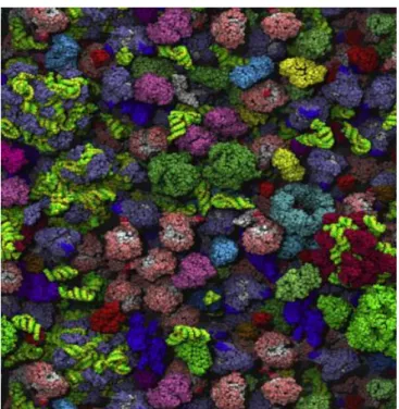
While a protein of interest may be dilute in the interior of a cell, it is naturally surrounded by a variety of other soluble macromolecules that together are present at high concentrations. For this reason, the intracellular compartment is usually described as highly crowded (Figure 1.4), with the distance between neighbouring macromolecules being of a similar magnitude as their diameter.[84,85] The E. coli cytoplasm contains approximately 400 g/L of proteins and RNA,
while mammalian cells can have protein and nucleic acid concentrations ranging from 70-300 g/L depending on the cell type.[86] Therefore, prokaryotes exhibit higher concentrations of
Figure 1.4– Schematic representation of the crowded Escherichia coli cytoplasm. This figure was prepared following computer dynamics simulation of the crowded E. coli intracellular environment.[85]
Crowding may affect the thermodynamic and kinetic behaviour of proteins by distinct mechanisms, including excluded volume and intermolecular interactions.
The excluded volume effect intuitively arises by realizing that two molecules can never occupy the same space in solution. Because of this steric hindrance, the fraction of volume that a macromolecule can occupy is significantly lower than the total volume of the cell and the randomness of motion and entropy are remarkably reduced. Therefore, the excluded-volume effect increases the free energy and thermodynamic activity of a solute. Accordingly, due to Le
Chatelier’s principle it should be expected that volume exclusion promotes a more compact
state.[87] Indeed, the direct influence of crowding on protein folding has been demonstrated for
the small ribosomal protein S16. This study showed that the urea-unfolded ensemble of S16 is more compact, with faster folding rates and slower unfolding kinetics, in the presence of an artificial crowder.[88] Moreover, the effect of volume exclusion on the conformational properties
of proteins has been investigated within cells by the group of Pielak using FlgM. This intrinsically
disordered protein inhibits the transcription factor σ28 which regulates genes essential for
flagellar assembly. FlgM is only transiently structured in dilute solutions, but its C-terminal
region forms an α-helical structure at biological relevant solute concentrations and within the
intracellular compartment.[89] However, disorder in its N-terminal portion remains regardless of
macromolecular crowding and binding to its functional partner.[90]
crowding agents, while capable of interacting weakly with proteins through non-specific van der Waals and hydrophobic forces, mostly mimic the intracellular environment by imposing constraints on the volume that biomolecules can occupy.[91] In this context, the reduction of
accessible volume has been shown to stabilize proteins, increase their enzymatic activity and accelerate their aggregation.[92,93]
Although volume exclusion constitutes a validated experimental paradigm for describing the effects of crowding on many aspects of protein behaviour, evidence has demonstrated that other factors should also be considered. The intracellular environment is not solely crowded, but extremely inhomogeneous and compartmentalized into membrane-less dynamic assemblies of components in which diffusive molecular transport with the surrounding cytoplasm occurs.[94]
Since macromolecular crowding increases viscosity at distinct length scales within cells, the free diffusion of proteins is not only decreased but displays anomalous characteristics as well. It has been demonstrated using computer simulations that subdiffusion is associated with increased probability of finding a neighbouring target, thus enhancing cellular events that depend on the interaction between many functional partners in comparison with normal diffusion.[95]
Several techniques have been used to reveal the anomalous diffusion of macromolecules within crowded environments, including fluorescence spectroscopy and NMR. As an example, the diffusion of the chymotrypsin inhibitor 2 (CI2, 7.4 kDa) protein has been analysed by NMR-based methods in solutions of glycerol, synthetic uncharged polymers and proteins, such as bovine serum albumin (BSA, 66.5 kDa) and lysozyme (14.3 kDa). In glycerol, the decrease of both translational and rotational diffusion with increasing viscosity followed the Stokes-Einstein law (Dt=κT/6πηr) and the Stokes-Einstein-Debye law (Dr=κT/8πηr3), respectively (where Dt is the
translational diffusion coefficient, Dr is the rotational diffusion coefficient, η is the solution
viscosity, κ is the Boltzmann constant, and r is the radius of protein being studied). However, meaningful deviations from the expected behaviour were induced by macromolecular crowding. Synthetic polymers as crowding agents caused a negative deviation from the Stokes laws, which means that increased viscosity reduces diffusion less than expected, and affects translation more than rotation. On the other hand, protein crowders were shown to have the opposite effect, attenuating rotational more than translational diffusion and inducing a positive deviation from the Stokes laws. NMR relaxation experiments revealed that the origin of these observable differences in diffusion behaviour were due to weak and nonspecific interactions between the protein crowders and CI2. Because synthetic polymers and globular proteins induce such dissimilar effects on diffusion, the former might not constitute the best alternative for modelling the effects of intracellular milieu on the behaviour of proteins. Besides, since CI2 is invisible in
1H-15N HSQC spectra from in-cell samples, as well as those containing high concentrations of
proteins, these results suggest that interactions between proteins explain some of the difficulties in obtaining in-cell NMR data from globular proteins.[96]
and thus exacerbate the excluded volume effect. On the contrary, enthalpy-driven attractive interactions are destabilizing since they increase surface exposure and favour unfolding.[97] This
was shown when the CI2 protein was resuspended in PVP, BSA and lysozyme. Using NMR-detected amide proton-deuterium exchange, protein stability was then evaluated at residue level. It was observed that while PVP stabilizes CI2, the protein crowders have a destabilizing effect on most residues.[98] Moreover, another study found that E. coli lysate, a model crowding
agent, destabilizes CI2 in a concentration-dependent manner, which strongly indicates an attractive interaction between the cytosolic components and the protein that is even capable of overcoming the stabilizing entropy-driven repulsions arising from excluded volume effects. In addition, it was noticed that E. coli lysates exerted an even stronger destabilizing effect on CI2 than the homogenous protein crowders, which strengthens the biological implications of weak, nonspecific interactions.[99] In conclusion, interactions between proteins can enhance or
diminish the excluded volume effects.
Presumably, the chemical origin responsible for the destabilizing effects of physiological crowders arises from a variety of sources, such as complementary charge-charge interactions, hydrogen bonding between amide and carbonyl groups, and hydrophobic forces. To better understand the chemical nature behind protein-protein interactions within the intracellular environment, the research group of Pielak assessed the stability of a mutant variant of CI2 with an isoelectric point (pI) of 6.0 in anionic and cationic protein lysate fractions. Since the net charge of CI2 at neutral pH equals -1.0, the repulsion arising from negative charge-charge interactions with the anionic fraction should favour the compact state and thus increase protein stability. However, using amide proton-deuterium exchange NMR experiments, only destabilization was observed. Furthermore, the anionic fraction is almost as destabilizing as the total protein lysate. The authors concluded that noncomplementary electrostatic interactions are not strong enough to offset the nonspecific attractive interactions between protein surfaces.[100]
1.2.2 The model protein GB1 for probing interactions within biological settings
The biological significance of transient attractive interactions in which proteins participate
was first postulated in 1982 by McConkey, who then proposed the term “quinary structure” to
designate a fifth level of protein complexity. The existence of quinary structures was put forward following the observation that evolutionarily distant protein homologs are indistinguishable from each other based on isoelectric point and molecular weight.[101] This is unexpected if the
external surface of proteins is only required to be hydrophilic and suggests that the intracellular crowded environment is much more organized than previously thought. Because these soft interactions are present in fundamental cellular constituents, such as ribosomes and the cytoskeleton, studies have since attempted to characterize their chemical basis.
equilibrium and its 56 amino acids are arranged in a four-stranded β-sheet of two symmetrically
disposed antiparallel β-hairpins connected by an α-helix [4β+α] (Figure 1.5).[102] Its tightly packed
and buried hydrophobic core, with a central part comprising seven solvent inaccessible residues (Leu-5, Leu-7, Ala-26, Phe-30, Ala-34, Phe-52, Val-54), explains its exceptionally high thermal stability, with a reversible melting temperature of 87oC.[102,103]
Figure 1.5 – Schematic cartoon diagram representing the folding and secondary elements of GB1. This protein is composed of 56 amino acids that are arranged two symmetrical anti-parallel β sheets connected by an α-helix.[102]
GB1 constitutes the prototypical model protein to investigate the potential of quinary interactions within living cells. Indeed, GB1 is one of the few globular proteins to yield an in-cell
1H-15N HSQC spectrum and since it is not intrinsic to either E. coli or eukaryotes, the probability
of specific interactions with other cytoplasmic components is greatly reduced. So, rather than performing a functional role, this inert protein allows the monitoring of physicochemical properties involved in structure stabilization within biologically relevant environments without compromising the usefulness of NMR experiments due to binding events. For instance, Selenko and colleagues showed that chemical shifts in HSQC spectra of 15N-labelled GB1 in Xenopus
In most microorganisms, including E. coli, anionic proteins are significantly more abundant than cationic ones at physiological pH.[105] Because GB1 is negatively charged, the resulting
electrostatic repulsions also facilitate the acquisition of high-quality in-cell data by overcoming the attractive interactions with other components, thus leading to a more compact state and overall stabilization. The contribution of electrostatic interactions towards protein quinary structure was demonstrated by the group of Pielak. Using NMR-based amide proton-deuterium exchange experiments, a decrease in pH of the E. coli cytoplasm from 7.4 to 5.0, which increases the number of proteins with a net positive charge, was correlated with reduced stabilization of the yet negatively charged GB1.[106] Furthermore, by monitoring the intracellular pH through
mutagenesis of a lysine with an histidine at position 10 of the GB1 protein, another study evaluated the importance of quinary interactions on the quality of in-cell HSQC spectra. The decrease in intracellular pH to 5.0 severely reduces the number of observable cross-peaks due to line broadening. Since the latter is a consequence of faster transverse relaxation rates due to slower molecular tumbling, these results demonstrate the influence of electrostatic protein interactions for in-cell NMR experiments.[107]
In line with this rationale, the group of Pielak demonstrated that E. coli cytoplasm stabilizes GB1 in comparison with buffer alone at constant pH and temperature. However, when GB1 was resuspended in individual protein crowders, a contrasting behaviour was observed. In the presence of lysozyme, GB1 was destabilized to such an extent that retrieval of quantitative information was not possible. Since lysozyme displays a positive net charge at cytosolic pH, this result can be explained based on the existence of complementary electrostatic interactions with GB1, which lowers the free energy of the denatured state in regard to the folded ensemble. This interpretation does not account for the destabilization of GB1 with BSA, since the latter protein is also anionic at a physiological pH. In this scenario, the nonspecific attractive interactions between both proteins dominate the hard-core repulsions arising from charge-charge effects. Therefore, the choice of crowder markedly affects the type and strength of interactions in which proteins engage.[108] The ability of quinary interactions to modulate protein stability within cells
was further supported by a study which described that the mutation of an aspartic acid for a lysine at the surface of the GB1 protein led to its substantial destabilization inside E. coli.[109]