http://dx.doi.org/10.1590/S1678-9946201860068
1Universidade do Estado de Mato Grosso, Faculdade de Ciências da Saúde, Cáceres, Mato Grosso, Brazil
2Instituto Lauro de Souza Lima, Departamento de Pesquisa, Bauru, São Paulo, Brazil
Correspondence to: Denise Costa Boamorte Cortela
Universidade do Estado de Mato Grosso, Faculdade de Ciências da Saúde, Av. Santos Dumont, S/N, Cidade Universitária, CEP 78200-000, Cáceres, MT, Brazil Tel: +55 65 99989-1719
E-mail: denisecortela@hotmail.com
Received: 29 June 2018
Accepted: 4 October 2018
Inflammatory cytokines in leprosy reactions and periodontal diseases
Denise Costa Boamorte Cortela1, Maria Renata Sales Nogueira2, Ana Carla Pereira2, Alcione Lescano de Souza Junior1, Eliane Ignotti1
ABSTRACT
The inflammatory cytokines involved in the immune response to chronic periodontal disease (CPD) in the context of leprosy reactions (LR) were analyzed in 57 new cases of multibacillary leprosy (MBL). They were stratified by the presence of CPD and LR. Messenger RNA (mRNA) expression of inflammatory mediators was determined by qRT-PCR using skin biopsy and by ELISA using serum samples, maintaining 5% of significance level in ANOVA and correlation analyses. Twenty-three (40.4%) patients presented the first LR, whereas 22 (45.0%) patients presented CPD. IL-4 and IL-6 serum levels were significantly lower in patients with CPD and LR than in patients without CPD but with LR; IFN-γ serum levels were higher in patients with CPD and LR than in patients with no CPD and no LR; IL-4 serum levels were negatively correlated with TNF-α gene expression, while IL-6 serum levels were positively correlated with IFN-γ gene expression, in the skin of subjects with CPD and LR. The presence of DPC in individuals with LR immunoregulated IL-6, IFN-γ, and IL-4 concentrations. The presence of DPC decreased serum levels of IL-6 and IL-4 in reactional individuals. CPD concomitant to LR resulted in increased IFN-γ serum levels.
KEYWORDS: Multibacillary leprosy. Reactions. Odontogenic infections. Cytokines. Chronic periodontal diseases.
INTRODUCTION
The leprosy reactions (LR) Type 1 (T1R), reverse reaction and type 2 (T2R) and erythema nodosum leprosum (ENH) are the most common clinical forms of LR observed in 20–50% of the cases of leprosy. They require immediate treatment as they cause disabilities and physical deformities1.
There are evidences that oral coinfections are among the predisposing factors for LR2. However, there are gaps in the literature regarding which types of odontogenic infections could influence the occurrence of reactional episodes.
The chronic periodontal disease has been identified as a risk factor to cardiovascular disease, stroke and diabetes by increasing systemic inflammatory cytokines, among them IFN-γ, TNF-α, IL-63-6. The periodontium, when affected, may persist as a reservoir of bacteria, bacterial products and inflammatory cytokines, favoring their interaction with structures or organs distant from the oral cavity. Such products and/or mediators may induce or perpetuate systemic effects7,8.
Appropriate innate and adaptive immune response interactions can influence the clinical outcome of leprosy, and recent studies suggest that higher serum IFN-γ concentrations are related to the development of leprosy reactions in individuals with the multibacillary form10,11.
Considering the plausibility of the relationship between CPD and LR, this study aimed to analyze the relationship between inflammatory cytokines involved in the immune response to CPD and LR. In terms of practical implications, the relationship between CPD and LR could lead to prioritized dentistry assistance for leprosy patients, consequently reducing the risk of physical disabilities.
METHODS
Fifty-seven new cases of leprosy were selected by the non-probabilistic sampling of convenience type. All the selected subjects were multibacillary (MB) according to bacilloscopic examination and were diagnosed as new cases by specialized physicians in reference centers from three endemic municipalities from Mato Grosso State, Brazil, from August the 1st, 2012 to January the 31st, 2014 (18 months).
Group I constituted new cases experiencing the first onset of LR (n=23); Group II was composed of new leprosy cases without LR during their first month of leprosy treatment (n=34). They were classified as LR type 1 (T1R) or reverse reaction and type 2 (T2R) or erythema nodosum leprosum (ENH).
The following patients were excluded from the study: those who were being treated for LR, were LR recurrent, used other medication for the last 6 months besides the multidrug therapy (WHO/MDT), or who presented any comorbidity during medical evaluation.
Data related to dental and gingival-periodontal conditions were collected at the first dental appointment in a specialized dentistry center. CPD was defined in patients who presented at least one (1) site with clinical insertion loss equal to 3 mm and probing depth equal to 4 mm12.
Blood, skin and gum tissues were collected. Blood samples collection and skin biopsy were performed on the day of leprosy diagnosis or the first LR.
Blood samples (10 mL) were obtained by venous puncture and collected in a vacuum tube containing a coagulation inhibitor for the serological analysis of inflammatory cytokines, such as TNF-α, IFN-γ, IL-1β, IL-6, IL-4, IL-10 and IL-12p70. After collection, the tubes were placed in an upright position at room temperature for clot retraction (approximately 60 min) and centrifuged (400 x g for 10 min). The obtained serum (2 mL) was stored in cryogenic tubes at -80 °C.
Qualified professionals performed punch skin biopsy of 5.0 mm diameter of tissue from the edge of the lesion. The gingival tissue biopsy was performed 7 and 14 d after blood collection and skin biopsy. The site for the gingival tissue collection represented the worst clinical condition after performing the periodontal clinical examination. The anatomical region adjacent to the dental element, i.e., the interproximal wedge, was used for removal of the gingival tissue. In case of patients who needed to undergo dental extraction or cystic removal, the biopsy sample was collected at the time of the surgical procedure.
RNA from the skin and gingival tissue samples was stored in a cryogenic tube and frozen at -80 °C prior to analysis of gene expression of inflammatory cytokines, TNF-α, IFN-γ, IL-1β, IL-6, IL-4, IL-10 and IL-12p70. Quantitative reverse transcription- polymerase chain reaction (qRT-PCR) was performed for mRNA analysis. Total RNA extraction was performed using TRIzol (Thermo Fisher Scientific, Carlsbad, CA, USA), according to the manufacturer’s protocol. The yield and purity of extracted RNA were evaluated using the NanoDrop spectrophotometer (Thermo Fisher Scientific, Carlsbad, CA, USA). Samples with appropriate quantities of RNA, above 100 ng/µL, and purity between 1.7 and 1.9 were considered for further analysis. The cDNA synthesis was performed from 1 µg of mRNA with SuperScript III (Invitrogen, Carlsbad, CA, USA), according to the manufacturer’s instruction. Gene expression was calculated by the relative standard curve method using the GAPDH gene expression as the endogenous control. Quantification of the target gene and the GAPDH gene expression was obtained from their Ct (threshold cycle) on the standard curve. Thus, the relative expression was calculated normalizing the target data by the GAPDH data in each sample.
The qPCR reactions were performed using TaqMan Gene Expression Assays (Applied Biosystems, Foster City, CA, USA), according to the manufacturer’s recommendations in a StepOne Plus real-time PCR equipment (Applied Biosystems, Foster City, CA, USA). Serum levels of inflammatory cytokines was measured by ELISA, using a Quantikine® ELISA Kit (R&D Systems, Minneapolis, MN 55413, USA) and a microplate reader (MultiSkan FC; ThermoScientific®) at 450 nm or 490 nm,
according to the manufacturer’s protocol, specific for each investigated cytokine.
The Spearman correlation coefficient was used for exploratory analysis. Data of the mediators resulting from blood samples and biopsies were evaluated regarding the distribution of normality by the Shapiro-Wilk test, normalized by a logarithmic function. The Student’s
was carried out in case of variables with more than two comparison groups using the Prism software version 5, at 5% significance level.
Ethical considerations
This research was approved by the Ethics Committee in Research of the Hospital Universitario Julio Müller of the Federal University Foundation of Mato Grosso, Protocol Nº 930/CEP-HUJU/2010. After explaining the purpose, procedure and importance of the study, written informed consent was obtained from all patients examined. For participants under the age of 18 years written informed consent was systematically obtained from the family heads or guardians. All study participants received instructions on oral hygiene, prevention of caries and periodontal disease. When necessary, they underwent dental and surgical treatment.
RESULTS
From the total of 57 multibacillary subjects studied, 23 (40.4%) new cases were evaluated to have their first
LR, of which 5 (21.7%) presented T1R and 18 (78.3%) presented T2R; 34 (59,6%) were new cases without LR; 22 (45.0%) patients presented CPD; 34 were (59.6%) male patients; the average age of the study group was 40.0 years (male: SD = ±18.2; female: SD = ±20.63); 29 (50.9%) patients declared themselves as African descendants; 28 (49.1%) attended high school or had a college degree; 47 (82.5%) lived in urban areas; 41 (71.9%) reported a family income inferior or equal to 2 minimum wages (US$ 602). Considering the clinical characteristics, 25 (43.9%) were BT; 10 (17.5%) BB; 5 (8.8%) BV and 17 (29.8%) were virchowian13; 31 (53.6%) patients showed no neural impairment, 24 (46.2%) had grade 1 physical disability, and none of the patients had grade 2 physical disability.
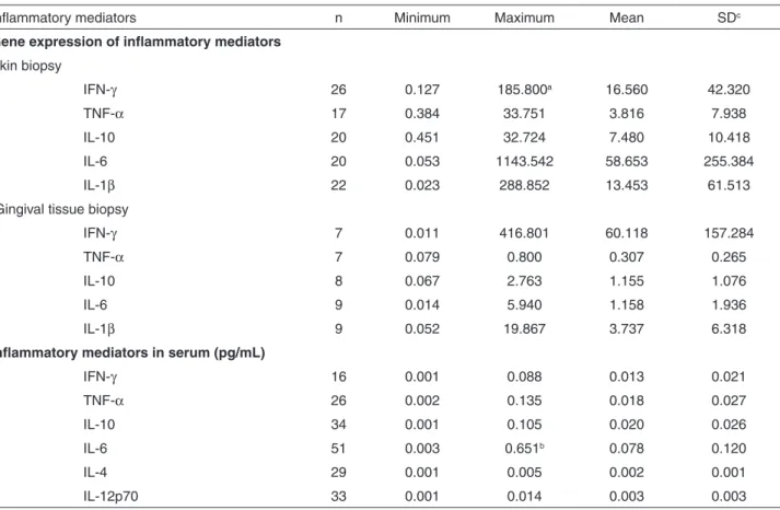
Results of the relative mRNA expression analysis of IFN-γ, TNF-α, IL-10, IL-6 and IL-1β from the skin biopsies of MB patients revealed a higher than average expression of IL-6, IFN-γ and IL-1β; in gingival tissue biopsies of subjects with gingival-periodontal impairment, mRNA expression of IFN-γ and IL-1β reached higher than average values. In serum samples, the level of the mediator IL-6 was found to be the highest, followed by IL- 10 and TNF- α (Table 1).
Table 1 - Descriptive analysis of inflammatory mediators in the skin and gingiva of multibacillary leprosy cases according to variables related to gene expression and serum quantitation (pg/mL)
Inflammatory mediators n Minimum Maximum Mean SDc
Gene expression of inflammatory mediators
Skin biopsy
IFN-γ 26 0.127 185.800a 16.560 42.320
TNF-α 17 0.384 33.751 3.816 7.938
IL-10 20 0.451 32.724 7.480 10.418
IL-6 20 0.053 1143.542 58.653 255.384
IL-1β 22 0.023 288.852 13.453 61.513
Gingival tissue biopsy
IFN-γ 7 0.011 416.801 60.118 157.284
TNF-α 7 0.079 0.800 0.307 0.265
IL-10 8 0.067 2.763 1.155 1.076
IL-6 9 0.014 5.940 1.158 1.936
IL-1β 9 0.052 19.867 3.737 6.318
Inflammatory mediators in serum (pg/mL)
IFN-γ 16 0.001 0.088 0.013 0.021
TNF-α 26 0.002 0.135 0.018 0.027
IL-10 34 0.001 0.105 0.020 0.026
IL-6 51 0.003 0.651b 0.078 0.120
IL-4 29 0.001 0.005 0.002 0.001
IL-12p70 33 0.001 0.014 0.003 0.003
In skin biopsies, IL-4 and IL-12p70 were not detected. In case of patients with CPD and LR, there was a negative correlation between serum IL-4 and TNF-α expressed in the skin (r = 0.83; p < 0.05); a positive correlation between IL-4 and IFN-γ present in serum (r = 0.89; p < 0.05) and between serum IL-6 and IFN-γ expressed in the skin (r = 0.79; p < 0.05) (Table 2).
Higher concentrations of IL-6 were found in serum samples of patients with LR (p = 0.036). There was no significant difference in the expression of IFN-γ, TNF-α, IL-10, IL-6 and IL-1β in the skin and results found the gingiva biopsies of patients with and without LR, even though the gene expression in skin biopsies was observed among the cases with LR. In case of gingiva, only IL-1β gene expression was high (Table 3).
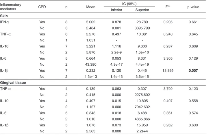
Patients with CPD and LR presented higher IL-1β gene expression in skin than those without CPD (p = 0.007)
(Table 3). No statistically significant difference was
observed for other mediators (Table 4).
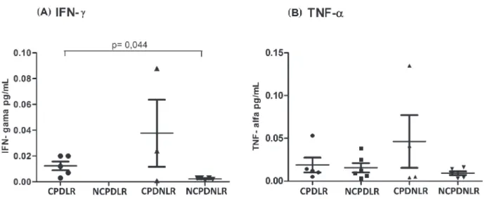
In patients with CPDLR, serum levels of IL-4 were significantly lower than those in patients with no CPDLR. On the contrary, these cases showed higher levels of IL-4 than patients with no CPD and no LR (Figure 1A).
Levels of IL-6 in patients with CPDLR were significantly lower than those in patients with no CPDLR (Figure 1B). In contrast, the levels of IL-6 in patients with no CPDLR were higher than those in patients with no CPD and no LR
(Figure 1B).
Serum levels of IFN-γ in patients with CPDLR were significantly higher than those in patients with no CPD and
no LR. However, no significant difference in serum levels of TNF-α was found among the groups (Figure 2B).
DISCUSSION
Accumulating evidence suggests the involvement of similar inflammatory cytokines in the development of PD and of LR9, and in this study, it was demonstrated that patients with CPD have a modified serum profile of IL-4, IL-6 and IFN-γ.
It is known that CD8+ cells, along with Th2 cells, produce predominantly IL-4. IL-4 has an antagonistic effect on IFN-γ and its main activity is to suppress the immune response mediated by cells owing to no optimization of TLR1 activation, as well as downregulation of the expression of these receptors on the surface of monocytes14. Contrary to the action mechanism of IL-4 on the cell activity, IL-6, IFN-γ and TNF-α promote the activation of the immune response mediated by Th1 cells15.
In this study, IL-4 detected in serum samples showed a negative correlation with TNF-α gene expression in skin biopsies of patients with CPDLR. Additionally, low and significant levels of IL-4 were found in serum samples of patients with CPDLR as compared to those in patients without PD, and higher levels of IFN-γ were observed in serum samples of patients with CPDLR than in serum samples of patients with no CPD and no LR. These results suggest an immunoregulatory role of PD in these conditions and a possibility of Th1 cell-mediated activation of mediators in the skin, which would be consistent with
Table 2 - Spearman correlation coefficient between inflammatory mediators (IFN-γ, TNF-α, IL-10, IL-6, IL-4 and IL-12p70) in the skin and those in serum samples of multibacillary leprosy cases with chronic periodontal disease and leprosy reaction
Skin biopsy Serum
IFN-γ TNF-α IL-10 IL-6 IL-1β IFN-γ TNF-α IL-10 IL-6 IL-4 IL-12p70
Skin
IFN-γ 1
TNF-α 0.60 1
IL-10 0.68 0.66 1
IL-6 0.60 0.50 0.30 1
IL-1β 0.00 0.20 0.49 1
Serum
IFN-γ 0.10 0.00 -0.50 -0.10 1
TNF-α 0.80 -0.50 0.40 0.20 0.56 1
IL-10 -0.30 0.40 0.60 0.76 0.95 1
IL-6 0.79* -0.20 0.36 0.40 0.04 0.61 0.50 -0.03 1
IL-4 0.00 -0.83* -0.41 0.00 0.00 0.89* 0.63 0.34 0.55 1
IL-12p70 0.80 0.60 -0.40 0.50 0.20 0.40 0.30 0.45 1
fostering of LR and impairment of the patient’s general condition16.
Santos et al.10 observed that reactional multibacillary patients presented highest concentrations of serum IFN-γ. Likewise, Motta et al.17 observed that the odontogenic
infection in subjects with leprosy could increase the pro-inflammatory response mediated by IFN-γ, while the contrasting effect would occur on the immunoregulatory activity of IL-4, triggering exacerbation of the inflammatory reaction.
Table 3 - Descriptive analysis of inflammatory mediators in the skin, gingiva and serum samples in cases of multibacillary leprosy according to leprosy reactions (Yes/No)
Inflammatory mediators
Leprosy
reactions N Mean
IC* (95%)
F** p-value
Inferior Superior
Skin
IFN-γ Yes 13 3.808 1.040 13.736 0.063 0.804
No 13 2.981 0.554 15.959
TNF-α Yes 8 2.059 0.726 5.870 0.645 0.434
No 9 1.305 0.571 3.004
IL-10 Yes 10 3.390 1.336 8.499 0.031 0.861
No 10 3.046 1.105 8.331
IL-6 Yes 8 1.789 0.139 22.874 3.331 0.085
No 12 0.290 0.122 0.691
IL-1β Yes 10 0.485 0.083 2.829 2.314 0.144
No 12 0.142 0.083 0.307
Gingival tissue
IFN-γ Yes 5 0.297 0.024 3.633 1.734 0.245
No 2 8.577 0.000 2.3e+22
TNF-α Yes 6 0.199 0.085 0.468 1.209 0.322
No 1 0.519 -
-IL-10 Yes 6 0.570 0.092 3.525 0.004 0.949
No 2 0.622 0.002 228.149
IL-6 Yes 7 0.467 0.071 3.096 1.251 0.300
No 2 2.578 0.038 177.683
IL-1β Yes 7 1.379 0.239 7.925 0.016 0.904
No 2 1.154 0.021 64.715
Serum (pg/mL)
IFN-γ Yes 10 0.008 0.003 0.024 4.125 0.062
No 6 0.002 0.001 0.004
TNF-α Yes 12 0.012 0.007 0.020 0.371 0.548
No 14 0.009 0.005 0.018
IL-10 Yes 15 0.010 0.005 0.022 0.006 0.939
No 19 0.010 0.006 0.017
IL-6 Yes 20 0.082 0.022 0.304 4.668 0.036
No 31 0.023 0.014 0.038
IL-4 Yes 12 0.002 0.001 0.003 1.547 0.224
No 17 0.001 0.001 0.002
IL-12p7 Yes 13 0.003 0.002 0.005 1.271 0.268
No 20 0.002 0.002 0.003
Both, LR and PD, independently present a complex network of immune activity that culminates with the participation of various cell types and inflammatory
cytokines, both in serum samples and tissues affected by the disease9.
The coexistence of infections, such as leprosy and
Figure 1- Comparison between serum levels of (A) IL-4 and (B) IL-6 among patients with CPD and those with no CPD with LR and no LR. Mann-Whitney test; 5% significance level. CPDLR (chronic periodontal disease and leprosy reaction); NCPDLR (no chronic periodontal disease and leprosy reaction); CPDNLR (chronic periodontal disease and no leprosy reaction); NCDPNLR (no chronic periodontal disease and no leprosy reaction).
Table 4 - Descriptive analysis of inflammatory mediators in the skin and gingiva of multibacillary leprosy cases with leprosy reaction in the presence of chronic periodontal disease (CPD: Yes/No)
Inflammatory
mediators CPD n Mean
IC (95%)
F** p-value
Inferior Superior
Skin
IFN-γ Yes 8 5.002 0.878 28.789 0.205 0.661
No 3 2.484 0.001 3395.799
TNF-α Yes 6 2.270 0.497 10.381 0.240 0.645
No 1 1.051 -
-IL-10 Yes 7 3.221 1.116 9.300 0.287 0.609
No 2 5.870 2.2e-9 1.5e+10
IL-6 Yes 5 0.664 0.053 8.331 3.305 0.129
No 2 43.380 4.3e-17 4.4e+19
IL-1β Yes 7 0.232 0.120 0.445 13.895 0.007
No 2 1.3e-13 1.4e-13 3.6e+15
Gingival tissue
TNF-α Yes 4 0.139 0.063 0.307 3.799 0.123
No 2 0.415 0.000 2275.602
IL-10 Yes 4 0.407 0.015 10.805 0.407 0.558
No 2 1.127 0.000 7942.632
IL-6 Yes 5 0.343 0.018 6.488 0.361 0.574
No 2 1.010 0.000 4865.866
IL-1β Yes 5 1.076 0.073 15.959 0.262 0.630
No 2 2.563 0.000 2.2e+4
PD, promotes a change in the pattern of inflammatory response by either activating, inhibiting, or enhancing these processes2,17,18. The same systemic condition was observed in diabetes and PD19.
The balance between Th1 and Th2 cells and a change in the mediator profile found in serum samples and of those expressed in skin, such as IFN-γ, TNF-α, IL-1β, IL-6 and IL-4, both during the occurrence of T1R and T2R, seems to be closely related to the clinical spectrum of the disease. In PD, although there is no consensus on the immunological standards involved in the pathophysiology, a combined function of Th1 and Th2 cells is possible20.
The complex association between the inflammatory cytokines and the immune cells system during LR and CPD manifestation has prompted researchers to conduct more specific studies on the cell type and cytokine determinants of these processes9, as well as on other cell types, such as keratinocytes and fibroblasts, as well as cytokine producers in response to bacterial stimulation or other cytokines16,21.
Particularly in the case of leprosy, in patients with borderline clinical features, the participation of other cell subtypes, such as the subpopulations of Th0 cells that seem to produce both IFN-γ and IL-4 in occurrence of T1R has been described. However, Th1 polarization along with IFN-γ production could also occur in the face of T1R recurrence22. Other cell subtypes, such as the regulatory T cells (Tregs), Th3 cells with an immunosuppressive profile and Th17 cells with a pro-inflammatory profile, may participate in the determination of the cell profile and the involved mediators23,24. However, the understanding of the association between molecular and cellular paths and the pathogenesis of CPD and LR is still incomplete.
Considering the involvement of IL-6 in the development of LR, the results of this study are consistent with those in the literature, in which patients with LR showed higher levels of IL-6 in serum samples than the levels in patients with no LR. Recognized as a potential inflammatory marker of LR, IL-6 stands out due to its multifunctional profile9,18,25.
In case of patients with CPD and reactional states, a positive correlation between IL-6 in the serum and IFN-γ gene expression in the skin was found. Therefore, an increased serum level of IL-6 may contribute to an increase in gene expression of IFN-γ in the skin, which could justify the intensification of Th1 type cell response with triggering or maintenance of the LR. However, in case of patients with CPD or reactional episodes, the serum level of IL-6 was lower than that in patients without CPD and reactional episodes, suggesting an IL-6 downregulation in the presence of PD. On the contrary, as described previously, other cell types, such as Th0 cells, Tregs, and keratinocytes, may influence the inflammatory mechanisms.
The influence of PD on the manifestation of LR was strengthened by the clinical improvement of reactional symptoms after subjects with odontogenic infections and LR received dental treatment18. Additionally, the probability of occurrence of LR may increase when the patient presents poor oral health26.
biopsy was performed, or even due to the sample size. Although this represents a study limitation, it should be emphasized that blood collection was performed on the same day of skin biopsy, before initiation of the reactional treatment, and in the initial weeks of leprosy treatment.
Considering the gingival tissue, IFN-γ and IL-1β showed higher mean values for gene expression, although not statistically significant, than that of other mediators studied. This is in agreement with the literature, since both IFN-γ and IL-1β are important mediators involved in bone resorption during the development of PD and the extent and severity of these diseases9. It is possible that treatment of reactional episodes or even polychemotherapy, favor the reduction of pathogenic microorganisms responsible for the development of CPD, and consequently, the expression of inflammatory cytokines in the gingiva. More specific studies are necessary to clarify this approach.
As an original study on CPD and LR, it could be stated that the results suggested an interaction between CPD and immunoregulatory mechanisms involved in the triggering, exacerbation and maintenance of LR. These interactions can promote the expression of TNF-α and IFN-γ in the skin, while downregulating IL-4 and IL-6 in the serum.
Considering the previous approaches, questions about the association between molecular and cellular paths and the pathogenesis of these diseases suggest the need of future investigations.
CONCLUSION
The presence of DPC in individuals with LR immunoregulated the concentrations of IL-6, IFN-γ and IL-4. The presence of DPC decreased serum levels of IL-6 and IL-4 in reactional individuals. CPD concomitant to LR resulted in increased IFN-γ serum levels.
ACKNOWLEDGMENTS
We are grateful to the dentist Glaucia Larroyed de Oliveira for providing dentistry assistance to the participants. The authors also gratefully acknowledge the patients, without whom this study would not have been possible. The study was supported by Fundação de Amparo
à Pesquisa do Estado de Mato Grosso, Brazil (FAPEMAT)
and CNPq (Process Nº 477727/2011).
REFERENCES
1. Scollard DM, Martelli CM, Stefani MM, Maroja MF, Villahermosa L, Pardillo F, et al. Risk factors for leprosy reactions in three endemic countries. Am J Trop Med Hyg. 2015;92:108-14.
2. Motta AC, Pereira KJ, Tarquínio DC, Vieira MB, Miyake K, Foss NT. Leprosy reactions: coinfections as a possible risk factor. Clinics (Sao Paulo). 2012;67:1145-8.
3. Grudyanov AI, Tkacheva ON, Avraamova TV. Correlation of chronic periodontal disease and cardiovascular disease. Stomatologiia (Mosk). 2017;96:4-7.
4. Mesia R, Gholami F, Huang H, Clare-Salzler M, Aukhil I, Wallet SM, et al. Systemic inflammatory responses in patients with type 2 diabetes with chronic periodontitis. BMJ Open Diabetes Res Care. 2016;4:e000260.
5. Leira Y, Seoane J, Blanco M, Rodríguez-Yáñez M, Takkouche B, Blanco J, et al. Association between periodontitis and ischemic stroke: a systematic review and meta-analysis. Eur J Epidemiol. 2017;32:43-53.
6. Escobar-Arregoces F, Latorre-Uriza C, Velosa-Porras J, Roa-Molina N, Ruiz AJ, Silva J, et al. Inflamatory response in pregnant women with high risk of preterm delivery and its relationship with periodontal disease: a pilot study. Acta Odontol Latinoam. 2018;31:53-7.
7. Hirschfeld J, Kawai T. Oral inflammation and bacteremia: implications for chronic and acute systemic diseases involving major organs. Cardiovasc Hematol Disord Drug Targets. 2015;15:70-84.
8. Van Dyke TE, van Winkelhoff AJ. Infection and inflammatory mechanisms. J Clin Periodontol. 2013;40 Suppl 14:S1-7. 9. Cortela DC, Souza Junior AL, Virmond MC, Ignotti E.
Inflammatory mediators of leprosy reactional episodes and dental infections: a systematic review. Mediators Inflamm. 2015;2015:548540.
10. Santos MB, de Oliveira DT, Cazzaniga RA, Varjão CS, Dos Santos PL, Santos ML, et al. Distinct roles of Th17 and Th1 cells in inflammatory responses associated with the presentation of paucibacillary leprosy and leprosy reactions. Scand J Immunol. 2017;86:40-9.
11. Fonseca AB, Simon MV, Cazzaniga RA, Moura TR, Almeida RP, Duthie MS, et al. The influence of innate and adaptative immune responses on the differential clinical outcomes of leprosy. Infect Dis Poverty. 2017;6:5.
12. Gomes Filho IS, Macedo TC, Cruz SS, Soledade KR, Trindade SC, Sarmento VA. Comparação de critérios que determinam o diagnóstico clínico da doença periodontal. Rev Odont Cienc. 2006;21:77-81.
13. Ridley DS, Jopling WH. Classification of leprosy according to immunity: a five-group system. Int J Lepr Other Mycobact Dis. 1966;34:255-73.
14. Modlin RL. The innate immune response in leprosy. Curr Opin Immunol. 2010;22:48-54.
15. Walker SL, Lockwood DN. The clinical and immunological features of leprosy. Br Med Bull. 2006;77-78:103-21. 16. Teles RM, Moraes MO, Geraldo NT, Salles AM, Sarno EN,
in the epidermis of leprosypatients. Arch Dermatol Res. 2002;294:355-62.
17. Motta AC, Simão JC, Furini RB, Ferreira MA, Palma PV, Komesu MC, et al. Oral coinfection can stress peripheral lymphocyte to inflammatory activity in leprosy. Rev Soc Bras Med Trop. 2013;46:73-8.
18. Motta AC, Furini RB, Simão JC, Ferreira MA, Komesu MC, Foss NT. The recurrence of leprosy reactional episodes could be associated with oral chronic infections and expression of serum IL-1, TNF-α, IL-6, IFN- and IL-10. Braz Dent J. 2010;21:158-64.
19. Chapple IL, Genco R. Diabetes and periodontal diseases: consensus report of the Joint EFP/AAP Workshop on Periodontitis and Systemic Diseases. J Clin Periodontol. 2013;40 Suppl 14:S106-12.
20. Souto GR, Queiroz-Junior CM, de Abreu MH, Costa FO, Mesquita RA. Pro-inflammatory, Th1, Th2, Th17 cytokines and dendritic cells: a cross-sectional study in chronic periodontitis. PLoS One. 2014;9:e91636.
21. Preshaw PM, Taylor JJ. How has research into cytokine interactions and their role in driving immune responses impacted our understanding of periodontitis? J Clin Periodontol. 2011;38 Suppl 11:60-84.
22. Verhagen CE, Wierenga EA, Buffing AA, Chand MA, Faber WR, Das PK. Reversal reaction in borderline leprosy is associated with a polarized shift to type 1-like Mycobacterium leprae T cell reactivity in lesional skin: a follow-up study. J Immunol. 1997;159:4474-83.
23. Venturini J, Soares CT, Belone AF, Barreto JA, Ura S, Lauris JR, et al. In vitro and skin lesion cytokine profile in Brazilian patients with borderline tuberculoid and borderline lepromatous leprosy. Lepr Rev. 2011;82:25-35.
24. Abdallah M, Emam H, Attia E, Hussein J, Mohamed N. Estimation of serum level of interleukin-17 and interleukin-4 in leprosy, towards more understanding of leprosy immunopathogenesis. Indian J Dermatol Venereol Leprol. 2013;79:772-6.
25. Stefani MM, Guerra JG, Sousa AL, Costa MB, Oliveira ML, Martelli CT, et al. Potential plasma markers of type 1 and type 2 leprosy reactions: a preliminary report. BMC Infect Dis. 2009;9:75.