ABSTRACT | Purpose: To evaluate the anatomical and functional outcomes of surgical treatment of retinal detachment
secondary to ocular toxoplasmosis. Methods: A retrospective
analysis of data from patients who had undergone vitreoretinal surgery for retinal detachment secondary to ocular toxoplasmosis was conducted. The parameters that were analyzed include surgical procedures, anatomical outcomes, visual acuity, and
postoperative complications. Results: This study included 22
patients, of which 13 were female (59.1%). The mean age was 28.5 years (SD ± 14.5, range 12-78 years) and the follow-up period varied from 1 to 163 months (mean 64 months). The mean baseline best-corrected visual acuity (BCVA) was 2.0 logMAR (SD ± 1.0). A total of 31 surgeries were performed, and the retina was reattached in 15 patients (68.2%) immediately after the first surgery and in 20 patients (90.9%) at a later point. The mean postoperative BCVA improved to 1.3 logMAR (SD ± 0.9) (p<0.05). Nineteen patients (86.4%) underwent cataract surgery with intraocular lens implant, and 12 patients (60.0%) underwent silicone oil removal. Five patients (22.7%) exhibited elevated intraocular pressure, and 1 patient (4.5%) developed hypotonia. Conclusion: Surgical treatment of retinal detachment secondary to ocular toxoplasmosis resulted in considerable anatomical and functional improvement. Although PPV with silicone oil injection demonstrated the best outcomes, it is not reasonable to conclude that this is the best surgical approach given the small number of patients included in this study.
Keywords: Toxoplasmosis, ocular; Retinal detachment; Vitrec-tomy; Scleral buckling
RESUMO | Objetivo: Avaliar os resultados anatômicos e fun-cionais após o tratamento do descolamento de retina secundário
à toxoplasmose ocular. Métodos: Análise retrospectiva de
dados de um banco de dados validado, que incluiu registros de pacientes submetidos à cirurgia vitreorretiniana para descolamento de retina secundário a toxoplasmose ocular. Foram analisados procedimentos cirúrgicos, sucesso anatômico,
acuidade visual e complicações pós-operatórias. Resultados:
Foram avaliados 22 olhos de 22 pacientes. Treze eram do sexo feminino (59,1%) e a idade média era de 28,5 anos (DP ± 14,5, intervalo de 12 a 78 anos). O período de acompanhamento variou de 1 a 163 meses (média de 64 meses). A melhor acuidade visual corrigida (BCVA) foi 2,0 logMAR (SD ± 1,0). Em geral, entre retinopexia (RSB) e vitrectomia pars plana (PPV) utilizando injeção de óleo de gás ou de silicone (SO), realizaram-se 31 cirurgias. A retina foi considerada colada em 15 olhos (68,2%) na primeira cirurgia e em 20 olhos (90,9%) ao final do estudo. A BCVA pós-operatória média melhorou para 1,3 logMAR (SD ± 0,9) (p<0,05). Dezenove olhos (86,4%) foram submetidos à cirurgia de catarata com implante de lente intraocular e 12 olhos (60,0%) tiveram remoção de óleo de silicone. Cinco olhos (22,7%) desenvolveram pressão
intraocu-lar elevada e 1 (4,5%) desenvolveu hipotonia. Conclusão: A
abordagem cirúrgica no descolamento de retina secundária a toxoplasmose ocular permitiu importante melhora anatômica e funcional. Embora a PPV com injeção de óleo de silicone tenha demonstrado melhores resultados, não é viável afirmar que é a melhor técnica cirúrgica, devido ao pequeno número e às particularidades dos olhos tratados.
Descritores: Toxoplasmose ocular; Descolamento retiniano; Vi-trectomia; Recurvamento da esclera
INTRODUCTION
The prevalence of toxoplasmosis, a disease caused by the obligate intracellular protozoan Toxoplasma gondii,
Surgical outcomes of rhegmatogenous retinal
detachment associated with ocular toxoplasmosis
Resultados cirúrgicos do descolamento regmatogênico de retina
associado à toxoplasmose ocular
Francisco Virmond Moreira1, Andressa Moreira Iwanusk1, Augusto Radünz do Amaral1, Mário Junqueira Nóbrega1,2, Fernando José De Novelli2
1. Universidade da Região de Joinville, Joinville, SC, Brazil. 2. Hospital de Olhos Sadalla Amin Ghanem, Joinville, SC, Brazil.
Submitted for publication: June 13, 2017 Accepted for publication: February 8, 2018
Funding: No specific financial support was available for this study.
Disclosure of potential conflicts of interest: None of the authors have any potential conflict of interest to disclose.
Corresponding author: Francisco V. Moreira.
varies with a number of socioeconomic, geographic, climatic, and cultural factors(1,2). In Brazil, 50-83% of the
population is seropositive for T. gondii, and the infection is considered to be one of the most common causes of posterior uveitis(3,4).
Toxoplasmosis can be acquired through the handling or ingestion of undercooked or raw meat such as pork and lamb; ingestion of oocysts present in the water and on unwashed food, or excreted by infected felines; or congenitally through the placenta of an infected preg-nant woman(5,6). The disease is usually self-limited in
immunocompetent individuals but can lead to serious visual complications affecting the posterior pole, the vitreoretinal interface, and the anterior segment(1,7-9).
Rhegmatogenous retinal detachment (RRD) is one of the leading causes of blindness associated with ocular toxoplasmosis. It originates from the posterior vitreous detachment secondary to intraocular inflammation and requires surgical treatment(8,10,11). Various techniques
for retinal repositioning, including retinopexy with scleral buckle (RSB) and pars plana vitrectomy (PPV) associated with gas or silicone oil (SO) injection, have been proposed over time. The postoperative results may vary depending on the condition of the macula, the detachment time, and the presence of proliferative vi-treoretinopathy.
The main objective of this study was to analyze the ana tomical and functional outcomes of patients with retinal detachment secondary to ocular toxoplasmosis treated in a single reference center in southern Brazil.
METHODS
Participants and setting
This non-comparative study was a case-series inclu-ding patients with rhegmatogenous retinal detachment secondary to ocular toxoplasmosis. All examinations and surgical treatments were performed by two expe-rienced surgeons (MJN, FJDN) between April 2000 and February 2013 at the Hospital de Olhos Sadalla Amin Ghanem, Joinville, Brazil.
The medical charts of patients who had undergone vitreoretinal surgery for retinal detachment secondary to ocular toxoplasmosis were reviewed, and individuals with a history of proliferative diabetic retinopathy, pa-thological myopia, pseudophakia, penetrating or blunt ocular trauma, or other causes of uveitis were excluded from the study. The patient data extracted from the
re cords include age, gender, operated eye, baseline best-corrected visual acuity (BCVA), surgical procedu-res undergone, number of operations, cataract surgery with intraocular lens implant, and silicone oil removal. Overall retinal reattachment and improvement in BCVA were considered to be the anatomic and functional ou-tcomes, respectively. Presence of postoperative compli-cations such as keratopathy, elevated intraocular pres-sure (IOP >21 mmHg), and hypotonia (IOP <8 mmHg) were also recorded.
This study was approved by the Ethics and Research of Human Beings Committee of the University of Joinville Region. It was carried out in accordance with the Regu-latory Standards and Guidelines for Research involving human subjects (Resolution 466/2012 of the National Health Board of Brazil). All patients received a detailed description of the surgical procedures to be undertaken and the risk of potential complications and were asked to sign an informed consent form thereafter.
Surgical procedure
Surgical techniques included scleral buckling, pars plana vitrectomy, or a combination of the two procedu-res. Scleral buckling was performed using an encircling silicone band (2.5 mm wide) with or without a seg-mental silicone tire (7 mm wide). Pars plana vitrectomy was carried out by using a 20 or 23-gauge system to apply an infusion of 15% perfluoropropane (C3F8) gas or 5000-centistoke silicone oil into the vitreous cavity. Phacoemulsification with an intraocular lens implant was carried in case of significant lens opacity, followed by a posterior segment surgery afterwards.
Data analyses
The quantitative variables were presented as mean and standard deviations (SD), while the qualitative variables were presented as absolute and relative fre-quencies (%). BCVA was evaluated using the Snellen scale and converted into logarithm units of the minimal angle of resolution (logMAR). Functional improvement or worsening was defined as a difference of more than 0.2 logMAR between baseline and final BCVA, and Friedman’s analysis of variance on ranks was used to compare preoperative and postoperative BCVA.
RESULTS
This study included a total of 22 patients, of which 13 were females (59.1%). The mean age of the patients was 28.5 years (SD 14.5), and the mean preoperative BCVA was logMAR 2.0 (SD 1.0) (Snellen 20/2000). Initial asses-sment showed that 5 patients (22.7%) had proliferative vitreoretinopathy (PVR), three patients had posterior PVR C1, and two patients had PVR C2. Two patients did not show PVR and there was no registration about PVR for the other 15 patients. Twelve patients (54.5%) presented with detachment of the macula.
Macular detachments were caused by one break per eye in eleven patients (50%), and two or more breaks per eye in six patients. Five patients had no records of how many breaks had caused the macular detachment.
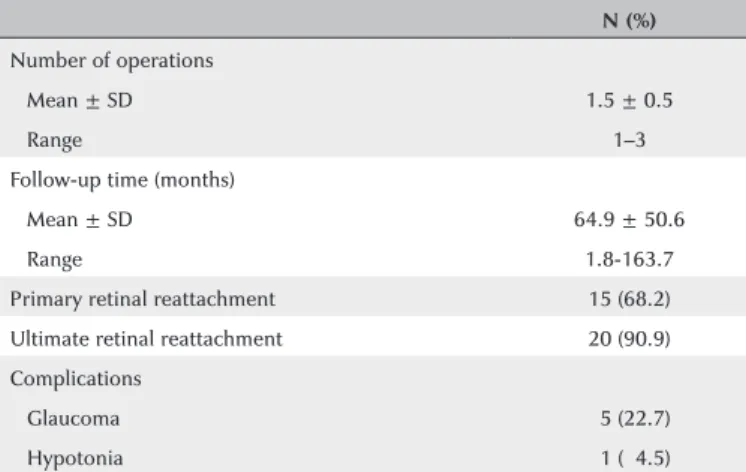
A total of 31 surgeries were performed and the mean follow-up period was 64.9 months (range 1.8-163.7 months). Primary retinal reattachment was achieved in 15 cases (68.2%) and ultimate reattachment was accomplished in 20 patients (90.9%). Post-surgical com-plications included increased intraocular pressure in 5 patients (22.7%), and hypotonia in one patient (4.5%) (Table 1).
Five different isolated or combined procedures were performed, and the most common combination pro-cedure was RSB, PPV, and SO (n=10 patients; 45.4%), as shown in table 2. The technique that achieved the highest rate of primary retinal reattachment was PPV and SO (4 patients; 100.0%), and all study participants who underwent RSB alone (4) or PPV with gas infusion (1) exhibited functional improvement.
Seven patients (31.8%), all of whom had undergone PVR, presented with recurrent RRD after the first surgery
and required subsequent procedures. Primary surgical procedures that required another operation included RSB (3 patients; 75%); PPV and GAS combined (1 patient; 100%); RSB, PPV and GAS combined (1 patient; 33.3%); and RSB, PPV and SO combined (2 patients; 20%). Of the recurrent cases, three patients who initially underwent RSB were treated using PPV and SO; one patient ini-tially treated with PPV and GAS underwent RSB, PPV and SO combined upon recurrence; one patient who was initially treated with RSB, PPV and GAS combined subsequently underwent PPV and SO; and two patients initially treated with RSB, PPV and SO combined un-derwent PPV and SO upon recurrence.
Silicone oil was injected in the eyes of 20 patients and removed from the eyes of 12 patients (60.0%). Nineteen patients (86.4%) underwent cataract surgery with intrao-cular lens implant, and the time elapsed between retinal and cataract surgery ranged from zero to 63 months (mean: 16.2 months) (Table 2).
Functional examination showed a mean BCVA im-provement of 1.3 logMAR (SD±0.9) after treatment (Table 3), and 15 patients (68.2%) exhibited final BCVA values that were equal to or more than +1.0 logMAR (≤20/200). Final postoperative evaluation showed an overall statistically significant improvement (p<0.05) in BCVA in all treated eyes (Figure 1). Specifically, 14 patients (63.6%) exhibited improvement of BCVA, 6 patients (27.3%) presented with stabilization of BCVA, and 2 patients (9.1%) showed worsening of their BCVA values. A severe decrease in visual acuity was observed in one patient (Figure 2).
DISCUSSION
Brazil has a high prevalence of toxoplasmosis. The current case-series study focused on the importance of surgical treatment of rhegmatogenous detachment associated with infection. The current study sample was similar to those described in previous studies, except for a slight predominance of females(2,5,10) and an earlier age
of onset of detachment (mean age 28.5 years)(3,4,7,8,10).
Previous studies have reported that the risk factors for RRD include age, myopia, severe inflammation, and positive family history, as well as a history of trauma, ca-taract surgery, and diabetes(2,5,10). Bouts of ocular
toxoplas-mosis (OT) may also predispose posterior vitreous detach-ment and, more rarely, RRD. A previous study conducted by Bosch-Driessen et al. reported a prevalence of 6% for RRD following OT(10).
Table 1. Overall anatomical and functional outcomes
N (%)
Number of operations
Mean ± SD 1.5 ± 0.5
Range 1–3
Follow-up time (months)
Mean ± SD 64.9 ± 50.6
Range 1.8-163.7
Primary retinal reattachment 15 (68.2)
Ultimate retinal reattachment 20 (90.9)
Complications
Glaucoma 05 (22.7)
Hypotonia 01 (04.5)
With regard to the anatomical outcomes, 90.9% of the study patients exhibited reattached retinas at the time of the last follow-up visit, and this finding was in agreement with previous evidence(3,4,7).
One of the initial objectives of this study was to try and identify the best surgical technique for the treatment of RRD associated with OT. The results presented in ta-ble 2 suggest that RSB alone and PPV with gas infusion were the best surgical approaches in terms of functional outcomes (BCVA). However, upon using these techniques, only one patient was seen to exhibit complete primary reti-nal attachment, suggesting that visual improvement could be achieved through successful secondary interventions. Simple operations would demonstrate better outcomes in case of recently developed RRD, while combined proce-dures that were associated with higher risk of poor results could be indicated for more complex cases of RRD with
Table 2. Anatomical and functional improvement by surgical technique
N (%) Primary retinal attachment Final retinal attachment Functional improvement
First surgical procedure
RSB 04 (18.2) 1 (025.0) 4 (100.0) 4 (100.0)
PPV+GAS 01 (04.5) 0 (000.0) 1 (100.0) 1 (100.0)
PPV+SO 04 (18.2) 4 (100.0) 4 (100.0) 2 (050.0)
RSB+PPV+GAS 03 (13.6) 2 (066.7) 3 (100.0) 2 (066.7)
RSB+PPV+SO 10 (45.5) 8 (080.0) 9 (090.0) 5 (050.0)
PHACO with IOL implant 19 (86.4) … … …
Silicone oil removal 12 (60.0) … … …
N (%)= number and percentage of patients; RSB= retinopexy with scleral buckle; PPV= pars plana vitrectomy; GAS= gas infusion; SO= silicone oil infusion; PHACO= phacoemul-sification; IOL= intraocular lens.
Table 3. Baseline and postoperative BCVA
Baseline BCVA Postoperative BCVA
Mean logMAR ± SD 2.0 ± 1.0 1.3 ± 0.9
Mean Snellen scale 20/2000 20/400
logMAR (Snellen scale) N (%) N (%)
0.48 (20/60) or better 02 (09.1) 05 (22.7)
<0.48 (20/60) - 1.30 (20/400) 05 (22.7) 06 (27.3)
<1.30 (20/400) 15 (68.2) 11 (50.0)
BVCA= best-corrected visual acuity; logMAR= logarithm of minimal angle of resolution; SD= standard deviation; N(%)= number and percentage of patients.
Figure 1. Variation of BCVA at baseline and postoperatively.
proliferative vitreoretinopathy. However, the small sam-ple size of the current study did not allow examination of any significant differences in the results associated with various surgical procedures.
Previous studies have also examined various sur-gical techniques using small sample sizes, Adan et al. included 8 patients with RRD who were treated using SB and PPV. Of these, five underwent long-acting gas endotamponade and 3 underwent SO endotamponade(3).
Papadopoulou et al. examined 2 RRD patients trea-ted with PPV and gas injection and a third one who underwent PPV and gas injection associated with SB(4),
while Bosch-Driessen et al. included 6 patients with RRD in their study, of which 4 patients treated by SB, one was treated using PPV and SO injection combined, and one was treated using cryocoagulation(10).All of
these studies reported improvements in the functional outcomes, although no statistically significant differen-ces between techniques were observed.
Overall, five patients (22.7%) exhibited preoperative PVR and nine patients (40.9%) presented with recurrent RRD due to PVR. This was in agreement with the study conducted by Papadopoulou et al. who reported multiple recurrences of RRD in OT, requiring several subsequent surgeries(4). Lucena et al. reported recurrence in one
patient (10%), while Bosch-Driessen et al. reported re-currence in two (33%)(10).
The impacts of macula detachments were not eva-luated as the necessary data were not available for all patients.
The findings of this study also showed a reduction of 18.2% in the number of patients with BCVA values of 20/400 or worse after surgical treatment (Table 3). Nevertheless, 68% of the patients presented final BCVA values ranging from +1.0 to +3.0 logMAR (20/200 to 20/20000). Adán et al.(3) and Bosch-Driessen(10)
repor-ted slightly better functional results, with 50% of the patients included in their study exhibiting final BCVA values that were ≤20/200. Conversely, Lucena et al.(7)
and Papadopoulou et al.(4) reported worse results, with
70% and 100% of patients exhibiting BCVA values that were ≤20/200, respectively.
PPV with SO resulted improved anatomic results but no improvement in functional outcomes when compa-red to the other procedures. This was in agreement with Adán et al. who reported better functional results with this technique in association with SB. The results of their study showed 2 patients whose functional outcomes improved from just hand motions to BCVA values of 20/80, and 1 patient whose BCVA score improved from
20/400 to 20/200(3). Conversely, Bosch-Driessen et al.
reported one patient who presented with a stable final BCVA value of 20/200 following treatment using PPV, SO injection, and SB(10).
At the time of the final postoperative visit, elevated intraocular pressure was observed in 5 patients (22.7%), and this was higher than the results reported by Adan et al. who observed one patient (12.5%) with postope-rative glaucoma(3). This finding could be attributed to a
higher frequency of intraocular surgeries performed in the present series.
Therefore, surgical techniques for the treatment of retinal detachment associated with ocular toxoplasmo-sis resulted in considerable anatomic and functional improvement overall. Although PPV with silicone oil injection demonstrated the greatest improvement in anatomic outcomes, it is not reasonable to conclude that this is the best surgical approach given the small number of patients included in this study. One limitation of the present study was the lack of information on the various risk factors of RRD that could have confounded the severity of the patient’s prognosis, including myo-pia, pseudophakia, and peripheral retinal degeneration.
REFERENCES
1. Foster CS, Vitale AT. Diagnosis and treatment of uveitis. 2nd ed. New Delhi: Jaypee Brothers. 2013. Chapter 35;Toxoplasmosis; p.543-64. 2. Kovačević-Pavićević D, Radosavljević A, Ilić A, Kovačević I,
Djurković-Djaković O. Clinical pattern of ocular toxoplasmosis
treated in a referral centre in Serbia. Eye (Lond). 2012;26(5):723-8. 3. Adán A, Giralt J, Alvarez G, Alforja S, Burés-Jesltrup A, Casaroli-Ma-rano RP, et al. Pars plana vitrectomy for vitreoretinal complications of ocular toxoplasmosis. Eur J Ophthalmol. 2009;19(6):1039-43. 4. Papadopoulou DN, Petropoulos IK, Mangioris G, Pharmakakis
NM, Pournaras CJ. Pars plana vitrectomy in the treatment of se-vere complicated toxoplasmic retinochoroiditis. Eur J Ophthalmol. 2011;21(1):83-8.
5. Kianersi F, Beni AN, Ghanbari H, Fazel F. Ocular toxoplasmosis and retinal detachment: five case reports. Eur Rev Med Pharmacol Sci. 2012;16 Suppl 4:84-9.
6. Kahawita S, Simon S, Gilhotra J. Flashes and floaters - a practical approach to assessment and management. Aust Fam Physician. 2014; 43(4):201-3.
7. Lucena DR, Ribeiro JA, Lucena DR, Jorge R. Roturas retinianas em retinocoidite por toxoplasmose: série de casos. Arq Bras Oftalmol. 2009;72(6):829-31.
8. Iwahashi-Shima C, Sato T, Bando H, Ikeda T, Emi K.Anatomic and functional outcomes of 25-gauge vitrectomy for repair of eyes with rhegmatogenous retinal detachment complicated by proliferative vitreoretinopathy. Clin Ophthalmol. 2013;7:2043-9.
9. Buttler NJ, Furtado JM, Winthrop KL, Smith JR. Ocular toxoplas-mosis II: clinical features, pathology and management. Clin Exp Ophthalmol. 2013;41(1):95-108.