98
Coroidopatia no lúpus eritematoso sistêmico
Choroidopathy in systemic lupus erythematosus
Aristófanes Mendonça Canamary Jr
1; Jacqueline Martins de Sousa
1; Gabriel Costa de Andrade
1; Heloisa Moraes do
Nascimento
11Uveal and AIDS Sector, Department of Ophthalmology and Visual Sciences, Escola Paulista de Medicina, Universidade Federal de
São Paulo, São Paulo, SP, Brazil.
A
BSTRACTSystemic lupus erythematosus (SLE) is an autoimmune disease in which can affect the eye in different ways. Lupus choroidopathy is rare and include retinal pigment epithelium (RPE) detachment and/or serous retinal detachment and pigment epitheliopathy. Most cases are associated with systemic disease activity and can be considered a factor of gravity and need for intense immunosuppression. Usually has good visual prognosis with proper treatment of SLE, although some cases may have irreversible damage to the outer retina and RPE. We describe a case of choroidopathy secondary to SLE during its multisystem activity with good clinical outcome after treatment with systemic immunosuppression.
Keywords: Systemic lupus erythematosus; Retinal detachment; Retinal pigment epithelium; Choroid; Fluorescein angiography; Case reports
R
ESUMOO Lúpus Eritematoso sistêmico (LES) é uma doença autoimune que pode afetar o olho de diversas formas. A coroidopatia lúpica é rara e apresenta-se com descolamento seroso de retina, descolamento do epitélio pigmentar da retina (EPR) e epiteliopatia pigmentar. A maioria dos casos está associada à atividade sistêmica da doença, podendo ser considerada um fator de gravidade e necessidade de imunossupressão intensa. Geralmente apresenta bom prognóstico visual com o tratamento adequado do LES, apesar de alguns casos evoluírem com danos irreversíveis na retina externa e EPR. Descrevemos um caso de coroidopatia secundaria ao LES com atividade multisistêmica com boa evolução após tratamento clínico com imunossupressão sistêmica.
Descritores: Lúpus eritematoso sistêmico; Descolamento de retina; Epítelio pigmentado da retina; Coróide; Angiofluorescei-nografia; Relatos de caso
RELATO DE CASO
Recebido para publicação em 08/06/2016 - Aceito para publicação em 28/08/2016. Os autores declaram não haver conflito de interesses.
Rev Bras Oftalmol. 2017; 76 (2): 98-100
99
I
NTRODUCTIONS
ystemic lupus erythematosus (SLE) is an autoimmune disea-se which can affect many organs, including the eyes.(1,2)Ocu-lar involvement occurs in about 30% of cases and may affect eyelids, cornea, retina and also the optic nerve.(2,3) Choroidopathy
with neurosensory retinal detachment is rare and usually occurs in patients with severe or hypertensive disease.(4) Although lupus
choroidopathy usually present with good prognosis, irreversible vision loss can occur.(2) We present a case of SLE choroidopathy
(a rare manifestation), that developed serous retinal detachment (RSD) and pigmentary changes of the retinal pigment epithelium (RPE). A good communication between the ophthalmologist and the rheumatologist; quickly, appropriate and intensive systemic treatment were conditions for a good prognosis.
Case report
Female, 22 year-old, in hospital treatment for SLE in cutaneous, articular, renal, hematologic and neurologic unset; complained about low visual acuity (VA): counting fingers in both eyes (OU). Negatives serology, FAN (homogeneous nuclear pattern 1/1280), rheumatoid factor, anti-DNA (1/320), ANCA, anti-Ro and anti-La antibodies positive, full complement and C2 decreased, increased ESR and CRP. The patient was treated with intravenous high-dose corticosteroids and cyclophosphamide, with systemic and ocular disease improvement. After discharge,
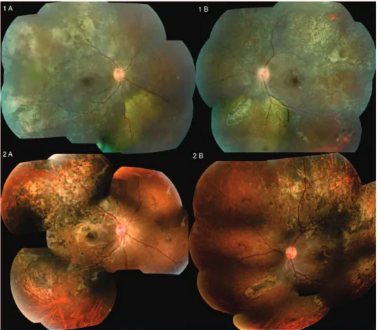
presented VA was 20/200 in the right eye (OD) and 20/50 in the left eye (OS), normal biomicroscopy and intraocular pressure OU. The fundus exam showed mild hyperemic and swollen optic disc, SRD in periphery, hyper and hypopigmentation areas of the RPE and vascular attenuation OU. (Figure 1 A and B) Fluores-cein angiography showed areas of hyper and hypofluorescence by RPE mottling and areas of poor peripheral perfusion OU and hyperfluorescence by leakage in the optical disc in left eye. After systemic treatment (oral corticosteroids at 1 mg/kg/day and immu-nosuppression with monthly cyclophosphamide pulses) there was ocular improvement. Fundoscopy showed normal disk, regression of SRD and of poor perfusion areas and accentuated RPE hyper and hipopigmentations. (Figure A and B). VA improved to 20/25 OU after 8 months. Currently the patient is in use of azathioprine 150mg/kg/day for systemic control.
D
ISCUSSIONSystemic lupus erythematosus (SLE) is a multisystem chro-nic inflammatory disease of unknown cause and usually affect young women.(1,2) Tissues and cells are affected by autoantibodies
and pathogenic immune complexes causing vasculitis, occlusion of small vessels and multiple organ dysfunction. Ocular involvement occurs in about a third of cases involving lid (mucocutaneous disease), keratoconjunctivitis sicca, retinal vascular disease and affecting the optic nerve.(2,3)
Lupus choroidopathy with exudative retinal detachment
Figure 1 A and B. Retinography of the right and left eye respectively showing mild hyperemic optic disc, serous retinal detachment in the periphery, hyper and hypopigmentation areas of RPE and vascular attenuation.
Figure 2 A and B. Retinography of the right and left eye respectively showing normal optic disk, accentuated hyper and hypopigmentation areas of the RPE and normal vessels
Coroidopatia no lúpus eritematoso sistêmico
100
is a rare ocular manifestation, with less than 40 cases reported in scientific literature until 2012.(2) Affects mainly women, 68%
bilateral and may be associated with lupus retinopathy.(5,6) The
pa-thogenesis is multifactorial, although uncontrolled hypertension, immune complex deposits in the choriocapillaris and anti-retinal pigment epithelium antibodies may contribute to its development.
(2) Our patient started ocular unset during acute phase of SLE at
22 years of age, with involvement of different organs. She initially presented cutaneous, articular, renal, hematologic and neurologic activity including hypertension, therefore, featuring important risk factors for lupus choroidopathy development. The most common clinical manifestations of lupus choroidopathy include exudative retinal detachment (36%), RPE detachment (32%) and retinal pigment epitheliopathy (21%).(6) In our case, there was not retinal
pigment epithelium detachment, however the other main mani-festations were present. Initially, the serous retinal detachment was bigger, but it disappeared during systemic treatment and the associated retinal pigment epitheliopathy became easier to be seen. Choroidal ischemia may also be present, manifesting as subretinianal hypopigmented spots and ischemia areas on fluo-rescein angiography.(6)
Imaging tests are important for evaluation and monitoring choroid and retina diseases secondary to SLE.(6) Fluorescein
angiography helps to identify optic nerve inflammation, retinal vascular disease, retinal ischemia, macular edema and search of subclinical signs. In our case, fluorescein angiography was impor-tant to verify RPE lesions, poor peripheral perfusion areas and its improvement after treatment. Green indocyanine evaluates the choroidal vasculature and can identify choroidopathy not seen in angiofluoresceinographic examination.(6) Optical coherence
tomo-graphy allows non-invasive structural, intra and subretinal fluid and detachment of the retinal pigment epithelium evaluation.(6,7)
Lupus choroidopathy is usually seen in patients with active disease, especially central nervous system vasculitis, nephropathy and uncontrolled blood pressure. It has been considered as a sys-temic disease activity score.(2,5,8) The presence of choroidopathy is
an indication of the need for aggressive and prolonged immunosu-ppression.(2,8) Our case accords to the scientific literature, showing
severe systemic involvement during lupus choroidopathy manifes-tation, as well as the importance of intensive immunosuppressive treatment. Ocular therapy such as focal laser photocoagulation or photo-dynamic therapy (PDT), may be instituted when there is insufficient systemic diseases control and poor resolution for lupus coroidopathy with immunosuppression therapy, particular-ly in acute phase.(9) Late ocular interventions may not improve
significantly vision due to prior damage to the RPE and photo-receptors, despite intra and subretinal fluid improvement.(9) As in
most cases, there was resolution of the lupus choroidopathy with
immediate and appropriate systemic treatment in our patient, who developed good visual acuity despite persistent and significant pigment changes in fundus OU.
C
ONCLUSIONRetinopathy and choroidopathy are SLE manifestations, especially during its active phase. Lupus choroidopathy is an indicator of severity and the patient may present poor ocular and systemic prognosis if not correctly treated. Therefore, good communication between the ophthalmologist and the rheuma-tologist is indispensable for the management and treatment of these patients.
R
EFERENCES1. Klejnberg T, Moraes Junior HV. Alterações oculares nos pacientes portadores de lúpus eritematoso sistêmico em acompanhamento ambulatorial. Arq Bras Oftalmol. 2006;69(2):233-7.
2. Palejwala NV, Walia HS, Yeh S. Ocular manifestations of systemic lupus erythematosus: a review of the literature. Autoimmune Dis. 2012;2012:290898.doi:10.1155/2012/290898.
3. EL-Shereef RR, Mohamed AS, Hamdy L. Ocular manifestation of systemic lupus erythematosus. Rheumatol Int. 2013;33(6):1637–42. 4. Gharbiya M, Bozzoni-Pantaleoni F, Augello F, Balacco-Gabrieli C.
Indocyanine green angiographic findings in systemic lupus erythe-matosus choroidopathy. Am J Ophthalmol. 2002;134(2):286-90. 5. Edouard S, Douat J, Sailler L, Arlet P, Astudillo L. Bilateral
choroido-pathy in systemic lupus erythematosus. Lupus. 2011; 20(11):1209–10. 6. Silpa-archa S, Lee JJ, Foster CS. Ocular manifestations in systemic
lupus erythematosus. Br J Ophthalmol. 2016;100(1):135-41. 7. Ozturk B, Bozkurt B, Karademir Z, Kerimoglu H. Follow-up of
lupus choroidopathy with optical coherence tomography. Lupus. 2011;20(10):1076–8.
8. Nguyen QD, Uy HS, Akpek EK, Harper SL, Zacks DN, Foster CS. Choroidopathy of systemic lupus erythematosus. Lupus. 2000;9(4):288-98.
9. Cho HY, Nasir HH, Sobrin L. Focal laser photocoagulation and pho-todynamic therapy for lupus choroidopathy. Lupus. 2014;23(4):412–6.
Correspondence: Aristófanes Jr
Visual Science Department
Rua Botucatu, 821 – Vila Clementino CEP: 04023-062 – São Paulo – SP - Brasil E-mail: [email protected]
Canamary Jr; AM, Sousa JM; Andrade GC; Nascimento HM