C
a s eR
e p o Rt2 6 3 Arq Bras Oftalmol. 2017;80(4):263-5 http://dx.doi.org/10.5935/0004-2749.20170064
Choroidal neovascularization silent on optical coherence tomography: report of two cases
Neovascularização de coroide silente a tomograia de coerência ótica: relato de dois casos
Felipe placeres Borges1, ruBens camargo siqueira1, ingrid ursula scott2, José augusto cardillo1, rodrigo Jorge1
Submitted for publicattion: October 26, 2016 Accepted for publicattion: April 12, 2017
1 Department of Ophthalmology, Faculdade de Medicina de Ribeirão Preto, Universidade de São
Paulo, Ribeirão Preto, SP, Brazil.
2 Departments of Ophthalmology and Public Health Sciences, Penn State College of Medicine,
Hershey, PA, USA.
Funding: No specific financial support was available for this study.
Disclosure of potential conflicts of interest: None of the authors have any potential conflict of interest to disclose.
Corresponding author: Felipe Placeres Borges. Hospital das Clínicas da Faculdade de Medicina de Ribeirão Preto. Avenida Bandeirantes, 3.900 - Ribeirão Preto, SP - 14049-900 - Brazil E-mail: [email protected]
ABSTRACT
Herein, we report two cases of vision loss after successful cataract surgery, associated with drusenoid retinal pigment epithelial detachment without fea-tures of choroidal neovascularization on optical coherence tomography along with angiographic examinations suggestive of choroidal neovascularization in which anatomical and functional improvements were achieved with intravitreal injections of anti-vascular endothelial growth factor.
Keywords: Choroidal neovascularization; Macular degeneration; Tomography, optical coherence; Intravitreal injections; Fluorescein angiography; Humans; Case reports
RESUMO
Relatamos dois casos de baixa visual após cirurgia bem sucedida de catarata, associada a descolamento drusenóide do epitélio pigmentar da retina (DPED) sem achados de neovascularização de coroide a tomografia de coerência óptica OCT (CNV silente ao OCT) e com exames angiográficos sugestivos de neovascularização da coroide (CNV), nos quais melhoras anatômicas e funcionais foram obtidas com aplicações intravítreas de anti-VEGF.
Descritores: Neovascularização da coroide;Degeneração macular; Tomografia de coerência óptica; Injeções intravítreas; Angiofluoresceinografia; Humanos; Relatos de casos
INTRODUCTION
Pigment epithelial detachment (PED) is a pathological process in which the retinal pigment epithelium (RPE) separates from the underlying Bruch’s membrane (BM) as a result of the presence of
blood, serous exudate, drusen, or a neovascular membrane(1,2).
Dru-senoid retinal pigment epithelial detachment (DPED) is a type of
PED that evolves from confluent and large, soft drusen(3,4), which are
extracellular deposits that accumulate between the basal lamina of the RPE and the inner collagenous layer of BM(5), and is considered to be a
feature of non-neovascular age-related macular degeneration (AMD)(6).
Historically, this fundus lesion has been distinguished from other PEDs, such as fibrovascular PEDs and hemorrhagic PEDs, by clinical appearance, fluorescein angiography (FA) findings, histopathology,
and a better short-term visual prognosis(4-8). In addition, DPEDs are
associated with intermediate AMD, unlike other types of PED, which
are generally associated with advanced neovascular AMD(6).
DPED can lead to choroidal neovascularization (CNV)(6,9). However,
in the early stages of DPED-associated CNV, diagnosis may be diffi cult because spectral-domain optical coherence tomography (SD-OCT) may not show characteristic signs of CNV (OCT-silent CNV). In this situation, FA and indocyanine green angiography
(ICGA) may be able to identify evidence of CNV(10). We report two
cases of DPED with OCT-silent CNV that responded to treatment with intravitreal anti-vascular endothelial growth factor (anti-VEGF) therapy.
CASE REPORTS
C
ASE1
A 69-year-old white man presented decreased central vision in the right eye for 5 days, 4 months after cataract surgery. The patient underwent radial keratotomy about 20 years previously, and had been informed, 9 years before undergoing cataract surgery, that he had drusen. His medical history was unremarkable.
ranibi-Ch o r o i d a ln e o va s C u l a r i z at i o ns i l e n to no p t i C a lC o h e r e n C et o m o g r a p h y: r e p o rto ft w oC a s e s
2 6 4 Arq Bras Oftalmol. 2017;80(4):263-5
zumab 0.5 mg for a presumed low-flow choroidal neovascular membrane. At 6 weeks post-injection, the BCVA in the right eye had improved to 20/63, with no changes observed on OCT. Following two additional intravitreal injections of ranibizumab 0.5 mg (performed 6 and 12 weeks after the initial injection, res pectively), the BCVA had improved to 20/50 with no change observed on OCT. After the fourth injection, which was performed 18 weeks after the initial injection, improvements in both the BCVA and the anatomy were observed; the PED progressively decrea-sed in size, leaving some hyperreflective foci above a normoreflec-tive inner segment ellipsoid line, along with a nearly flat RPE on OCT after the fifth injection (given 8 weeks after the fourth injec-tion). After the fifth injection, at 10 weeks, the BCVA had improved to 20/32. A sixth injection was administered, and the inter-injection interval after the fifth injection was extended to 12 weeks.
C
ASE2
A 79-year-old white man presented after 15 days of decreased central vision in the left eye 1 month after cataract surgery in that eye. He had smoked cigarettes for over 30 years, and denied other ocular surgeries, pathologies, and medication use.
Examination demonstrated a BCVA of 20/32 in the right eye and 20/100 in the left eye. Intraocular pressure was 16 mmHg in the right eye and 14 mmHg in the left eye. Slit-lamp examination was
nota-ble for a nuclear sclerotic cataract in the right eye and a well-po-sitioned posterior chamber intraocular lens in the left eye. Dilated fundus examination exhibited irregular foveal pigmentation with medium-sized drusen in the right eye and a foveal DPED in the left eye. FA showed a hyperfluorescent point inferonasal to the fovea in the left eye that increased in the late phases of the FA, and did not correspond to the site of the DPED (Figure 2). ICGA showed a hypo fluorescent dot in the region of the PED and normal retinal and choroidal vasculature. SD-OCT (hyperfluorescent lesion centered 20° line, 512 A-scans per B-scan, automatic real-time setting of 15) findings were consistent with the fundus examination findings. Based on the FA findings, an inferonasal CNV was suspected, and the patient was treated with three intravitreal ranibizumab 0.5 mg injections administered at 6-week intervals. The lesion gradually faded throughout the treatment course, leaving a flat BM with a disrupted external retina on OCT; the BCVA 8 weeks after the third injection was 20/40. At this time, a fourth injection was administe-red, and the BCVA improved to 20/32 12 weeks after.
DISCUSSION
DPED is part of the clinical spectrum of AMD(5,6). A postulated
me-chanism of the development of DPED with a small accumulation of fluid without the classical signs of CNV is that the confluence of soft drusen is associated with a buildup of lipidic membranous debris that
Figure 1. (Case 1): A) Early phase pre-treatment. Left: luorescein angiography (FA) with discrete foci of parafoveal hypoluorescence as a result of drusen blockage. Right: indocyanine green angiography (ICGA) demonstrates a hyperluorescent spot in the foveal region (*). B) Late phase pre-treatment. Left: FA shows staining of the drusen. Right: ICGA demonstrates increased hyperluorescence and better delineation of the foveal lesion (*). C) Pre-treatment optical coherence tomography (OCT) demonstrates an epiretinal membrane and a double-humped PED. D) Early phase post-treatment. Left: FA shows early hyperluorescence from the drusen. Right: ICGA no longer shows the hyperluorescent lesion. E) Late phase post-treatment. Left: FA shows staining of the drusen. Right: ICGA demonstrates absence of the hot spot visualized on the pre-treatment ICGA. F) Post-treatment OCT shows that the PED has lattened.
A B
D E
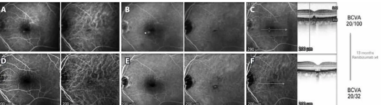
Figure 2. (Case 2): A) Early phase pre-treatment. Left: FA shows a small round hyperluorescent lesion inferonasal to the fovea. Right: ICGA demonstrates a hypoluorescent foveal dot in the PED. B) Late phase pre-treatment. Left: FA shows an increased area of luorescein leakage from the lesion. Right: ICGA demonstrates no late changes. C) Pre-treatment multimodal FA/OCT. Of note, the hyperluorescent dot observed on FA does not correspond to the foveal PED. D) Early phase post-treatment. Left: FA shows absence of the hyperluorescent dot seen on the pre-treatment FA. Right: ICGA demonstrates trans-mission hyperluorescence. E) Late phase post-treatment. Left: No late hyperluorescent dot is observed on FA. Right: ICGA demonstrates transtrans-mission hyperluorescence. F) Post-treatment FA/OCT demonstrates involution of the PED with atrophy of the outer retina.
A
D E F
B C
C
Bo r g e s FP, e ta l.
2 6 5
Arq Bras Oftalmol. 2017;80(4):263-5
creates a hydrophobic barrier in BM, resulting in the accumulation of
fluid and enlargement of the DPED(11).
We have described two patients with DPED without the characte-ristic signs of CNV on OCT, whose vision improved after anti-VEGF therapy. FA and ICGA examination revealed suspected CNV without definitive evidence, which is probably due to low-flow CNV. Evidence for this diagnosis was found after prompt clinical response, which resulted in improved visual acuity following anti-VEGF therapy, being less likely to be related to the natural involution of the PEDs.
Interestingly, both cases occurred after cataract surgery, which may have served as an inflammatory stimulus for CNV development. No previous studies have reported that cataract surgery can increase
the conversion rate from dry to wet AMD(12,13); however, in patients
that already had CNV, the number of injections tended to increase(14).
It is possible that silent CNV was already present in our patients and that their development was triggered by surgical intervention.
The current report is the first to demonstrate the anatomical and functional responsiveness of OCT-silent CNV to an anti-VEGF agent in the setting of DPED with progressive worsening of vision. Type 1 CNV without exudative findings have been demonstrated in asymptomatic patients. Roisman et al. and Querques and Souied looked for CNV in the eyes of asymptomatic patients with
interme-diate AMD using OCT angiography(15,16), and found neovascular
nets in all eyes with plaques on ICGA. Nevertheless, because no worsening of vision was seen, the question was raised whether we
should treat these lesions, even with evidence of CNV growth(16).
In these scenarios of vision loss and suspected neovascularization in dry drusenoid lesions, FA and ICGA still appear to have roles in the diagnosis and follow-up of AMD, and anti-VEGF therapy may be a potential therapeutic option. New technologies such as OCT angio-graphy(17), which was not yet available at the time that these patients
were treated, may provide additional information regarding the early signs of CNV in patients with AMD, thus enabling earlier diagnosis.
REFERENCES
1. Gass JD. Pathogenesis of disciform detachment of the neuroepithelium. Am J Oph-thalmol. 1967;63(3):Suppl:1-139.
2. Gass JD. Pathogenesis of tears of the retinal pigment epithelium. Br J Ophthalmol. 1984;68(8):513-9.
3. Casswell AG, Kohen D, Bird AC. Retinal pigment epithelial detachments in the elderly: classification and outcome. Br J Ophthalmol. 1985;69(6):397-403.
4. Hartnett ME, Weiter JJ, Garsd A, Jalkh AE. Classification of retinal pigment epithelial detachments associated with drusen. Graefes Arch Clin Exp Ophthalmol. 1992;230(1): 11-9.
5. Sarks JP, Sarks SH, Killingsworth MC. Evolution of soft drusen in age-related macular de generation. Eye. 1994;8:269-83.
6. Cukras C, Agrón E, Klein ML, Ferris FL 3rd, Chew EYç, Gensler G, Wong WT Age-Related Eye Disease Study Research Group. Natural history of drusenoid pigment epithelial detachment in age-related macular degeneration: Age-Related Eye Disease Study Report No. 28. Ophthalmology. 2010;117(3):489-99.
7. Bressler NM, Silva JC, Bressler SB, Fine SL, Green WR. Clinicopathologic correlation of drusen and retinal pigment epithelial abnormalities in age-related macular degene-ration. Retina. 1994;14(2):130-42.
8. Mrejen S, Sarraf D, Mukkamala SK, Freund KB. Multimodal imaging of pigment epi-thelial detachment: a guide to evaluation. Retina. 2013;33(9):1735-62.
9. Ouyang Y, Heussen FM, Hariri A, Keane PA, Sadda SR. Optical coherence tomography-ba-sed observation of the natural history of drusenoid lesion in eyes with dry age-related macular degeneration. Ophthalmology. 2013;120(12):2656-65.
10. Querques G, Srour M, Massamba N, Georges A, Bem Moussa N, Rafaeli O, et al. Functional characterization and multimodal imaging of treatment-naive “quiescent” choroidal neovascularization. Invest Ophthalmol Vis Sci. 2013;54(10):6886-92. 11. Roquet W, Roudot-Thoraval F, Coscas G, Soubrane G. Clinical features of drusenoid
pigment epithelial detachment in age-related macular degeneration. Br J Ophthalmol. 2004;88(5):638-42.
12. Dong LM, Stark WJ, Jefferys JL, Al-Hazzaa S, Bressler SB, Solomon SD, et al. Progres-sion of age-related macular degeneration after cataract surgery. Arch Ophthalmol. 2009;127(11):1412-9.
13. Hooper CY, Lamoureux EL, Lim L, Fraser-Bell S, Yeoh J, Harper CA, et al. Cataract surgery in high-risk age-related macular degeneration: a randomized controlled trial. Clin Exp Ophthalmol. 2009;37(6):570-6.
14. Saraf SS, Ryu CL, Ober MD. The effects of cataract surgery on patients with wet macular degeneration. Am J Ophthalmol. 2015;160(3):487-92.
15. Roisman L, Zhang Q, Wang RK, Gregori G, Zhang A, Chen CL, et al. Optical Coherence Tomography angiography of asymptomatic neovascularization in intermediate age-related macular degeneration. Ophthalmology. 2016;123(6):1309-19.
16. Querques G, Souied EH. Vascularized drusen: slowly progressive type 1 neovascu-larization mimicking drusenoid retinal pigment epithelium elevation. Retina. 2015; 35(12):2433-9.