Braz. J. of Develop., Curitiba, v. 5, n. 12, p.33248-33260 dec 2019 . ISSN 2525-8761
Evaluation of the effects of low intensity laser in proliferation of dental
pulp stem cells
Avaliação dos efeitos do laser de baixa intensidade na proliferação de
células-tronco de polpa dentária
DOI:10.34117/bjdv5n12-374
Recebimento dos originais: 10/11/2019 Aceitação para publicação: 27/12/2019
Patrícia Yanne de Oliveira
Mestra em Clínica Odontológica pela Universidade Federal de Juiz de Fora Instituição: Universidade Federal de Minas Gerais
Endereço: Rua Doutor João Penido Filho, 362/502 – Bom Pastor - Juiz de Fora – MG. E-mail: [email protected]
Leandro Marques de Resende
Doutor em Odontologia pela Universidade de Ribeirão Preto Instituição: Universidade Federal de Juiz de Fora
Endereço: Universidade Federal de Juiz de Fora, Faculdade de Odontologia - Rua José Lourenço Kelmer, s/n – CEP: 36036-900 Campus Universitário – Bairro São Pedro - Juiz de
Fora – MG.
E-mail: [email protected]
Carolina de Oliveira Lima
Mestra em Clínica Odontológica pela Universidade Federal de Juiz de Fora Instituição: Universidade Federal do Rio de Janeiro
Endereço: Rua Rogério Karp, 310 – Recreio dos Bandeirantes - Rio de Janeiro - RJ, Brasil E-mail: [email protected]
Mariane Floriano Lopes Santos Lacerda
Doutora em Endodontia pela Universidade Estácio de Sá
Instituição: Universidade Federal de Juiz de Fora – Campus Governador Valadares Endereço: Rua Manoel Byrro, 241 -Bairro Vila Bretas – Governador Valadares, MG, brasil
E-mail: [email protected]
Carlos Magno da Costa Maranduba
Doutor em Biotecnologia pela Universidade de São Paulo Instituição: Universidade Federal de Juiz de Fora
Endereço: Universidade Federal de Juiz de Fora, Faculdade de Odontologia - Rua José Lourenço Kelmer, s/n – CEP: 36036-900 Campus Universitário – Bairro São Pedro - Juiz de
Fora – MG.
Braz. J. of Develop., Curitiba, v. 5, n. 12, p.33248-33260 dec 2019 . ISSN 2525-8761
Antônio Marcio Resende do Carmo
Doutor em Odontologia pela Universidade Estadual Paulista Júlio de Mesquita Filho Instituição: Universidade Federal de Juiz de Fora
Endereço: Universidade Federal de Juiz de Fora, Faculdade de Odontologia - Rua José Lourenço Kelmer, s/n – CEP: 36036-900 Campus Universitário – Bairro São Pedro - Juiz de
Fora – MG.
E-mail: [email protected]
ABSTRACT
Objective: In this study, the effects of the low intensity laser were evaluated in the proliferation of human dental pulp stem cells (DPSCs). Design: These cells were irradiated every 12 hours for 72 hours or at 0 and 48 hours only, with a Red-InGaAlP laser (660nm, 30mW and 0.5 or 1J/cm2) for 16 or 33 seconds and their proliferation was assessed by the MTT assay. In
addition, the Trypan Blue assay was used to analyze the viability of DPSCs in the best parameter recorded by MTT. Results: It was observed that the lowest dose of the laser (0.5J/cm2) in applications at 0 and 48 hours obtained the higher proliferation rates then all the
other groups. Finally, through the Trypan Blue assay, we observed that the viability of the stem cells was not affected by the low intensity laser (0.5J/cm2). Conclusions: Our data corroborate with other data from the literature and therefore suggest that the low intensity laser can be used in order to improve cell proliferation. However, further studies should be carried out in order to evaluate if these parameters can be used in other cell lines.
Key-words: Dental pulp stem cells, low level laser, cell proliferation, laser therapy, stem cells.
RESUMO
Objetivo: Neste estudo, os efeitos do laser de baixa intensidade foram avaliados na proliferação de células-tronco da polpa dental humanas (DPSCs). Métodos: Estas células foram irradiadas a cada 12 horas por 72 horas ou apenas às 0 e 48 horas, com um laser Red-InGaAlP (660nm, 30mW e 0,5 ou 1J/cm2) por 16 ou 33 segundos e sua proliferação foi avaliada pelo ensaio MTT. Além disso, o ensaio Trypan Blue foi utilizado para analisar a viabilidade das DPSCs no melhor parâmetro registrado pelo MTT. Resultados: Observou-se que a menor dose do laser (0,5J/cm2) nas aplicações de 0 e 48 horas obteve as maiores taxas de proliferação que os demais grupos. Através do ensaio Trypan Blue, observou-se que a viabilidade das células-tronco não foi afetada pelo laser de baixa intensidade (0,5J/cm2).
Conclusões: Nossos dados corroboram com outros dados da literatura e, portanto, sugerem que o laser de baixa intensidade pode ser utilizado para melhorar a proliferação celular. No entanto, mais estudos devem ser realizados para avaliar se esses parâmetros podem ser utilizados em outras linhagens celulares.
Palavras-chave: Células-tronco da polpa dentária, laser de baixa intensidade, proliferação celular, terapia a laser, células-tronco.
Braz. J. of Develop., Curitiba, v. 5, n. 12, p.33248-33260 dec 2019 . ISSN 2525-8761
1 INTRUDUCTION
Low intensity laser (LIL) has been used as adjunctive therapy or as an isolated therapy in several areas in dentistry [9]. A number of experimental and clinical studies suggest that laser therapy modulates cellular metabolic processes, increasing the regenerative potential of biological tissues [5, 7].
LIL has been used to stimulate the proliferation of various cell types. However, biostimulation is not always observed, due to a variety of factors that influence the process [8, 10]. In this sense, in vitro biostimulation depends on parameters related to the laser, such as wavelength, dosage, power and irradiation time, type of irradiated cell, and physiological characteristics of cells at the moment of irradiation [11, 12]. As a consequence of these factors, the interaction of laser light with cells and tissues may stimulate or inhibit cell proliferation [8].
In dentistry, researches with dental pulp stem cells has been the object of increasing interest in different areas with the aim of replace conventional therapeutic modalities [1,2,3]. These cells have become so important and interesting because they have self-renewal properties and they are also capable of differentiating into one or many different specialized cell types under controlled conditions in vitro.
The major challenge in the propagation of these cells has been a strong contamination of the cell culture, since it requires long periods of cultivation to obtain them. Thus, techniques aimed at increasing cell proliferation have been evaluated [1, 8, 14, 16, 19]. One of these techniques concerns to LIL. This tool has been shown to be positive in increasing the proliferation of DPSCs. However, there is still no consensus as to what parameters should be used.
Thus, the present work aimed to evaluate if the laser affects the proliferation and viabilityof the stem cells of the pulp of permanent teeth.
2 MATERIALS AND METHODS
This project was approved by the Ethical Committee of the School of Dentistry of the University of Juiz de Fora (CAEE 44551615.0.0000.5133).
Braz. J. of Develop., Curitiba, v. 5, n. 12, p.33248-33260 dec 2019 . ISSN 2525-8761 • Cell Culture
Dental pulp stem cells (DPSCs) were kindly provided by Genetec Biobank of the Department of Biology of the Federal University of Juiz de Fora. These cells were cultured in DMEM-F12 supplemented with 10% fetal bovine serum (FBS)(HyClone), 100 U/mL penicillin and 100 μg/mL streptomycin, 2 mM L-glutamine and 0.01 mM non-essential amino acids (Invitrogen, CA). in humid atmosphere, 5% CO2 at 37°C. The cells were incubated for
a period of 24 hours for adhesion to the bottom of the culture T25 cm2 (Corning, USA). The
culture medium was changed at intervals of 2 to 3 days, until the cells reached 80 to 95% confluency. The experiment was performed at passage 11 (P11). After plating, the medium was supplemented with 2% FBS and it was stimuled with LIL.
• Laser Irradiation
In the eleventh passage (P11), cell cultures were irradiated with InGaAIP diode laser (Photon Lase I DMC, Brazil) in continuous mode using 30mW power, 660nm wavelength and doses of 0.5 or 1.0J/cm2. Each well treated at doses of 0.5 or 1.0J/cm2 was irradiated for 16 or 33 seconds at 0 and 48 hours (groups 1 and 2 respectively) or every 12 hours for 72 hours (groups 3 and 4). The control group (group 5) was not irradiated.
After the first laser irradiation, the cells were analyzed at 24, 48 and 72 hours. For laser irradiation, the probe was directed perpendicularly to each plate, with a focal distance of 0.5cm from the plate. The cells were incubated leaving a well, between the seeded empty, to prevent intentional dispersion of light between the wells during laser irradiation. The control group remained in the 96-well plate at greater distance from the irradiated groups.
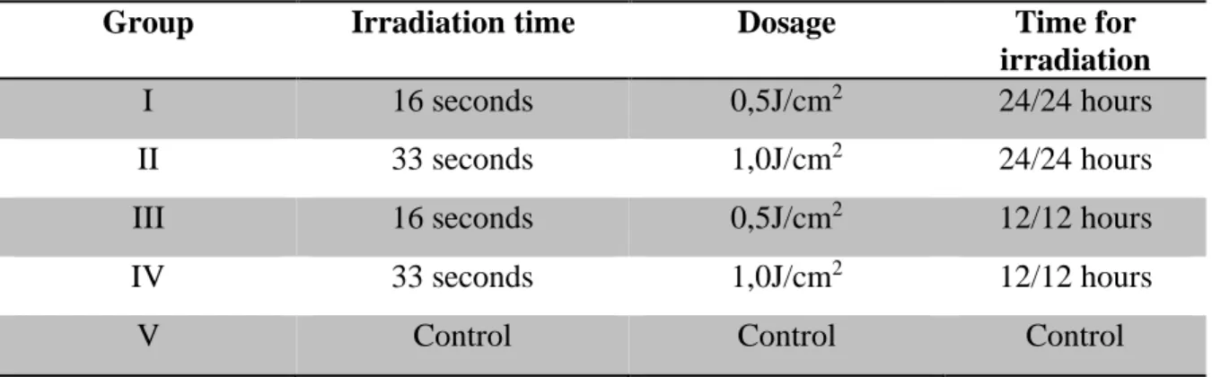
Table 1: Experimental design for the MTT assay.
Group Irradiation time Dosage Time for
irradiation
I 16 seconds 0,5J/cm2 24/24 hours
II 33 seconds 1,0J/cm2 24/24 hours
III 16 seconds 0,5J/cm2 12/12 hours
IV 33 seconds 1,0J/cm2 12/12 hours
Braz. J. of Develop., Curitiba, v. 5, n. 12, p.33248-33260 dec 2019 . ISSN 2525-8761
• Cell growth
Growth curves were obtained through the MTT assay used to establish the proliferation of cells submitted to laser therapy. For analysis of the proliferation pattern of the obtained lineage, 1000 cells per well were seeded (day 0) in 96-well plates. The plates were divided into control and another four test groups, so that the control group was cultivated in the absence of low intensity laser irradiation, while the test groups were influenced by the laser throughout the experiment. For each group, 12-plicates were performed. After 24, 48 and 72 hours, the culture medium was removed and then culture medium was added with 10% of a previously prepared solution (5mg/ml) of the MTT reagent (Thiazole Blue Tetrazolium, Sigma, code M2128). The plates were then incubated in an oven at 37°C with 5% CO2 for 4 hours. After
that time, the MTT medium was removed and 200μL of the isopropanol-0.04M HCl acid solubilizer was added. The plates were then incubated for one hour. The wells were read in the spectrophotometer at 570nm using as white three wells with 200μL of the isopropanol-acid.
• Trypan Blue analyses
The integrity of the cell membrane and the direct count of the living and dead cells were evaluated by the Trypan Blue. This dye does not enter living cells but passes through the membranes of dead cells. Thus, the stem cells of the dental pulp plaque were exposed to the best parameter of the low-level laser and Trypan Blue was performed after 24 hours. The medium was removed from the wells and cells were washed with 200μL of PBS. Cells were separated by addition of 100μl of trypsin /EDTA 0.5%. RPMI-1640 supplemented with 10% FBS (50μL) and 0.5% trypan blue (50 μL) (Merck, Germany) were added additionally to each well and the plates were incubated for 5 minutes. Subsequently, a 20μL aliquot was removed and placed in a Neubauer Hemocytometer. The number of viable and non-viable cells was finally counted under the microscope. The number of viable cells was calculated according to the following formula:
Braz. J. of Develop., Curitiba, v. 5, n. 12, p.33248-33260 dec 2019 . ISSN 2525-8761 Viable cell count × Dilution × 104
_____________________________________
N
Where n is the number of hemocytometer squares that have been counted. The
percentage of viability was calculated as:
Viable Cell Count
___________________________________ x100
Total of counted cells
Statistical Analysis
The results were statistically analyzed by the IBM SPSS Statistics program. The data
measured by absorbance were submitted to analysis of variance ANOVA complemented by
the Tukey test with significance level of 5% (p <0.05).
3 RESULTS
• Cell growth
Our data showed that the absorbance in the 0.5J treatment (24-hour laser application)
is higher than in the 0.5J treatments (12-hour laser application) (Table 1). Since the F value is
significant for the blocks, there is a difference between the means of absorbance between at
Braz. J. of Develop., Curitiba, v. 5, n. 12, p.33248-33260 dec 2019 . ISSN 2525-8761 block is lower than in the 48 hour and 72 hour blocks while the absorbance in the 48 hour
block is longer than in the 72 hour block.
In the MTT block at 48 hours the absorbance in the 0.5J treatment (24-hour laser
application) is higher than in the 1J treatments (24-hour laser application), 0.5J (12-12 hours),
1J (laser application every 12 hours) and 10% SFB; The treatment 1J (laser application every
24 hours) is practically equal to treatments 0.5J (laser application every 12 hours) and 1J (laser
application every 12 hours) and greater than treatment with SFB 10%; The 0.5J treatment
(laser application every 12 hours) is practically equal to 1J (laser application every 12 hours)
and greater than in SFB10% treatment; In the treatment 0.5J (laser application every 12 hours)
is practically equal to that of the 1J treatment (laser application every 12 hours) and higher
than that of the 10% SFB treatment; In 1J treatment (12-hour laser application) is greater than
in SFB10% treatment.
Thus, the best result found after the application of the laser is 0.5J (graphic 1), which is applied every 24 hours. In order to perform the osteogenic differentiation, only this parameter was used.
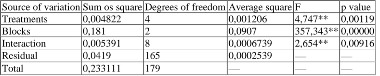
Table 2 Variance analysis
Source of variation Sum os square Degrees of freedom Average square F p value
Treatments 0,004822 4 0,001206 4,747** 0,00119
Blocks 0,181 2 0,0907 357,343** 0,00000
Interaction 0,005391 8 0,0006739 2,654** 0,00916
Residual 0,0419 165 0,0002539 ⎯ ⎯
Total 0,233111 179 ⎯ ⎯ ⎯
* Significant value for significance level of 5% ** Significant value for significance level of 1%
Table 3 Treatmens averages
Treatments 0,5J (24h) 0,5J (24h) 0,5J (24h) 0,5J (24h) 0,5J (24h) Average 0,15925 0,15308 0,14653 0,15292 0,14481
Braz. J. of Develop., Curitiba, v. 5, n. 12, p.33248-33260 dec 2019 . ISSN 2525-8761
Graph 1 Joint distributions of treatments and blocks. We can observe that the best parameter found after the application of the laser was 0.5J (24h).
• Viability test with Trypan Blue
The result of Trypan Blue reflects under the number of viable and non-viable cells after staining. At this stage, the test was performed only with the best MTT result (0.5J/cm2 application for 48 hours) and the control group (absence of the laser). After staining with Trypan Blue and statistical analyzes, we can conclude that although the test group presented a higher number of viable cells and a smaller number of cells with ruptured cell membrane, this value was not statistically significant (P <0.05) compared with the control group. The results were also able to show that the laser was in no way damaging the dental pulp stem cells.
Braz. J. of Develop., Curitiba, v. 5, n. 12, p.33248-33260 dec 2019 . ISSN 2525-8761
Graph 2 Effects of low intensity laser on the viability of DPSCs 4 DISCUSSION
LIL has been used in wound healing for the past 30 years. It is also widely applied in various branches of regenerative medicine (eg tissue regeneration) and dentistry, where it is used to improve healing processes. This therapy shows beneficial effects on a variety of pathological conditions, including pain relief, and inflammation [10,13]. Thus, laser is also an important tool for cell culture, since when employed at low intensity it has a biostimulatory effect on cell proliferation in vitro. This biostimulation, however, is dependent on the parameters of laser irradiation, such as wavelength, laser output energy, and energy density [1,14].
There are many studies that address the potential of low intensity laser on cell activity. However, few studies have been done on stem cells of dental pulp. Some authors affirm that only in the state of nutritional stress the biostimulatory effects of the low-intensity laser are observed [15]. However, in this study, the laser acted positively in both nutritional deficits (5% FBS) and ideal conditions (10% FBS), and irradiated dental pulp stem cells showed a higher proliferation when compared to control. Many studies point to the great effects of the laser under cell proliferation and these say that for this to occur, the medium must have 20% confluence and must always be changed. In addition, irradiation should occur in the dark and energy density should always vary according to the cell line worked [11]. In this study, the factors mentioned above were taken into account and the irradiation occurred in the dark and in a density of energy previously tested by other authors.
Braz. J. of Develop., Curitiba, v. 5, n. 12, p.33248-33260 dec 2019 . ISSN 2525-8761 The choice of some of the laser irradiation parameters in the cell culture in this study was based on previous in vitro studies of other authors, such as [1,8,16,17, 20, 21], all these studies also performed on stem cells. InGaAlP laser with 660nm was used and generated results in all the studies cited above and also in the present study, considerably increasing the proliferation of DPSCs in the test groups when compared to the control groups (non-irradiated).
Previously used parameters of density energy were used in this study, but new parameters of application of these energies were added (application every 12 hours). After the tests, however, we observed that this new parameter generated less proliferation of the DPSCs when compared to the group already tested by other authors [8,21]. The best energy density found in this study was 0.5J/cm2 in an application of the InGaAlP laser once a day for 48
hours, which does not corroborate previous research by others authors [8,16], who observed that the dosage of 1J/cm2 after 48 hours and 1J/cm2 after 72 hours of irradiation respectively obtained better results when compared with the control and 0.5J/cm2 groups. However, our results resemble the work of Hou et al. [18] that considered the energy density 0.5 J/cm2 as optimal.
In relation to the viability analysis by Trypan Blue, no statistically significant difference between the test and control groups was observed. These results also corroborate with the results of Barboza et al. (2014). However with our results we can see that although the laser did not increase viability and cell differentiation by this tests, the laser was not harmful to the cells.
Therefore, the results of the present study are promising. Laser phototherapy can significantly influence the proliferation of stem cells, which leads to better tissue healing. However, in order to verify if this therapy can contribute to the optimal fixation and functional improvement of the cells after the implant, as well as reduce the time of tissue healing, future studies are necessary to evaluate its potential in bone and muscle neoformation after the implantation of stem cells into injured tissues.
In short, this in vitro study suggests that DPSCs respond positively to laser phototherapy at a wavelength of 660nm. Thus, the association of laser phototherapy and DPSC culture can be of importance for tissue engineering and future regenerative medicine. In addition, it opens the possibility of greater use of laser phototherapy to improve the growth of other types of stem cells in dentistry.
Braz. J. of Develop., Curitiba, v. 5, n. 12, p.33248-33260 dec 2019 . ISSN 2525-8761
ACKNOWLEDGMENTS
We are grateful to Federal University of Juiz de Fora for the continuous support.
REFERENCES
Eduardo FP, Bueno DF, Freitas PM, Marques MM et al. Stem Cell Proliferation Under Low Intensity Laser Irradiation: A Preliminary Study. Lasers Surg Med. 2008:40:433–8.
Rodriguez-Losano FJ, Insausti CL, Iniesta F, Blanquer M et al. Mesenchymal dental stem cells in regenerative dentistry. Med Oral Patol Oral Cir Bucal. 2012:1:1062-67.
Silva FS, Almeida PN, Rettore JVP, Maranduba CP et al. Toward Personalized Cell Therapies by Using Stem Cells: Seven Relevant Topics for Safety and Success in Stem Cell Therapy. J Biomed Biotechnol. 2012:12:1-12.
Ulloa-Montoya F, Verfaillie CM, Hu WS. Culture Systems for Pluripotent Stem Cells. JBB. 2005:100:12-27.
Casagrande L, Cordeiro MM, Silva AN, Jacques EN. Dental pulp stem cells in regenerative dentistry. Society of The Nippon Dental University. 2001:99:1-7.
Demarco FF, Conde, MCM, Cavalcanti BN, Casagrande L et al. Dental Pulp Tissue Engineering. Braz Dent J. 2011:22:3-14.
Miyagi SPH, Moreira MSNA, Soares LF, Martins MM. Laserterapia e células-tronco na Odontologia. Rev Implantenews. 2012:9:53-57.
Barboza CAG, Ginani F, Soares DM, Henriques ACG et al. Laser de baixa intensidade induz à proliferação in vitro de células-tronco mesenquimais. Rev Einstein. 2014:12:75-81.
Braz. J. of Develop., Curitiba, v. 5, n. 12, p.33248-33260 dec 2019 . ISSN 2525-8761 Henriques ACG, Cazal C, Castro JFL. Ação da laserterapia no processo de proliferação e diferenciação celular. Revisão da literatura. Rev. Col. Bras. Cir. 2010:37:295-302.
Ebrahimi T, Rokn AR, Heidaris M, Nokhbatolfoghahaie R et al. The Influence of Low-Intensity Laser Therapy on Bone Healing. J Dent (Tehran). 2012:9:238-48.
Emelyanov AN, Kiryanova VV. Photomodulation of Proliferation and Differentiation of Stem Cells By the Visible and Infrared Light. Photomed Laser Surg. 2015:33:164-74.
Lin F, Josephs SF, Alexandrescu D, Ramos F, Bogin V et al. Lasers, stem cells, and COPD. J Transl Med. 2010:8:1-10.
Alghandi KM, Kumar A, Moussa NM. Low-level laser therapy: a useful technique for enhancing the proliferation of various cultured cells. Lasers Med Sci. 2012:1-13.
Azevedo LH, Eduardo FP, Moreira MS, Eduardo CP et al. Influence of different power densities of LILT on cultured human fibroblast growth - A pilot study. Lasers Med Sci. 2006: 21:86-9.
Almeida-Lopes L, Rigau J, Zangaro RA Guidugli-Neto J et al. Comparison of the low level laser therapy effects on culturehuman gingival fibroblasts proliferation using different irradiance and same fluence. Lasers Surg Med. 2001:29:179-84.
Zaccara IM, Ginani F, Mota-Filho HG, Henriques ACG et al. Effect of low-level laser irradiation on proliferation and viability of hum an dental pulp stem cells. Lasers Med Sci. v. 30, n. 9, p. 2259-2264, 2015.
Pereira LO, Longo JPF, Azevedo RB. Laser irradiation did not increase the proliferation or the differentiation of stem cells from normal and inflamed dental pulp. Arch Oral Biol. 2012:57:1079-85.
Braz. J. of Develop., Curitiba, v. 5, n. 12, p.33248-33260 dec 2019 . ISSN 2525-8761 Hou JF, Zhang H, Yuan X, Li J et al. In Vitro Effects of Low-Level Laser Irradiation for Bone Marrow Mesenchymal Stem Cells: Proliferation, Growth Factors Secretion and Myogenic Differentiation. Lasers Surg Med. 2008:40:726–33.
Leonida A, Paiusco A, Rossi G, Carini F et al. Effects of low-level laser irradiation on proliferation and osteoblastic differentiation of human mesenchymal stem cells seeded on a three-dimensional biomatrix: in vitro pilot study. Lasers Med Sci. 2012:1-8.
Moura-netto, C, Ferreira LS, Maranduba CM, Volpi AC et al. Low-intensity laser phototherapy enhances the proliferation of dental pulp stem cells under nutritional deficiency. Braz Oral Res. 2016:1-6.
Ginani F, Soares DM, Rocha AO e Barboza CAG. Laser de baixa intensidade promove
proliferação de células-tronco derivadas de tecido adiposo criopreservadas. Rev Einstein. 2017:334-338.