UNIVERSIDADE DA BEIRA INTERIOR
Ciências da Saúde
Neuroinflammation and neurogenesis in the
choroid plexus
Nádia Patrícia Amaral Morete
Dissertação para obtenção do grau de mestre em
Ciências Biomédicas
(2
ºCiclo de estudos)
Orientador: Professora Doutora Cecília Santos
Coorientador: Mestre Joana Tomás
“The price of success is hard work, dedication to the job at hand, and the determination that whether we win or lose, we have applied the best of ourselves to the task at hand.”
Acknowledgments
It is extremely difficult to express all my gratitude, my feelings, my respect and also my deep admiration to all of you that during these five years in Covilhã, directly or indirectly, help me along my way. I just want to say thanks for everything!
To Cecília Santos, I wish to express my sincere thanks for your scientific support, for your supervision, for your constructive criticism. I wish to express my sincere and honest thanks for the opportunity to work in your group research.
To Joana Tomás I want to express my thankfulness. More than a supervisor she was my “research mother” with whom I had learn not only, I hope, to be a good scientist, but also to be a simple human being always ready to chase my dreams without giving up.
To Graça Baltazar and Liliana Bernardino, I would like to thank for all the scientific and logistic support provided.
Also, I thanks to the entire lab group, “Neurons & Neurons”, for their friendship, thank you for helpful advice and a great work environment.
I thanks to my friends, especially Ana Sofia, Raquel, Telmo, and Lena for their brotherhood, I spent some of the best moments of my life with you!! Thanks for your friendship, for always being there.
And the most importantly, I would also like to express my sincere thanks to my parents. Thanks for the unconditional support and for always believing in me when I doubted myself. Thank You, thank You, thank you!!! I love you more than myself.
Abstract
The choroid plexus (CP), located within the cerebral ventricles, are composed of a highly vascularized stroma surrounded by a tight layer of epithelial cells that restrict cellular and molecular traffic between the blood and the cerebrospinal fluid (CSF). Stroma is surrounded by dendritic cells, collagen fibbers, fibroblasts and B and T cells. Some of these cells are innate immune cells whose main function is antigen presentation which suggest that CP may play an active role in immunological brain protection. CP can be seen as a first line of defense in brain against harmful stimulus, whether they are molecules, cells or pathogens, due to the presence of tight junctions (TJ). TJ control the paracellular permeability across lateral intracellular spaces preventing solutes and water from passing freely through the paracellular pathway and form a boundary between the apical and basolateral plasma membrane domains by preventing diffusion of proteins between the membrane compartments. On the other hand, recent studies suggest that the CP cells can differentiate into neurons and astrocytes. So we proposed to evaluate the effect of an acute neuroinflammatory insult, lipopolysaccharide (LPS), in CP neurogenesis. The reactive oxygen species (ROS) and nitric oxide (NO) production alterations in protein content of extracellular medium was analyzed in vitro with the establishment of a primary culture of choroid plexus epithelial cells (CPEC). The inflammatory response of CP to LPS was analyzed in vivo in C57BL/6 mice injected with LPS and sacrificed 7h after and ex vivo in CP explants from newborn rats incubated with LPS. For that we studied neuroinflammatory markers such as: CD11b, a microglia marker; NeuN, a mature neurons marker and GFAP, an astrocytes marker, by the whole mount technique. We also studied the effect of LPS in CP integrity as a barrier by studying the differential expression of TJ proteins. The effect of LPS in Occludin, E-cadherin, ZO-1 and claudin-1 expression was quantified in vitro by immunocytochemistry (ICC), occludin and E-cadherin expression was also analyzed ex vivo, by whole mount and in vivo by Western blot. LPS triggered an increase in ROS production, NO and in total protein content of extracellular medium in CPEC. The neuroinflammatory response of CP to LPS resulted in an over expression of CD11b and GFAP, a decrease in NeuN expression but no changes in TTR expression were observed. Our findings suggest an increase in iNOS production. Lastly, we observed that CP kept its barrier proteins despite minor alterations in occludin expression. In conclusion, the work developed shows that CP responded to acute neuroinflammation by increasing the expression of proteins involved in immunological brain protection. Morphologically CP keeps its integrity as a barrier but the alteration on proteins that encode TJ indicates that a disturbance on this CP function may be compromised.
Keywords
Resumo alargado
Sendo parte integrante do sistema nervoso central, os plexos coroides (CP) são estruturas ramificadas, altamente vascularizadas, com uma camada de células epiteliais secretoras que projetam numerosas vilosidades nos quatro ventrículos do cérebro. Os CP são constituídos por uma monocamada externa de células epiteliais coroidais (CPEC), cuja membrana apical está em contacto direto com o líquido cefalorraquidiano, enquanto que a membrana basal das CPEC se localiza sobre o estroma. O estroma é rodeado por células dendríticas, fibras de colagénio, fibroblastos e células B e T. Neste conjunto, encontram-se células imunitárias inatas cuja função é apresentar antigénios, moléculas que são capazes de iniciar resposta imune, o que sugere que o CP pode desempenhar um papel ativo na proteção imune do cérebro. Durante muito tempo, pensava-se que o cérebro era uma estrutura “imunologicamente privilegiada”, isto é, que o cérebro se encontrasse separado fisicamente do sistema imunitário periférico através da barreira hematoencefálica, não havendo trocas entre o cérebro e o restante organismo. A barreira hematoencefálica em conjunto com a barreira formada pelas CPEC protege o sistema nervoso central contra agentes patogénicos através das junções intercelulares que são compostas por proteínas de integridade de membrana. Estas proteínas permitem ao CP ter capacidade imunitária, uma vez que controlam a permeabilidade celular, impedindo os solutos de água de passar livremente, formando uma fronteira entre os domínios de membrana plasmática apicais e basolaterais, impedindo a passagem de proteínas entre os compartimentos da membrana. Não obstante, a permeabilidade da barreira pode ser influenciada por uma neuroinflamação permanente. A neuroinflamação é caracterizada pela ativação da microglia, stress oxidativo e expressão dos principais mediadores inflamatórios sendo um processo ativo de defesa contra vários insultos, lesões traumáticas e metabólicas, infeções e doenças neurodegenerativas tais como a doença de Alzheimer e de Parkinson. Outro “dogma” das neurociências durante muitos anos foi a não existência de neurogénese, isto é, as células do sistema nervoso não se regeneravam. Contudo, estudos recentes afirmam que o CP, quando sujeito a uma agressão, tem capacidade de formar novas células e estas por sua vez são capazes de se diferenciarem. Assim, neste trabalho propusemo-nos a avaliar o efeito de um insulto neuroinflamatório agudo provocado pelo lipopolissacarídeo (LPS), na neurogénese e na neuroinflamação do CP. Mais especificamente, a resposta inflamatória induzida pelo LPS no CP foi avaliada in vivo, ex vivo e in vitro através da análise de marcadores inflamatórios tais como CD11b, iNOS, expressão de TTR e produção de espécies reativas de oxigénio (ROS) e de óxido nítrico (NO). Avaliámos também o efeito do LPS na neurogénese in vivo e ex vivo através do estudo da presença de diferentes marcadores: NeuN, CD11b e o GFAP. Por fim, verificámos se o LPS teria algum efeito na integridade da membrana. A resposta inflamatória foi analisada: in vivo em CP de ratinhos C57BL/6 injetados com 1,0 mg/kg de LPS e sacrificados 7h depois; explantes de CP
de ratos recém-nascidos e de animais com 21 dias que foram posteriormente incubados durante 48h com 0,1 µg/mL de LPS. A produção de ROS, NO e alterações no teor de proteínas do meio extracelular foi avaliada em CPEC e verificou-se um aumento na produção de ROS e NO e um incremento no teor de proteínas totais do meio extracelular, em CPEC. Foi possível verificar, em explantes de CP, um aumento na expressão de CD11b, um marcador de microglia, e GFAP que é utilizado como um marcador de astrócitos, uma diminuição na expressão de NeuN, um marcador de neurónios maduros e não se verificou alterações na expressão de TTR, a principal proteína decretada pelo CP. Foi realizado Western blot para verificar se o CP libertaria TTR para o meio de cultura e se a quantidade desta libertada no meio de cultura das CPEC variava ao longo do tempo, apesar de ter ocorrido libertação de TTR par o meio a sua quantidade manteve-se constante ao longo do tempo. Realizou-se Western blot para verificar se havia alterações na secreção de iNOS por parte do CP, tendo sio possível verificar um aumento significativo da enzima nos plexos incubados com LPS comparados com os controlos. O efeito do LPS na integridade do CP como barreira foi analisado através do estudo da expressão das proteínas ocludina, claudina-1, e-caderina e ZO-1 in vitro em CPEC, a expressão de ocludina e e-caderina foi também estudada ex vivo e in vivo. As imagens de imunocitoquimíca referentes às proteínas de integridade de membrana: ocludina, claudina-1, e-caderina e ZO-1, foram adquiridas num microscópio confocal (Zeiss) e a fluorescência emitida foi quantificada. Os resultados revelaram que as CPEC mantiveram a expressão das suas proteínas de integridade de membrana, depois de um insulto com LPS, contudo a expressão da ocludina e da ZO-1 apresentaram uma redução significativa. Por sua vez os resultados ex vivo não mostraram nenhuma alteração na expressão de ocludina e e-caderina. Os ensaios in vivo confirmaram a expressão de ocludina e e-caderina em CP. Assim, podemos concluir que o CP tem uma resposta ativa à neuroinflamação induzida pelo LPS, o que se comprova pelo aumento das proteínas relacionadas com a resposta imunitária e também com um aumento do stress oxidativo a que as células são sujeitas. Verificou-se também que um insulto neuroinflamatório compromete a diferenciação das células do plexo, havendo decrescimento da neurogénese em todas as experiencias realizadas. Finalmente, outra das conclusões que foi possível conjeturar com este estudo foi, que morfologicamente o CP mantém a integridade da barreira, embora não nos seja possível inferir se esta fica ou não comprometida.
Palavras-chave
Table of contents
Chapter 1:Introduction ... - 1 -
1. Introduction ... - 2 -
1.1 Nervous System ... - 2 -
1.2 Choroid Plexus... - 2 -
1.2.1 Choroid plexus functions ... - 4 -
1.2.2 Brain barriers: Endothelial Versus Epithelial Cells of CP ... - 4 -
1.3 Neuroinflammation ... - 6 -
1.3.1 Indicators of neuroinflammation ... - 8 -
1.4 The choroid plexus in neuroinflammation ... - 9 -
1.5 Neurodegenerative diseases ... - 9 -
1.5.1 Alzheimer Disease ... - 9 -
1.5.2 Parkinson Disease ... - 10 -
1.6 Neurogenesis ... - 11 -
1.7 Relationship between neurogenesis and neuroinflammation ... - 11 -
Chapter 2: Aim ... Erro! Marcador não definido.-133 Chapter 3: Materials and Methods ... - 15 -
3.1 Animals ... - 16 -
3.1.1 Primary Cultures of rat Choroid Plexus epithelial cells ... - 16 -
3.2 Effect of LPS in CP ... - 17 -
3. 2.1 Effects of LPS in Choroid Plexus Epithelial Cells. ... - 17 -
3. 2. 2 Effects of LPS in neurogenesis and inflammatory markers in Choroid Plexus explants. ... - 18 -
3.2.3 Inflammatory response of CP to a LPS insult, in vivo ... - 18 -
3.3 Assessment of ROS production, inflammatory response and neurogenesis markers . - 19 - 3.3.1.1 ROS production in CPEC ... - 19 -
3.3.1.2 ROS production in CP explants ... - 19 -
3.3.3 Protein extraction from CP ... - 20 -
3.3.4 SDS-PAGE and Immunoblot ... - 20 -
3.3.5 Immunocytochemistry ... - 21 -
3.3.6 Whole Mount ... - 22 -
3.4 Statistical analysis ... - 23 -
Chapter 4: Results ... - 25 -
4.1 Effects of LPS in Choroid Plexus Epithelial Cells. ... - 26 -
4.1.1 ROS production ... - 26 -
4.1.2 NO production ... - 26 -
4.1.3 Extracellular Protein ... - 27 -
………...
4.1.4 The effect of LPS in CPEC integrity... - 28 -
4.2. Effect of LPS in CP explants ... - 33 -
4.2.1 ROS production ... - 33 -
4.2.2 Whole Mount ... - 34 -
4.2.3 Western Blot ... - 39 -
4.2.4 The effect of LPS in CPEC integrity... - 41 -
4.3 Study of the response of CP in vivo to a LPS insult ... - 42 -
4.3.1 TTR ... - 43 -
4.3.2 iNOS ... - 44 -
4.3.3 E-cadherin and occludin ... - 44 -
4.4 Study of neurogenesis in CP in vivo in response to a LPS insult ... - 45 -
4.4.1 TTR ... - 45 -
4.4.2 CD11b ... - 47 -
4.4.3 NeuN ... - 48 -
4.4.4 GFAP ... - 50 -
Chapter 5. Discussion and Conclusion ... - 53 -
Chapter 6. Future perspectives ... - 59 -
Chapter 7. References ... -Erro! Marcador não definido. Chapter 8. Appendix ... - 68 -
………-61
List of Figures
Figure 1: Schematic illustration of choroid plexus as it resides within the lateral ventricle and
as defined by its morphological appearance ... - 3 -
Figure 2: Schematic representation of tight junctions and adherens junctions complexes in choroidal epithelial cells ... - 5 -
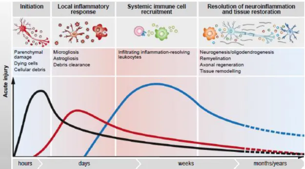
Figure 3: Local immune cell response to acute and chronic Central Nervous System. ... - 8 -
Figure 4: Schematic representation of the procedure to study the production of ROS, NO and protein extracellular in the culture medium, in vitro. ... - 17 -
Figure 5: Schematic representation of the procedure performed to study the effects of the LPS insult in tight junctions by immunocytochemistry ... - 17 -
Figure 6: Schematic representation of the procedure performed to study neurogenesis in CP explants upon an inflammatory insult with 0.1 µg/mL of LPS ex vivo. ... - 18 -
Figure 7: Schematic representation of the experimental layout to analyze the response of CP to 1.0 mg/kg of LPS inject in vivo. ... - 18 -
Figure 8: Schematic representation of the procedure performed to assess neurogenesis upon LPS insult in vivo.. ... - 19 -
Figure 9: Time-course ROS production response to 0.1 µg/mL of LPS, in CPEC. ... - 26 -
Figure 10: Time-course of oxide nitric production in response to 0.1 µg/mL of LPS in CPEC medium. ... - 27 -
Figure 11: Quantification of extracellular proteins released from CPEC to the culture medium after incubation with 0.1 µg/mL of LPS for 24 hours. ... - 27 -
Figure 12: WB of TTR in extracellular medium during a time line of incubation with LPS 0.1 µg/mL. ... - 28 -
Figure 13: Quantification of TTR in CPEC medium after incubation with 0.1 µg/mL of LPS.. .. - 28 - Figure 14: Effect of LPS incorporation in occludin integrity. ... - 29 -
Figure 15: Effect of LPS in CPEC occludin expression ... - 29 -
Figure 16: Effect of LPS incorporation in claudin-1 integrity ... - 30 -
Figure 17: Effect of LPS in CPEC claudin-1 expression.. ... - 30 -
Figure 18: Effect of LPS incorporation in ZO-1 integrity.. ... - 31 -
Figure 19: Effect of LPS in CPEC ZO-1 expression. ... - 31 -
Figure 20: Effect of LPS incorporation in E-cadherin integrity. ... - 32 -
Figure 21: Effect of LPS in CPEC E-cadherin expression. ... - 32 -
Figure 22: ROS production after 2 hours of 0.1 µg/mL of LPS incorporation ... - 33 -
Figure 23: ROS production after 2 hours of 0.1 µg/mL of LPS incorporation in newborn and 21 days rats ... - 34 -
Figure 24: Representative confocal images of LPS effects on CP TTR expression ... - 35 -
Figure 26: Representative confocal images of LPS effects on CP CD11b expression. ... - 36 -
Figure 27: Effect of LPS on CP CD11b expression.. ... - 36 -
Figure 28: Representative confocal images of LPS effects on CP NeuN expression.. ... - 37 -
Figure 29: Effect of LPS on CP NeuN expression.. ... - 37 -
Figure 30: Representative confocal images of LPS effects on CP GFAP expression ... - 38 -
Figure 31: Effect of LPS in CP ex vivo GFAP expression ... - 38 -
Figure 32: Representative WB image of TTR.. ... - 39 -
Figure 33: Quantification of expression of TTR in explants of 21 days rats incubated with 0.1 µg/mL of LPS. ... - 39 -
Figure 34: Representative WB image of iNOS ... - 40 -
Figure 35: Quantification of expression of iNOS in explants of rats with 21 days incubated with 0.1 µg/mL of LPS.. ... - 40 -
Figure 36: Representative confocal images of LPS effects on CP occludin expression ... - 41 -
Figure 37: Effect of LPS in CP occludin expression.. ... - 41 -
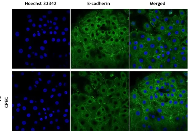
Figure 38: Representative confocal images of LPS effects on CP E-cadherin expression. .. - 42 -
Figure 39: Effect of LPS in CP E-cadherin expression. ... - 42 -
Figure 40: Representative WB image of TTR. ... - 43 -
Figure 41: Quantification of TTR expression in explants collected from 1, 3 and 9 months mice injected with 1.0 mg/kg of LPS. ... - 43 -
Figure 42: Representative WB image of iNOS.. ... - 44 -
Figure 43: Representative WB image of E-cadherin. ... - 44 -
Figure 44: Representative WB image of occludin ... - 45 -
Figure 45: Representative confocal images of LPS effects in vivo in CP TTR expression ... - 46 -
Figure 46: Effect of LPS in CP TTR expression.. ... - 46 -
Figure 47: Representative confocal images of LPS effects on CP CD11b expression ... - 47 -
Figure 48: Effect of LPS in CP in vivo CD11b expression. ... - 48 -
Figure 49: Representative confocal images of LPS effects on CP NeuN expression. ... - 49 -
Figure 50: Effect of LPS in CP in vivo NeuN expression. ... - 49 -
Figure 51: Representative confocal images of LPS effects on CP GFAP expression. ... - 50 -
Figure 52: Effect of LPS in CP in vivo GFAP expression ... - 51 -
List of Tables
Table 1: Primary antibodies used in Immunofluorescence and Western analysis and their respective isotype, dilution, molecular weight, company and technique were where used.- 22 -
Table 2: Secondary antibodies used in Immunofluorescence and western analysis and their respective isotype, dilution, company and technique were where used. ... - 23 -
Abbreviations
A Beta-amyloid
AD Alzheimer Disease
AJ Adherens Junctions
Ara-C Arabinoside
BBB Blood Brain Barrier
BCSFB Blood-Cerebrospinal Fluid Barrier
BSA Bovine Serum Albumin
CAPS 3-(Cyclohexylamino)-2-hydroxy-1-propanesulfonic acid
CNS Central Nervous System
CP Choroid Plexus
CPEC Choroid Plexus Epithelial Cells
CSF Cerebrospinal Fluid
DCFH-DA Dichloro-dihudro-fluorescein-diacetate
DHE Dihydroethidium
ECF Enhanced Chemifluorescence
EGF Epidermal Growth Factor
DMEM Dulbecco’s Modified Eagle Medium
EDTA Ethylenediaminetetraacetic acid
EGTA Ethylene glycol tetraacetic acid
FBS Fetal Bovine Serum
GFAP Glial Fibrillary Acidic Protein
ICC Immunocytochemistry
IL Interleukin
iNOS inducible NOS
LPS Lipopolysaccharide
ND Neurodegenerative Disease
NeuN Neuronal Nuclear Protein
NO Nitric Oxide
NOS Oxide Nitric Synthase
PBS Phosphate Buffered Saline
PBS-T Phosphate Buffered Saline with Tween
PD Parkinson Disease
PDVF Polyvinylidene difluoride
PFA Paraformaldehyde
PNS Peripherical Nervous System
ROS Reactive Oxygen Species
RNS Reactive Nitrogen Species
SDS Sodium Dodecyl Sulfate
SEM Standard Error of the Mean
TBS Tris Buffered Saline
TBS-T Tris Buffered Saline with Tween
TNF Tumor Necrosis Factor
TJ Tight Junctions
TLR Toll Like receptor
TTR Transthyretin
WB Western Blot
WM Whole Mount
1. Introduction
1.1 Nervous System
The human nervous system is highly integrated and can be divided in two parts: the central nervous system (CNS) and the peripheral nervous system (PNS). The CNS includes the brain and spinal cord and it is the integrating and commanding centre of the nervous system. It interprets sensory input and dictates motor responses based in memory (past experience), reflexes and current conditions. The PNS, the part of the nervous system outside the CNS, consists mainly of nerves that extend from the brain and spinal cord. It serves as communication thread that links all parts of the body to the CNS(1). CNS consists of neurons and glial cells. Among glial cells, astrocytes constitute nearly 40% of the total CNS cell population in the adult human brain while microglia cells are derived from myeloid cells and comprise approximately 12% of cells in the brain (2).
Astrocytes guide the development and migration of neurons during brain development, production of growth factors, maintenance of the integrity of the blood-brain barrier (BBB), and participate in the immune and repair responses to diseases and brain injury (3, 4). Microglial cells represent resident brain macrophages and can be transformed into activated immunocompetent antigen-presenting cells during the pathological process.
The cell-cell interactions between glial cells and neurons may be important in the regulation of brain inflammation and neurodegeneration (2).
1.2 Choroid Plexus
The choroid plexus (CP) arises from the ependymal lining of the brain ventricles and protrude into the ventricles of the brain (5).
In the brain there are four CP, with tissues in each lateral, third and fourth ventricles (6). The lateral plexus are thin leaf-like undulated structures arising from the inner ventricular surface, the third plexus protrudes from the roof of the third ventricle, and the fourth plexus is formed caudally and ventrally to the cerebellum: the CP in the third and forth ventricles are branched villi that protrude into the ventricle (6).
CP tissues comprise a continuous monolayer of cuboidal (7) and columnar epithelial cells localized over connective tissues, underneath the basement membrane (8).
These ciliated epithelial cells are characterized by numerous mitochondria spread throughout the cytoplasm, a central nucleus, and an abundant Golgi apparatus located laterally and toward the ventricular lumen (8). Depending on the physiological or pathological conditions, the epithelial cells lie on an epithelial basement membrane surrounding a thin stroma with numerous collagen fibbers, dendritic cells, macrophages, fibroblasts, B and T cells, and large capillaries with a fenestrated endothelium (9, 10). Some of these cells are innate immune cells whose main function is antigen presentation which suggests that the CP may play an active role in immunological brain protection (10, 11). CP are richly innervated, receiving adrenergic, cholinergic, peptidergic and serotoninergic fibers. The distribution of nervous fibers varies widely according to species (11). CP cells are connected by junctional complexes at the luminal membrane, consisting of tight junctions (TJ), adherens junctions (AJ) and desmosomes (Figure 1).
Beneath the epithelial basement membrane is a network of fenestrated capillaries that are surrounded by connective tissue composed by fibroblasts and immune cells. The fenestration of capillaries are sealed with thin diaphragms, which permit ions, water and small molecules, as nutrients or vitamins, to pass without difficulty into the interstitial fluid of each plexus(8).
Figure 1: Schematic illustration of choroid plexus as it resides within the lateral ventricle (left) and as
defined by its morphological appearance (right). CSF – Cerebrospinal fluid. (Adapted from Emerich et. al., 2004 (12)).
1.2.1 Choroid plexus functions
Due to its many functions, relying primarily in the unique structure of the epithelial cells, CP is considered a multipurpose organ (7). The best known function is cerebrospinal fluid (CSF) formation, which not only regulates homeostasis in the CNS, providing buoyancy for the brain and spinal cord, but also participates in neural stem cell renewal, neuroprotection, sleep/awake cycles and in several neurological disorders, as Alzheimer Disease (AD) and Parkinson Disease (PD) (13). In humans, CSF volume is 0.37 mL/min, almost 500 mL/day (10). CSF is produced mainly by active secretion, with water entering the CSF from the blood along an osmotic gradient or by specific water channels such as aquaporins. The epithelial CP cells secrete CSF by moving sodium, chloride and bicarbonate (the bicarbonate can also be synthesized in the epithelial cells using the carbonic anhydrase enzyme) from the blood to the ventricles to create the osmotic gradient that drives the secretion of H2O. The CSF is a clear
fluid, with few cells and little protein, and has a lower pH and concentration of glucose, potassium, calcium, bicarbonate and amino acids compared with blood plasma. On the other hand the sodium, chloride and magnesium content is greater in CSF than in plasma (10). CP participates not only in the synthesis, secretion and regulation of several biologically active compounds of the CSF but also in the maintenance of brain metal bioavailability and in the surveillance of toxic compounds in the CSF, thereby protecting the brain against neurotoxic insults (14).
CP is ideally located to distribute molecules both locally and globally to the brain due to its secretor capacity into the CSF. The CP possesses several specific transport systems, containing an array of receptors and also serves as a source of biologically active products. The CP epithelium is not only a target but also a source of neuropeptides, growth factors and cytokines for the CSF (15).
1.2.2 Brain barriers: Endothelial Versus Epithelial Cells of CP
The BBB and the blood-CSF barrier (BCSFB) protect the CNS from the changeable milieu of the blood stream to establish CNS homeostasis, which is a requirement for proper neuronal function. Whereas the BBB is located at the level of highly specialized endothelial cells within CNS microvessels, the BCSFB is formed by the epithelial cells of the CP (16).
The intercellular junctions of the BBB endothelial cells and of the CP epithelial cells are composed by TJ and AJ. The TJ are constituted by: claudins, occudins and juctional adhesions molecules and the AJ are constituted by cadherins (Figure 2).
1.3.1 Tight Junctions and Adherens junctions
Tight junctions form channels that allow permeation between cells, resulting in epithelial surfaces of different tightness (17). A TJ is an intercellular adhesion complex ensuring close contact between adjacent cells and are found in the apical region of the cell (18). TJ control the paracellular permeability across lateral intercellular spaces preventing solutes and water from passing freely through the paracellular pathway, and form a boundary between the apical and basolateral plasma membrane domains by preventing diffusion of proteins between the membrane compartments (18, 19). TJ are important components of numerous signaling pathways controlling gene expression, cell differentiation and proliferation.
Figure 2: Schematic representation of tight junctions and adherens junctions complexes in choroidal epithelial cells (adapted from Szmydynge-Chodobska et.al., 2007 (20)).
Occludin was the first TJ integral membrane protein identified. This ~64 kDa protein specifically located at TJs of epithelial and endothelial cells is incorporated into the network of TJ strands. Occludin consists of four transmembrane domains, two extracellular domains, and three cytoplasmatic domains (21), forming a homophillic dimmer (21). Occludin interacts with the actin cytoskeleton and several intracellular proteins. Its over expression increases electrical resistance, implying a pro-barrier phenotype and increases permeability to small molecule tracers (22). Occludin, as well as claudin-1, claudin-2, and claudin-11, has been reported to be expressed in choroidal epithelium (13).
Similar to occludin, claudins have four transmembrane domains, two extracellular loops and a short carboxyl intracellular tail (22). The claudin’s family consists of at least 24 members with molecular weight from 20 to 27 kDa. Claudins’ extracellular loops form the back none of TJ by forming dimmers and binding homotypically to other claudin molecules in adjacent endothelial cells.
Zonula Occludens (ZO) proteins are classified as members of the membrane associated guanylate kinase family. These proteins are thought to have both structural and signaling roles, and are characteristically defined by three protein-protein interaction modules. The way by which ZO proteins interact with the membrane proteins appears to be specific to each type of membrane protein (22).
E-cadherins comprise a member family of calcium - dependent adhesion molecules. The intracellular domain of E-cadherin binds to β-catenin, and to α-catenin, which in turn interacts with actin, and this catenin-mediated anchorage of E-cadherin to the actin cytoskeleton is required for strong cell-cell adhesion. E-cadherin, as a mature protein, have molecular weight between 80 and 120 kDa, but it is approximately 135 kDa if we considered the molecular weight of the precursor. E-cadherin is a protein that functions to mediate cell-cell binding critical to the maintenance of tissue structure and morphogenesis.
Occludin and the members of the family of claudin proteins are the major constituents of TJ, they are connected to the actin filaments through cytoplasmic adaptor proteins, ZO-1, members of the membrane-associated guanylate kinase family of proteins (23). Interestingly, ZO-1 not only binds to occludin and claudins, but is also able to interact with α-catenin (24). However, in epithelial cells bearing well-developed TJs, ZO-1 does not co-localize with AJ (25).
The permeability of BBB could be altered by the chronic inflammatory pain (26). The inflammatory process disrupts the integrity of BBB and is associated with decreasing of the expression of occludin, promoted by the disruption of occludin oligomeric assemblies (21). Some recent studies support that the expression of claudin-1 and E-cadherin in animals with or without inflammation showed similar results, meaning that the expression is not influenced by neuroinflammation (19, 26). Comparing the immunoreactivities for ZO-1 in the CP between healthy mice and infected mice, it was observed differences in immunoreactivity for the TJ proteins investigated. Importantly, even under inflammatory conditions all TJ associated proteins remained expressed properly (24). In conclusion, brain barriers are able to mount, at least, an initial response to peripheral inflammation, either in reaction to infiltration of inflammatory mediators to the CNS, or due to the effects of infiltration of activated peripheral immune cells, and this vascular inflammatory response may in itself contribute significantly to neuroinflammatory disease (19).
1.3 Neuroinflammation
Being physically separated from the peripheral immune system, the CNS is conventionally recognized as being “immunologically privileged”. Immune privilege in the CNS is partially dependent on the BBB, which is intended to limit the entry of solutes and ions into the CNS.
The precise mechanisms through which peripheral inflammation stimuli trigger brain are poorly understood (27).
Neuroinflammation is characterized by the activation of microglia and expression of major inflammatory mediators without typical features of peripheral inflammation such as edema and neutrophil infiltration. Neuroinflammation is an active defensive mechanism against several insults, metabolic and traumatic injuries, infections and neurodegenerative disease (ND) (22). Moreover, it causes cognitive impairment, even if is acutely stimulated by immunostimulatory component such as lipopolysaccharide (LPS).
Bacterial molecules such as LPS, a component of the external cell membrane of gram negative bacteria (28), have the capacity to activate different groups of cells in CNS: microglia, astrocytes and mast cells (29).
Activation of microglia is a characteristic found on the most CNS autoimmune disorders, which represent the main effector cells of the immune system in the CNS, and its chronic state contributes to progression of ND. Most of the relevant mechanistic studies that are related to neuroinflammation have been carried out in the context of AD and PD. However, a role for neuroinflammation in the pathogenesis of other ND is also emerging. Studies suggest a role for innate immune activation in the pathogenesis of frontotemporal dementia, amyotrophic lateral sclerosis and Huntington’s disease (30). The role of microglia in demyelingting disease is controversial and both protective and degenerative functions have been proposed (29, 31). Under acute injurious conditions microglial activation is among the first immune-related events at the lesion site (Figure 3), yet it seems that microglia either cannot acquire a resolving-phenotype, or fail to be skewed to this phenotype in a time-dependent manner.
Under physiological conditions, microglia stay as a quiescent population, but upon exposure to LPS, microglia are activated and produce pro-inflammatory mediators such as cytokines, chemokines, prostanoids, reactive oxygen species (ROS) and nitric oxide (NO). Microglia are the primary cellular source of pro-inflammatory cytokines, including tumor necrosis factor-α (TNF-α) and interleukin (IL)-1β, and is characterized by an increased proliferation and morphological changes (32).
Mast cells can be found within the brain and their functions include the attractant and activation of other immune cells by secreting pro-inflammatory cytokines, and chemoattractants (33). Finally, astrocytes also contribute to the immune response by liberating both pro- and anti-inflammatory cytokines, chemokines and complement components (4, 34).
Figure 3: Local immune cell response to acute and chronic Central Nervous System. (Adapted from
Schwartz et. al., 2014 (35))
1.3.1
I
ndicators of neuroinflammation
One indicator of neuroinflammation is oxidative stress which occurs when the homeostatic processes fail and free radical generation is much beyond the capacity of the body’s defenses, thus promoting cellular injury and tissue damage (36) .
ROS formation occurs continuously in the cells as a consequence of both enzymatic and non-enzymatic reactions (36). They participate in several physiological mechanisms such as cellular signaling, gene expression and pathogen elimination (37). However, at high levels, ROS can cause irreversible and detrimental cellular damage.
Closely related to ROS are the reactive nitrogen species (RNS). NO is produced by constitutive Nitric Oxide Synthase (NOS) during vasodilating processes or during transmission of nerve impulses. In the presence of insults, NO is produced by catalytic action of inducible NOS (iNOS) and is present at higher concentrations (38, 39). NO can cause damage to proteins, lipids and DNA either directly or after reaction with superoxide, leading to the formation of very reactive peroxynitrite anion. In a healthy brain, the level of iNOS is undetectable (2). In CNS, upregulation of iNOS in various cells types, including astrocytes and microglia, is proposed to be the leading source of NO production during neuroinflammation (2).
Neurodegenerative diseases are characterized by a “redox state” imbalance (representing a disparity between formation of ROS and RNS and elimination by various reducing or antioxidant systems) and chronic inflammation, a major cause of cell damage and death (2).
1.4 The choroid plexus in neuroinflammation
The CP can be seen as the first line of defense in the brain against harmful stimulus, whether they are molecules, cells or pathogens. The presence of the CP TJ prevents the passage of several toxic compounds into the CSF. The CP is also involved in the interaction between the peripheral immune system and the brain. In their studies, Engelhard et al. demonstrated that CP epithelial cells and CP stroma allow the communication between a peripheral inflammatory stimulus and the initiation and development of an innate and adaptive response in the brain (22). Following systemic activation of the immune system by the administration of LPS there is a rapid and transient induction, in the CP, of immune-modulators such as Il-1β and TNF-α (31). Activation of the CP may certainly spread throughout the brain indicating that the CP transmits information between the immune system and the brain through the BCSFB.
Structurally, the BCSFB lacks endothelial TJ or astrocytes-glial limiting membrane. This relative structural permissiveness for immune cell trafficking, and the fact that the cellular ventricular composition of the take to suggestion that T cells enter the CSF, in a regulated manner, via the CP. Unlike the BBB, the CP constitutively expresses adhesion molecules and chemokines, which support transepithelial leukocyte trafficking (40). Accessory molecules, important to leukocytes, are found expressed at low levels in CP epithelial cells but can also be up-regulated during inflammation.
Transthyretin (TTR) is a major protein synthesized by the epithelial cells of the CP by CSF, where its synthesis represents approximately 20% of the total protein synthesis (41). TTR is also synthesized by the liver to the peripheral circulation (42). Its main function is the transport of thyroid hormones and the indirect transport of retinol via its binding to plasma retinol-binding protein (43) that are essential for normal brain development (44). Some studies reported that TTR acts as an endogenous detoxifier of protein oligomers through the inhibition of amyloid fibril formation with potential neuroprotective effects in AD (41, 45).
1.5 Neurodegenerative diseases
Neurodegeneration, the progressive dysfunction and loss of neurons in the CNS, is the major cause of cognitive and motor dysfunction (32). Neuronal degeneration is well-known in AD and PD.
1.5.1 Alzheimer Disease
Alzheimer disease accounts for up to 70% of all diagnosed cases of dementia, an age-related condition which affects approximately 36 million people worldwide (46). AD is a progressive and irreversible ND associated with cognitive decline (47, 48), with the presence of senile plaques, neurofibrillary tangles, persistent neuronal loss deterioration (48) and proliferation of reactive astrocytes in the cortex, hippocampus, amygdala and associated areas of frontal,
temporal, pariental and occipital cortex. The observed symptoms are related to pariental dysfunction and difficulty in word-finding commonly develop fairly early in the course of the disease, and deficits in attention, work, memory and other aspects of executive functions (49).
Studies performed in AD field strongly support the hypothesis that amyloid beta (A deposition in the brain is the primary event in AD progression (50). Aprotein and oxidative stress are believed to play central roles in AD development preceding A pathology and neurofibrillary tangles (51).
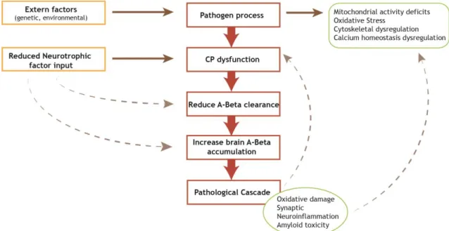
Figure 2: Proposed sequence of pathological processes involved in Alzheimer’s disease. Various
pathogenic processes contribute to the dysfunction of the choroid plexus which results in impaired Aβ processing. This and the resultant accumulation of Aβ can in turn feed back to enhance the pathogenic processes. CP-Choroid Plexus; Aβ-amyloid beta. (Adapted from (52))
1.5.2 Parkinson Disease
Parkinson disease is the most common neurodegenerative movement disorder, and the second most common ND, affecting nearly 4 million people worldwide. It is characterized by progressive hypokinesia, slowness of movement, rigidity, development of a resting tremor, and loss of postural reflex (53).
The hallmarks of neuroinflammation, in PD, are the presence of activated microglia and reactive astrocytes in the parenchyma (neurons, astrocytes, and endothelial cells) of the CNS, direct participation of the adaptive immune system, increased production of cytokines, chemokines, prostaglandins, a cascade of complement proteins, ROS and RNS, which in some cases can result in disruption of the BBB (54). The extent to which neuroinflammation and peripheral immune responses contribute towards the development of PD or modify its course is not exactly known (54, 55).
1.6 Neurogenesis
Before the 1990s, it was consensual that neurons in the adult brain were not regenerated. Consequently, it was thought that if neurons died during the adult life for any reason (e.g., oxidative stress, stroke, ND, head trauma, normal aging), they would never be replaced (56). The first evidence that neurogenesis occurs in the adult rodent brain was described by Altman and Das in 1965 (57), where they report that new neurons are continuous and spontaneously born in only two specific brain regions: the subgranular zone of the hippocampus, with cells migrating to the granule layer of the dentate gyrus and the subventricular zone with cells migrating to the olfactory bulb (56, 58). The hippocampus is critical to learning and memory as well as mood regulation, and adult neurogenesis necessary for normal function (58). Notably, the hippocampus is also particularly vulnerable in AD.
Due to the longstanding dogma that postnatal neurogenesis is non-existent in mammals, it took many years before the Altman and Das discovery was broadly accepted.
Multi-potent stem cells divide asymmetrically producing one daughter progenitor cell and one stem cell. The progenitor cell can then divide asymmetrically producing daughter cells that differentiate into either astrocytes or neurons and one progenitor cell retains the capacity to divide multiple times (56).
1.7 Relationship between neurogenesis and neuroinflammation
The effect of neuroinflammation in neurogenesis can have negative and positive consequences, which can result in development or inhibition of the process. The effect relies principally on how inflammatory cells are activated and for how long the inflammation occurs (59). We can distinguish two types of inflammation: acute and chronic inflammation. Acute inflammation comprises the immediate and early response to an injurious agent and is basically a defensive response that paves the way for repair of the damaged site being typically short-lived and unlikely to be detrimental to long-term neuronal survival. The chronic response occurs when the harmful stimulus persists over time, and contrary to the acute form, it is a long-standing and often self-perpetuating neuroinflammatory response which, in the end, results in detrimental consequences for neurons. In chronic inflammation, as AD, neuroinflammation has also been shown to induce a blockade in neurogenesis. The direct mechanism as how neuroinflammation is able to induce a disruption to neurogenesis has not yet to be fully elucidated. Some studies performed by Monje et al. revealed that peripheral administration of LPS, inducing an increase in central pro-inflammatory cytokine production, was sufficient to induce a 35% decrease in hippocampal neurogenesis (60). This disruption of neurogenesis by LPS was also shown to be able to induce spatial learning and memory deficits task, symptoms observed in AD. However, some aspects of the neuroinflammatory response result in beneficial outcomes for CNS. Among these benefits,neuroprotection phenomena, the maintenance of neurogenesis as a mechanism of brain repair, the mobilization of neural precursors for repair, remyelinization, and even axonal regeneration are included. The final result depends largely on how microglia, macrophages, and/or astrocytes are activated and the duration of the inflammation (59).
2. Aim
The main goal of this work was to evaluate the effect of an acute neuroinflammatory insult to the neurogenic potential, protein synthesis and barrier function of the CP. More specifically:
i) the inflammatory response induced by LPS in CP will be assessed in vivo, ex vivo and in vitro through analysis of inflammatory markers such as CD11b, iNOS, changes in TTR expression, and production of ROS and NO.
ii) the effect of LPS in CP neurogenesis in vivo and ex vivo will be analysed by studying the presence of the mature neuron marker, NeuN, the microgial marker ,CD11b and the astrocyte marker, Glial Fibrillary Acidic Protein (GFAP) in CP.
iii) the effect of LPS in CP integrity as a barrier will be analysed by assessing its effects on TJ proteins in vitro.
Methods
3. Materials and Methods
3.1 Animals
Animals were handled in compliance with the National Institutes of Health guidelines and the European Union rules for the care and handling of laboratory animals (Directive 2010/63/EU). Animals’ experiments were also performed according to the Portuguese law for animal well being and the protocol approved by the Committee on the Ethics of Animal Experiments of the Health Science Research Centre of the University of Beira Interior (DGV/2013). Beyond that, all efforts were made to minimize animal suffering.
Tissue sampling for in vitro and ex vivo experiments and from in vivo experiments was carried out in animals decapitated under anesthesia with intraperitoneal injection of ketamine hydrochloride (150 mg/kg) plus medetomidine (0.3 mg/kg) and transcardially perfused with cold saline. The brain was removed and CP from the lateral ventricles kept for further analysis.
3.1.1 Primary Cultures of rat Choroid Plexus epithelial cells
The method used for the establishment of primary culture of CEPC has been previously described by Martinho et al. (43). Briefly, postnatal rats, with 3-5 days, were sacrificed and CP were dissected under conventional light microscopy from lateral ventricles. CP was rinsed twice in Phosphate Buffered Saline (PBS) without calcium and magnesium and digested with 0.2% pronase (Fluka) in PBS at 37˚C for five minutes. Pre-digested tissues were recovered by sedimentation in Dulbecco’s Modified Eagle Medium – high glucose (DMEM, Sigma) with 10% fetal bovine serum (FBS, Biochrom AG) and 100 units/mL of penicillin/streptomycin (Sigma). Cells were pelleted by centrifugation and were resuspended in DMEM supplemented with 100 units/mL antibiotics, 10% FBS, 10 ng/mL, epidermal growth factor (EGF, Sigma), 5 µg/mL insulin (Sigma), 20 µM cytosine arabinoside (Ara-C, Sigma) and were seeded in 12 wells with a 15 mm lamella inside for immunocytochemistry (ICC) or in a 96 well plate (VWR) for ROS, NO and extracellular protein studies. Cells were grown in a humidified incubator at 37ºC with 5% CO2. The growth medium was changed 1 day after the initial seeding and every 2 days
3.2 Effect of LPS in CP
3.2.1 Effects of LPS in Choroid Plexus Epithelial Cells.
Newborn Wistar Han rats, between 3 and 5 days, were sacrificed, and CP from lateral ventricles were removed, for CPEC isolation.
Once differentiated CPEC were serum starved for four hours, and were incubated with culture medium containing 0.1 µg/mL of LPS (Sigma). Cells were recovered for further analysis after 1, 2, 4, 6, 8, 12, 18 or 24 hours of incubation with LPS, and analysed for ROS production, NO production, and quantification of extracellular medium protein was performed (Figure 4). TTR and iNOS expression were also analyzed in cells and in the extracellular medium by Western blot (WB), for different times of incubation with 0.1 µg/mL of LPS (Figure 4).
0h
Figure 4: Schematic representation of the procedure to study the production of ROS, NO and protein extracellular in the culture medium, in vitro. CPEC were incubated with culture medium with
0.1 µg/mL of LPS for different hours 1, 2, 4, 6, 8, 12, 18 and 24 hours, after. LPS- lipopolysaccharide; ROS- Reactive Oxygen Species, NO- Nitric Oxide
Impairments in CPEC barrier proteins (occludin, claudin-1, ZO-1 and E-cadherin) were evaluated by ICC. For this propose CPEC were serum starved 4 hours and incubated with 0.1 µg/mL of LPS during 24 hours (Figure 5). All experiments were performed independently by three times.
0h
Figure 5: Schematic representation of the procedure performed to study the effects of the LPS insult in tight junctions by immunocytochemistry. CPEC were incubated with 0.1 µg/mL of LPS during
24 hours after being serum starved for 4 hours. LPS- lipopolysaccharide ; ZO- Zonula Occludens.
Medium without serum
Fresh medium with 0.1 µg/mL of LPS
4hours before LPS Sampling at: 1,2,4,6,8,12,18 or 24 hours
ROS, NO, extracelular protein 4 hours before LPS Occludin Claudin-1 ZO-1 E-cadherin Medium without serum
Fresh medium with 0.1 µg/mL of LPS
3.2.2 Effects of LPS in neurogenesis and inflammatory markers in Choroid
Plexus explants
To study the effect of LPS in neurogenesis of CP explants, newborn Wistar Han rats (P3-P5) and 21 days rats were used, CP from lateral ventricles were extracted and placed directly in serum free culture medium (DMEM) during 4 hours followed by 48 hours incubation with LPS 0.1 g/mL at 37ºC in an atmosphere containing 5% of CO2. After incubation, CP explants were
analysed by whole mount (WM), using the following markers: NeuN, GFAP, CD11b and TTR. This experiment was repeated for protein extraction followed by WB (iNOS, TTR, E-cadherin and occludin), and for analysis of ROS production (Figure 6).
Figure 6: Schematic representation of the procedure performed to study neurogenesis in CP explants upon an inflammatory insult with 0.1 µg/mL of LPS ex vivo. LPS- lipopolysaccharide, TTR-
Transthyretin, NeuN- Neuronal Nuclear Protein, GFAP- Glial Fibrillary Acidic Protein.
3.2.3 Inflammatory response of CP to a LPS insult, in vivo
C57BL/6 mice with 1, 3 and 9 months old were randomly divided into two groups at the beginning of the experience: the control group and the experimental group (9 animals in each group). In the experimental group, a single intraperitoneal dose of LPS (1.0 mg/kg) dissolved in NaCl (0.9%, pH 4.5 -7.0) was administered. Control group received an intraperitoneal dose of NaCl (0.9%, pH 4.5 -7.0). To reduce the stress-induced changes in the hypothalamus and pituitary axis, animals were handled for one week before injections (Figure 7). CP were collected 7 hours after LPS injection directly to protein extraction buffer. Protein extracts from these CP were used to compare the expression of iNOS, TTR, E-cadherin and occludin, by WB upon an inflammatory acute insult during aging.
Figure 7: Schematic representation of the experimental layout to analyze the response of CP to 1.0 mg/kg of LPS inject in vivo. LPS- lipopolysaccharide, iNOS- inducible nitric oxid synthase, TTR-
transthyretin. 8 days 7 hours Fresh medium with 0.1 µg/mL LPS 24 hours Medium without serum 4hours TTR CD11B NeuN GFAP iNOS TTR E-cadherin Occludin Intraperitoneally injection with 1.0 mg/kg of LPS Handled 0 days
3.2.4 Neurogenesis in CP in vivo in response to a LPS insult
C57BL/6 mice, of different ages, 1 and 3 months old, were intraperitoneal injected with LPS (1.0 mg/kg) or with vehicle (0.9% NaCl, pH 4.5- 7.0). Mice were randomly divided into two groups at the beginning of the experience, with 9 animals each. To reduce the stress-induced changes in the hypothalamus and pituitary axis, animals were handled for one week before injections. CP were collected 28 days after LPS injection directly to 4% paraformaldehyde (PFA) and used for detecting Neuronal Nuclear Protein (NeuN), CD11b, GFAP and TTR by WM (Figure 8).
Figure 8: Schematic representation of the procedure performed to assess neurogenesis upon LPS insult in vivo. LPS- lipopolysaccharide, TTR- Transthyretin, NeuN- Neuronal Nuclear Protein, GFAP- Glial
Fibrillary Acidic Protein.
3.3 Assessment of ROS production, inflammatory response and
neurogenesis markers
3.3.1.1 ROS production in CPEC
ROS production was determined with dihydroethidium (DHE, Life technologies). In this assay, blue fluorescent DHE is dehydrogenated by superoxide to form red fluorescent ethidium bromide (61). At the end of the experiments described in 3.6.1, cells were incubated for 4 hours with DHE 5 µM. Finally, fluorescence was measured at 620 nm in a microplate reader
spectrofluorometer, Anthos 2020 (62).
3.3.1.2 ROS production in CP explants
Intracellular ROS was measured in CP explants by dichloro-dihudro-fluorescein-diacetate (DCFH-DA, Sigma) oxidation. DCFH-DA enters cells passively and is deacetylated by esterase to form non-fluorescent product DCFH, and DCFH reacts finally with ROS to form the fluorescent product, DCF (61, 63).
CP were incubated with 10 M DCFH-DA, dissolved in DMEM, for 20 minutes at 37ºC, in dark. Unincorporated DCFH-DA was removed by washing the tissues twice with PBS. To stain DNA, CP were incubated with Hoechst 33342 (1:1000) 20 minutes at 37ºC and after a final wash with PBS fluorescence was quantified, immediately, at wavelengths of 488 nm for excitation and 510 nm for emission under a LSM710 confocal laser scanning microscope (Carl Zeiss, Jena, Germany). 8 days 28 days 0 days TTR CD11B NeuN GFAP Intraperitoneally injection with 1.0 mg/kg of LPS Handled
3.3.2 Nitric oxide production in CPEC
The culture medium of CPEC from experiment 3.3.2 (50 µL) was collected to estimate NO concentration by measuring the amounts of nitrite secreted by CPEC into the culture medium, using a colorimetric reaction using the Griess reagent (Promega). This assay relies on a diazotization reaction that was originally described by Griess in 1879, and is based on the chemical reaction which uses sulfanilamide and NED (N-1-apthylethylenediamine dihydrochloride) under acidic conditions (62). The culture supernatants were mixed with an equal amount of the Griess reagent in a 96 well plates (Corning). The absorbance of the mixture was read at 542 nm using a microplate reader, Spectra MAX Gemini EM, Molecular Devices.
3.3.3 Protein extraction from CP
CP collected from animals in the experiments described in sections 3.2.2 and 3.2.3, were homogenized in protein extraction buffer (Tris-HCl 25mM Tris-HCl 25 mM pH 7.4, Ethylenediaminetetraacetic acid (EDTA) 2.5 mM, Ethylene glycol tetraacetic acid (EGTA) 2.5 mM, Triton X-100 2%, PMSF 1.0 mM and 10 µL/mL, Complete EDTA Free protease inhibitor cocktail (Roche) 10 µL/mL). After a centrifugation at 10,000xG for 10 minutes at 4ºC supernatants were collected and the total protein quantified.
Protein measurement was performed by the Bradford Method with the Bio-Rad protein assay
reagent (Bio-Rad) following the manufacturer’s instructions. This method is a colorimetric
assay which involves the addition of an acidic dye to protein solution, the absorbance maximum for an acidic solution of Coomassie® Brilliant Blue G-250 dye shifts from 465 nm to 595 nm when binding to protein occurs.
A set of standards (0-10 µg/mL) was prepared from a stock solution of Bovine Serum Albumin (BSA). The Bradford values obtained for the standard were then used to construct a standard curve and used to determine the concentration of our samples. Measurements were performed in 96 well plates after 15 minutes incubation at room temperature. Quantification of the proteins was performed with a microplate reader, Anthos 2020, at 595 nm. After quantification protein extracts were stored at -80ºC until use.
3.3.4 SDS-PAGE
and Immunoblot
WB is a method to detect proteins in a given sample of tissue or cells homogenate or extract. It gives information about the size of the proteins (by comparison with a size marker or ladder in kDa), and also gives information on the relative protein expression.
25 µL of samples from section 3.2.1 and 30 µg of each sample obtained from experience described in section 3.2.2 and 3.2.3 was denaturated at 100ºC for 10 min. Then denaturated samples were loaded in a 4.7% stacking gel (4.7% of acrylamide (Applichem), 1.25 M Tris-HCl (pH 6.8, Biorad), 10% Sodium Dodecyl Sulfate (SDS), 7.2 mL H2O) followed by a 12%
SDS-polyacrylamide gel (12% of acrylamide, 1.875 M Tris-HCL, 0.2% SDS, 5.95 mL H2O) with the
were blotted onto a polyvinylidene difluoride (PDVF) membrane (GE Healthcare). Therefore, a wet transfer was performed. For that PDVF membrane was previously activated in 100% methanol and equilibrated in transfer buffer (10 mM 3-(Cyclohexylamino)-2-hydroxy-1-propanesulfonic acid (CAPS) from Bio-Rad, 10% methanol).
To blot the samples into the PDVF membrane, a Bio-Rad system was used. The transfer sandwich consisted in a fiber pad, one filter paper soaked with transfer buffer the gel and the PDVF membrane. Two layers of wet filter paper and finally one fiber pad. Once completed the transfer sandwich was transferred to the gel holder cassette and put in the support. Blotting was performed for 30 minutes to TTR (14 kDa) and 1:30h to iNOS (130 kDa), E-cadherin (135 kDa), occludin (60-82 kDa), with a voltage of 750 mA. The difference in blotting times is due to the protein size.
Once protein transfer finished, the membrane was washed twice with Tris Buffered Saline (TBS) for 5 min at room temperature. Afterwards, membrane was blocked with 0.5% non-fat dry milk (Paturages) dissolved in TBS 0.1% Tween (TBS-T). After rinsed with TBS-T, the primary antibody against TTR, iNOS, E-cadherin and occludin was applied overnight at 4°C in the appropriate dilution in TBS-T (see tables 1 and 2). The membrane was washed three times with TBS-T 0.1% for 15 min at room temperature and incubated for 1 hour at room temperature with the correspondent secondary antibody alkaline phosphatase linked dissolved in TBS-T.
Protein detection was done after three washing steps of 15 minutes with TBS-T, immunoreactive bands were detected using the Enhanced Chemifluorescence (ECF) subtract, fluorescent substrate for alkaline phosphatase-based detection of protein blots (GE Healthcare) and images were acquired with the Molecular Imager FX Pro Plus MultiImager system (Bio-Rad). Proteins expression was quantified by densitometric analysis using the QuantityOne software from Bio-Rad. The -actin protein was chosen for normalize the data of protein expression.
3.3.5 Immunocytochemistry
Immunocytochemistry is a technique used to verify the presence of a specific protein or antigen in cells, using specific antibodies. ICC is a valuable tool to study the presence and sub-cellular location of proteins.
Cells were fixed with 4% PFA in 0.1 M PBS, pH 7.4, for 10 minutes, at room temperature. Subsequently cells were block with 20% FBS in PBS for 60 minutes followed by the primary diluted antibody incubation in 1% FBS in PBS, overnight at 4ºC (Table 1). The primary antibody was removed and cells were washed with PBS. Cells were incubated with the secondary antibody for one hour at room temperature in the dark (Table 2). For counter staining, cells were incubated with Hoechst 33342 (1:1000, Invitrogen) in PBS, for 10 minutes at room temperature in the dark. Afterward preparations were covered with a coverslip with a drop of
mounting medium Entellan (Merck). Slides were stored in the dark at -20ºC and analysed under a LSM710 confocal laser scanning microscope (Carl Zeiss, Jena, Germany).
3.3.6 Whole Mount technique
Whole Mount is used to determine the cellular location and distribution of a given protein in a tissue by the use of specific antibodies.
CP were fixed with 4% PFA for 45 minutes at room temperature. CP were then cryoprotected in 30% saccharose until CP reached the bottom (~5h) and then incubated for 4 hours with the blocking solution (PBS 2.5% BSA and 0.2% Triton). Explants were incubated overnight at 4ºC with the primary antibody GFAP, NeuN, CD11b and TTR, in the adequate dilution (Table 1). Antibodies were diluted in blocking solution. After six washes with PBS-T, the secondary antibodies were applied for three hours at room temperature in the dark. For nucleus staining, CP were incubated with Hoechst 33342 (1:1000) twenty minutes at room temperature in the dark. Finally, the coverslip was mounted with the mounting medium Entellan (Merck) and kept at -20ºC until observation under a LSM710 confocal laser scanning microscope (Carl Zeiss, Jena, Germany).
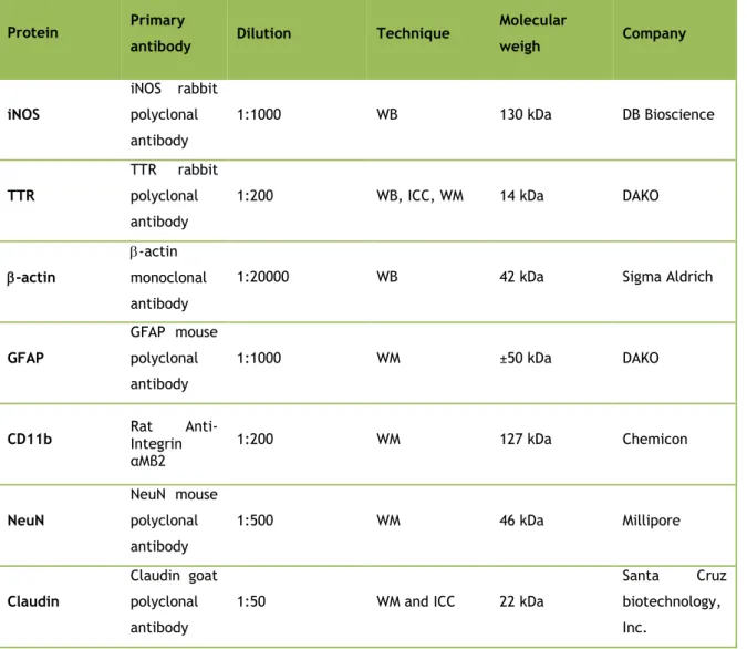
Table 1 : Primary antibodies used in immunofluorescence and Western analysis and their respective
protein, dilution, technique where were used, molecular weight and company. ICC – Immunocytochemistry, WM – Whole Mount, WB – Western Blot.
Protein Primary
antibody Dilution Technique
Molecular weigh Company iNOS iNOS rabbit polyclonal antibody 1:1000 WB 130 kDa DB Bioscience TTR TTR rabbit polyclonal antibody
1:200 WB, ICC, WM 14 kDa DAKO
-actin
-actin monoclonal antibody
1:20000 WB 42 kDa Sigma Aldrich
GFAP
GFAP mouse polyclonal antibody
1:1000 WM ±50 kDa DAKO
CD11b Rat Integrin
Anti-αMβ2 1:200 WM 127 kDa Chemicon NeuN NeuN mouse polyclonal antibody 1:500 WM 46 kDa Millipore Claudin Claudin goat polyclonal antibody
1:50 WM and ICC 22 kDa
Santa Cruz biotechnology, Inc.
E-cadherin rabbit polyclonal antibody 1:50 1:200 WM, ICC WB 135 kDa Santa Cruz biotechnology, Inc. ZO-1 ZO-1 goat polyclonal antibody
1:25 WM and ICC 220 kDa
Santa Cruz biotechnology, Inc. Occludin Occludin rabbit polyclonal antibody 1:50 1:200 WM, ICC WB 60-82 kDa Santa Cruz biotechnology, Inc.
Table 2 : Secondary antibodies used in immunofluorescence and Western analysis and their dilution,
technique were where used and company. ICC – Immunocytochemistry, WM – Whole Mount, WB – Western Blot.
Secondary antibody Dilution Technique Company Alkaline phosphatase conjugated anti-rabbit 1:20000 WB AmershamLife Sciences Alkaline phosphatase conjugate
anti-mouse 1:20000 WB AmershamLife Sciences
Mouse anti rat alexa
546 1:1000 WM
Invitrogen, Molecular probes
Goat anti-mouse Alexa
488 1:1000 WM, ICC
Invitrogen, Molecular Probes
Goat anti-rabbit Alexa
488 1:1000 WM, ICC
Invitrogen, Molecular Probes
Donkey anti-goat igG
CFL 488 1:200 WM and ICC
Santa Cruz
biotechnology, Inc.
3.4 Statistical analysis
Data are expressed as mean ± standard error of the mean (SEM) of at least three independent experiments, performed in triplicate. Differences between groups were analyzed by one-way ANOVA followed by Bonferroni's Multiple Comparison Test. Comparisons between two groups were done with Unpaired t-test. All calculations were made using GraphPah software 6.0 Demo (GraphPad Software Inc.). Statistical significance was set at p≤0.05.
4. Results
4.1 Effects of LPS in Choroid Plexus Epithelial Cells
In order to understand the response of CP to LPS in vitro, ROS and NO production were registered along a time course (0-24 hours) in CPEC and total protein content in the culture medium was evaluated.
4.1.1 ROS production
ROS production was evaluated at 1, 2, 4, 6, 8, 12, 18 and 24 hours after exposure to 0.1 g/mL LPS. A significant increase in ROS production was observed 2 hours after LPS
exposure (p≤0.05) (Figure 9) with a tendency to decrease afterwards, however without statistical significance.
Figure 9: Time-course ROS production response to 0.1 µg/mL of LPS, in CPEC. Cells were incubated
for 1, 2, 4, 6, 8, 12, 18 and 24 hours with 0.1 µg/mL of LPS. Fluorescence was measured by microplate reader spectrofluorometer at 620 nm. Data represent the mean ± SEM of three independent experiments performed in quadruplicate. Statistically significant differences among groups (*P≤0.05) were determined by one-way ANOVA and are indicated by asterisks. LPS- lipopolysaccharide, ROS- reactive oxygen species.
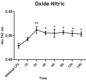
4.1.2 NO production
NO production was measured in the CPEC medium culture that was incubated for different times with 0.1 µg/mL of LPS. Our results suggest an increase of NO levels 2 hours after LPS exposure, with statistical significance when compared to the samples without LPS (Figure 10). These levels of NO were maintained till the end of the experiment.
Figure 10: Time-course of oxide nitric production in response to 0.1 µg/mL of LPS in CPEC medium.
Cells were stimulated for 1, 2, 4, 6, 8, 12 and 24 hours with 0.1 µg/mL of LPS. Data represent the Mean ± SEM of three independent experiments performed in quadruplicate. Statistically significant differences among groups were determined by one-way ANOVA, and are indicated by asterisks*p≤0.05, **p≤0.01, compared with control cultures (without LPS). Abs-Absorbance; LPS- lipopolysaccharide.
4.1.3 Extracellular Protein
We also assessed the levels of total protein in CPEC medium upon LPS incubation. After 1 hour of exposure to 0.1 g/mL of LPS we observed an increase of about two fold in the protein content of the growth culture medium of CPEC. This up-regulation remained constant along the 24 hours of exposition to LPS (Figure 11).
Figure 11: Quantification of extracellular proteins released from CPEC to the culture medium after incubation with 0.1 µg/mL of LPS for 24 hours. The experiment was done three times and performed
in quadruplicate. Statistically significant differences among groups (***p≤0.001) were determined by one-way ANOVA and are indicated by asterisks. LPS- lipopolysaccharide.