http://dx.doi.org/10.1590/1516-635x1702209-218
Author(s)
Chen YHI
Peh HCII
Roan SWII
I Associate Professor, PhD, Department of
Animal Science and Biotechnology, Tunghai University. 181 Taichung Harbor Road, Taichung 406, Taiwan, ROC
II Professor, PhD, Department of Animal
Science, National Chung Hsing University. 250, Kuo Kuang Road, Taichung 402, Taiwan, ROC
Mail Address
Corresponding author e-mail address Shii-Wen Roan
Department of Animal Science, National Chung Hsing University. 250, Kuo Kuang Road, Taichung 402, Taiwan, ROC Phone: +886 422840365#234, Fax: +886
422860265
E-mail: swroan@dragon.nchu.edu.tw
Keywords
Gompertz function, mule ducks, uropygial gland growth curve.
Submitted: February/2014 Approved: October/2014
ABSTRACT
Growth curves for the uropygial gland (UG) of white, 3-way crossed mule ducklings were established using the Gompertz function. In total, 144 ducklings were fed in 12 floor pens with 12 birds in each pen. Each pen contained an equal number of animals of each sex. Feed and water were supplied ad libitum throughout the entire experimental period. The weekly change in UG weight was recorded in males and females from hatch to 8 weeks of age. The weight and length of the UG, the width of the lobus glandulae uropygialis, the length and width of the pluma of the circulus uropygialis, and the index of the papilla uropygialis were measured once a week in individual ducklings in one pen. The average UG weight gain observed in white, 3-way crossed drakes was significantly higher than that of ducks of 21–56 days of age (P < 0.05). The UG length was 1.64–2.23 times the width of the left or right lobe, and the development of the UG was delayed from 3–4 weeks of age. The morphology of the UG changed from elliptical to elongated-elliptical with age. The right and left lobus glandulae uropygialis were symmetrical. The Gompertz growth functions of the UG in drakes and ducks were W=5.49e-e-0.675(t-1.955) and W=4.76e-e-0.685(t-1.936), respectively,
where t represents age in weeks. These equations indicated that the maximum growth rate for drakes occurred at 14.1 days of age and for ducks at 13.6 days of age.
INTRODUCTION
higher relative weight than that of non-water-bathing geese. Moreover, this gland is able to stimulate feather growth. Kozák (2011) showed that geese with access to water show an improved down content.
The secretions of the UG/preen gland contain esters and fatty acids that are important for waterfowl. The beneficial roles of the UG in waterfowl are: 1. water repellence: the gland plays a role in waterproofing the feathers when waterfowl preen and spread UG oil on them (Luttmann & Luttmann, 1978), and in addition of making the feathers flexible and water repellent (Jacob & Ziswiler, 1982), the gland helps keep young waterfowl from catching a chill; 2. prevention from drowning: when ducklings and goslings are soaked with water, young birds can quickly lose mobility, thus becoming waterlogged and, in some cases, even drown (Holderread, 1987); 3. increasing buoyancy: most types of geese are accustomed to breeding on the water, as it is very difficult for the larger geese to mount females on land, and increased buoyancy enables geese to bear the burden of the body weight of the gander (Luttmann & Luttmann, 1978); 4. protection of feathers: fatty acid waxes produced by the UG can inhibit the growth of dermatophytes and keratinophilic fungi (Blaxter & Trotter, 1969; Pugh & Evans, 1970a, 1970b; Jacob & Ziswiler, 1982; Bandypadhyay & Bhatttacharyya, 1996); and 5. provision of vitamin D: solar rays that reach the UG oil spread in a thin layer over the feathers and result in vitamin D production, which is ultimately consumed by the birds as they preen (Luttmann & Luttmann, 1978; Jacob & Ziswiler, 1982). The large quantity of oil secreted by the UG is consistent with a fully developed UG. However, meat ducks are generally sold when they reach 10-12 weeks of age, whether the UG is mature or not. Further research is required to understand this apparent contradiction.
The Gompertz function has been used to estimate growth rates in poultry (Anthony et al., 1986) and growing pig (Yoosuk et al., 2011, 2012a, 2012b). Duan-yai et al. (1999) derived the Gompertz function of the broiler growth curve from growth data on broilers. Growth curves have been established for the native Taiwanese chicken (Lee et al., 1997; Wang & Roan, 2002), Chinese geese (Chen et al., 2003a), White Roman geese (Chen et al., 2003a), and mule ducks (Chen & Roan, 2005). However, there is no information available concerning the growth curve of the UG in mule ducklings. Therefore, the purpose of the present study was to investigate the growth curve of the UG in mule ducklings to establish basal data for further research.
MATERIALS AND METHODS
Animals and management
A total of 144 1-day-old ducklings with similar body weights (BW) were randomly divided into 12 open pens (10 m × 6 m area) with a wire floor, with 6 males and 6 females being placed in each pen. The sex of each bird was determined via vent examination. Water was continuously dropped into dishes (50 cm × 40 cm × 10 cm volume) in each pen to supply the birds with water for drinking and playing. The diets provided to the birds at 0-3 and 4-8 weeks of age consisted of pellets containing crude protein (%)/metabolizable energy (kcal/kg) ratios of 19.3/2,900 and 16.1/2,950, respectively. The ducklings had their bills trimmed at 1 day of age and were brooded in an electric brooder from 0-2 weeks of age. Feed and water were supplied ad libitum. The average room temperature was 25.7°C throughout the experimental period (0-8 weeks of age). Studies by Shen (1988) and the NRC (1994) were consulted for the formulation of the experimental diets.
Sampling and analytical parameters
Uropygial gland measurement
One duckling from each of the 12 pens was randomly selected for the measurement of BW once a week from 1-56 days of age. The ducklings’ uropygial glands were surgically removed according to the methods previously described (Chen & Tsang, 2003; Chen et al., 2001b). The body weight and UG weight of the individual ducklings were measured with an electronic scale. The length and width of each lobus glandulae uropygialis (LGU) and the papilla uropygialis (PU) as well as the length of the pluma of the circulus uropygialis (PCU) were determined with a digital calliper. The widths of the right and left LGU were measured at the midpoint of their length (Figure 1). The LGU index and PU index were calculated according to Jacob & Ziswiler (1982), i.e., as LGU length/LGU width and PU height/PU width, respectively.
Uropygial gland growth curve
The average weekly UG weights of ducklings were entered into the Gompertz growth curve model, W = ae-e-b(t-t*)(Duan-yai et al., 1999), where W indicates the
Allometric growth ratio
The allometric growth ratio is described with the following equation (Rose, 1997):
y = log (a) + k log (x)
where x = body weight (g); y = the weight of a body part (g); k = the allometric growth ratio (slope of line); and a = a constant (intercept).
Statistical analysis
The data were analysed via analysis of variance using the general linear model procedure. All statistical analyses were conducted using the SAS software package (SAS Institute, 1996). Correlations between the average UG weight of both sexes and age were determined with the NLIN (Non-Linear) procedure, followed by derivation of the regression equation. Correlations between the BW and UG measurements were determined using the PROC CORR procedure.
RESULTS
Body weight and uropygial gland growth measurements
The BW and UG weights of the white, 3-way crossed mule ducklings from 1–56 days of age are presented in Table 1. There were no significant differences in BW detected between the sexes during the entire experimental period (p > 0.05). However, the differences in UG weight and relative UG weight between sexes were significant either from 21–56 days of age or throughout the experimental period, except at 35, 42 and 56 days of age. The UG weights in the female and male mule ducks increased with age and reached a plateau at approximately 4.76 and 6.23 g (49 days of age), respectively.
The LGU length and width and the index of the right and left LGU in the white, 3-way crossed mule ducklings from 1–56 days of age are presented in Table 2.
Table 1 - Body weight (BW), uropygial glandweight (UGW) and relative uropygial glandweight (RUGW) recorded in the white, 3-way crossedmule ducklings from 1–56 days of age.
Variable Sex Day of age Significant
1 7 14 21 28 35 42 49 56 L Q C
--- g
---BW F 49a 181b 446c 793d 1063e 1478f 1780g 1940g 2199h *** ** *
M 48a 184b 436c 844d 1120e 1558f 1894g 1913g 2299h *** *** NS
SEM 1 5 7 21 44 48 50 103 36
x
48a 182b 440c 818d 1091e 1518f 1837g 1926g 2246h *** NS ***
UGW F 0.12a 0.72a 1.99b 2.97c* 3.01d* 3.68d* 4.33de* 4.76e* 4.63 e* *** NS NS
M 0.15a 0.90b 2.30c 4.08d 4.05d 4.58e 5.30f 6.23g 6.16g *** ** NS
SEM 0.01 0.03 0.05 0.19 0.40 0.21 0.21 0.29 0.23
x
0.13a 0.80b 2.14c 3.50d 3.53d 4.13e 4.81f 5.49g 5.39g *** NS NS
RUGW (g/100g BW)
---F 0.24a* 0.40d* 0.45d* 0.38c* 0.29b* 0.25a 0.24a 0.25a* 0.21a *** *** ***
M 0.31a 0.49b 0.53b 0.48ab 0.34a 0.29a 0.28a 0.33a 0.27a *** *** ***
SEM 0.01 0.01 0.02 0.02 0.02 0.01 0.01 0.02 0.01
x
0.27a 0.44c 0.49d 0.43d 0.33b 0.27a 0.26a 0.29b 0.24a *** *** NS
a-h Means within the same row with different superscripts differ significantly (p < 0.05). *Differences between sexes at a given age are significantly different (p < 0.05). F: female; M: male.
The LGU length was significantly greater in male mule ducks than in females from 14–56 days of age (p < 0.05). The width of the right LGU was significantly greater in males from 21–56 days of age, except on day 28 (p < 0.05), and the width of the left LGU was similar at 21 and 49 days of age. The mean PCU length in the mule ducks increased with age, reaching a growth limit at
49 days of age. The shapes and the measurements of the right and left lobus glandulae uropygialis appeared to be asymmetrical (Figure 1 and Table 2), whereas the right and left lobus glandulae uropygialis length (LGUL) and lobus glandulae uropygialis width (LGUW) did not differ significantly throughout the entire experimental period (data not shown in Table 2).
Table 2 – The lobus glandulae uropygialis (LGU) length and width and LGU indices observed in the white, 3-way crossed mule ducklings from 1–56 days of age.
Variable Sex Day of age Significant
1 7 14 21 28 35 42 49 56 L Q C
mm
---LLGUL F 6.10a 12.74b 17.48c* 20.90d* 22.07 ade* 23.33de* 23.85e* 25.00e* 24.94e* *** *** NS
M 6.44a 13.74b 19.21c 24.07d 24.50d 26.01de 26.84e 28.76f 28.63f *** *** ***
SEM 0.17 0.24 0.36 0.60 0.53 0.58 0.65 0.73 0.68
x
6.27a 13.24b 18.35c 22.49d 23.28d 24.83e 25.34e 26.88f 26.78f *** *** NSLLGUW F 3.81a 7.10b 9.55c 10.42d* 10.78 d 10.83d* 10.97d* 12.14e* 12.11e* *** *** *
M 3.83a 7.27b 10.07c 11.61d 10.77 c 11.76 d 11.89 d 13.01 e 12.89 e *** *** ***
SEM 0.09 0.12 0.15 0.29 0.20 0.23 0.24 0.29 0.21
x
3.82a 7.19b 9.81c 11.01d 10.78d 11.29d 11.43d 12.58e 12.50 e *** *** NSRLGUL F 6.10a 12.56b 17.46c* 20.98d* 22.07d* 23.39e* 23.88e* 24.98 e* 24.88 e* *** *** NS
M 6.44a 13.76b 19.11c 24.06 d 24.50 d 26.01de 26.77e 28.56 f 28.34f *** *** ***
SEM 0.18 0.31 0.37 0.60 0.53 0.58 0.65 0.69 0.65
x
6.27a 13.16b 18.29c 22.52d 23.28d 24.86e 25.32e 26.77 f 26.61f *** *** NSRLGUW F 3.74a 6.97 b 6.93 b 10.52c* 10.52c 10.68c 11.01c 11.90 d* 12.12d *** *** *
M 3.77a 7.24b 9.63c 11.32d 10.58d 11.42d 11.72d 12.83e 12.83e *** *** ***
SEM 0.11 0.13 0.12 0.25 0.28 0.19 0.25 0.26 0.24
x
3.76a 7.10b 9.58 c 10.92 d 10.55 d 11.05 d 11.36d 12.36e 12.47e *** *** NSindex
---LLGUI1 F 1.61a 1.80b 1.83b 2.01c 2.05c* 2.21e 2.18de 2.06cd* 2.06cd* ** *** *
M 1.68a 1.89b 1.91b 2.08c 2.28d 2.21cd 2.26d 2.22d 2.22d ** ** NS
SEM 0.03 0.03 0.02 0.04 0.05 0.03 0.04 0.05 0.03
x
1.64a 1.84b 1.87b 2.04c 2.16d 2.21d 2.22d 2.14d 2.14d *** ** *RLGUI1 F 1.64a 1.80b 1.84b* 2.00c* 2.10c* 2.25d 2.18cd 2.10c 2.06c ** NS **
M 1.70a 1.91b 1.98b 2.13c 2.33c 2.28c 2.28c 2.23c 2.21c *** *** NS
SEM 0.03 0.05 0.03 0.04 0.05 0.03 0.05 0.03 0.04
x
1.67a 1.86b 1.91b 2.01c 2.22d 2.27d 2.23d 2.17d 2.13d *** ** **a-f Means within the same row with different superscripts differ significantly (p < 0.05).
*Differences between sexes at a given age are significantly different (p < 0.05).
L and RLGUW: left and right lobus glandulae uropygialis width; L and RLGUL: left and right lobus glandulae uropygialis length. F: female; M: male.
Figure 1 – The shape and measuring position glandulae uropygialis of white three--way crossed mule duck. LGU: lobus glandulae uropygialis; PU: papilla uropygialis; PCU: pluma of circulus uropygialis.
The PCU length, PU length and height, and PU index determined in the white, 3-way crossed mule ducklings from 1–56 days of age are shown in Table 3. The right and left LGU index and the PU length and height increased with age in both female and male mule ducks. The mule ducklings displayed maximum right and left LGU indices at 42 days of age; however, the PU index calculated for 1-day-old white, 3-way crossed mule ducklings was higher than at any other time in the sampling period.
Uropygial gland growth curve
The UG growth curves obtained for the white, 3-way crossed mule ducklings from 0–8 weeks of age are shown in Figure 2 (A, B, C). The Gompertz growth function, W = ae-e-b(t-t*), indicated that the ages at which
the maximum growth rates occurred in male and female ducks were 14.1 and 13.6 days, respectively. The maximum growth rate for the two sexes combined was observed at 13.7 days.
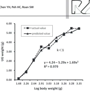
The allometric growth ratios of body parts are shown in Figure 3. The ratio of the allometric growth rate (k) of the UG to the total BW gain in the mule ducks was < 1; i.e., the growth rate of the UG was lower than the growth rate for BW.
Correlation between body weight and uropygial gland measurements
The correlation coefficients for UG weight, LGU length and width, PU length and height and PCU
length in mule ducklings are presented in Table 4. BW was significantly correlated with the UG measurements (p < 0.001), e.g., the correlation coefficient for BW and UG weight was 0.90 (p < 0.001). Moreover, UG weight, LGU length and width, PU length and height and PCU length were highly significantly correlated (P < 0.001).
Figure 2 – The growth curve of uropygial gland in white three-way crossed mule ducklings from 0 to 8 weeks of age. Means represent 12 birds per age. A. male mule ducklings; B. female mule ducklings; C. male and female mule ducklings combine.
DISCUSSION
Body weight and uropygial gland growth measurements
ducks, even though these ducks had 50% Muscovy descent. This result is in agreement with the findings of Chen & Roan (2005), who noted that the growth rate of male mule ducks is similar to that of female ducks. The relative UG weights in the mule ducks at 4 weeks of age ranged from 0.29-0.34 g/100 g BW (Table 1), while that of geese was found to be 0.22 g/100 g BW (Chen et al., 2003b). Moreover, at 5 weeks of age, the relative UG weights observed in the mule ducks in the present study and reported for chicks by Sandilands et al. (2004) were 0.25 and 0.14 g/100 g BW, respectively. Comparison of the relative UG values among ducklings, goslings and chickens of the same age shows that the development of the UG occurs at a faster rate in ducklings than in goslings and chickens. There was a difference in absolute UG weight detected between the male and female mule ducks, which is in agreement with
Table 3 – The papilla uropygialis length (PUL) and height (PUH), papilla uropygialis index (PUI) and pluma of the circulus uropygialis length (PCUL) observed in the white, 3-way crossed mule ducklings from 1–56 days of age.
Variable Sex Day of age Significant
1 7 14 21 28 35 42 49 56 L Q C
--- mm
---PUL F 2.70a 4.24b 6.93c 7.84cd 8.34d 8.22d 8.63de 9.01e 9.87ef *** *** NS
M 2.66a 4.89b 6.58c 8.11d 7.84d 8.32d 8.27d 8.18d 8.90d *** *** ***
SEM 0.07 0.16 0.16 0.18 0.20 0.31 0.17 0.43 0.39
x
2.68a 4.57b 6.76c 7.97d 8.09d 8.28d 8.48d 8.59d 9.39e *** *** NSPUH F 1.36a 3.49b 4.96c 5.64d 5.74d 6.29de 6.67e 6.60e 6.78e *** *** NS
M 1.29a 3.66b 4.24b* 5.19c 5.32c 6.17d 5.83cd* 5.61cd 6.84e *** *** ***
SEM 0.07 0.11 0.19 0.20 0.25 0.14 0.20 0.29 0.18
x
1.33a 3.57b 4.60c 5.41d 5.53d 6.23e 6.25e 6.10e 6.81f *** NS NSPCUL F 5.22a 6.33a 10.31b 15.80 c 18.73 d 18.71 d 19.11d 19.23 d 19.93d *** *** NS
M 4.63a 5.24a 10.17b 14.60c 17.74d 18.91de 19.68de 19.98e 18.97de *** *** NS
SEM 0.25 0.29 0.66 0.49 0.45 0.74 0.61 0.73 0.72
x
4.93a 5.79a 10.24b 15.20c 18.23d 18.81d 19.40d 19.60d 19.47d *** *** NS--- Index
---PUI1 F 2.02c 1.22a 1.42ab 1.39ab 1.50b* 1.32ab 1.28ab 1.37ab 1.47b * * **
M 2.10c 1.35ab 1.57ab 1.59b 1.54ab 1.35ab 1.41ab 1.47ab 1.30a *** NS ***
SEM 0.10 0.05 0.06 0.06 0.05 0.03 0.05 0.06 0.06
x
2.06 c 1.29a 1.49b 1.49b 1.52b 1.34a 1.35ab 1.42ab 1.38ab ** NS ***a-f Means within the same row with different superscripts differ significantly (p < 0.05).
*Differences between sexes at a given age are significantly different (p < 0.05). 1 PUI = PU length/PU height.
F: female; M: male.
L: linear; Q: quadratic; C: cubic. *p < 0.05; **p < 0.01; ***p < 0.001; NS: Not significant. Figure 3 – Allometric growth ratios of body parts.
the results of Brake et al. (1993), who found that the absolute UG values of male broilers were lower than those of female broilers. Hormones may be the main factor accounting for the differences in the absolute and relative UG weights between the sexes in mule ducklings. Castrated and non-castrated cockerels injected with testosterone propionate show an increase in both relative and absolute UG weights (Kar, 1947), and female mule ducks display lower blood testosterone concentrations than male mule ducks from 0–8 weeks of age (Peh et al., 1992).
The mean left LGU and right LGU indices of the female and male ducks were similar, ranging from 1.64–2.27 at 1–56 days of age; i.e., the length was 1.64–2.27 times the width of the left or right LGU. Chen et al. (2001a) observed that the LGU index of Muscovy ducks ranges from 2.20–2.32 at 1–3 weeks of age. Based on the LGU index values obtained here, the shape of the UG of mule ducks is similar to that of Muscovy ducks. Both the left and right LGU indices increased with age, indicating that the growth in LGU length was greater than the growth in LGU width. Therefore, the shape of the LGU changed from an ellipse to an elongated ellipse with age. The LGU and PU indices of 4-week-old White Roman goslings were found to be 1.86 and 2.00, respectively. In previous study Chen et al. (2003b) found for mule ducks of both sexes, and same age, values of 2.16–2.22 and 1.52 for those indices (Tables 2 and 3, respectively). Therefore, the morphology of the UG of the ducks is similar to that of geese, as in both species, the UG is elliptical. There were no significant differences in LGUL and LGUW observed for the right and left lobus glandulae
uropygialis throughout the entire experimental period. Therefore, the shape of the lobus glandulae uropygialis is symmetrical.
Uropygial gland growth curve
The age at which the maximum UG growth rate occurred was greater in the male mule ducks than in the females (14.1 vs. 13.6 days), in contrast to published results indicating that the age at which the maximum BW growth rate is observed is lower in male mule ducks than in females (28.1 vs. 28.5 days) (Chen & Roan, 2005). These results suggest that the UG maximum growth rate varies according to age and sex. The actual value of the UG growth curve was lower than the predicted value for 4-week old mule ducklings (Figure 2-A, B, C). Thus, it appeared that the growth rate of the UG was retarded. A significant amount of feather growth generally occurs from 4–6 weeks of age in ducklings. However, it is unclear whether this change in body physiology is the reason for the observed delay (i.e., the main nutrients in the body are used to feather growth at this time, rather than to UG development, and the UG may catch up when the duckling down is molted and the adult feathers grow) or other factors, e. g. some hormonal variation, resulted in the situation. Further research is required to explain this phenomenon.
Comparison of the allometric growth rate (0.689) of the UG obtained in the present study with the allometric growth rates of the liver and digestive tract (0.87 and 0.90) reported for the common duck (Rose, 1997) showed that the growth rate of the UG was slower than that of BW, as observed for the liver and digestive tract.
Table 4 – Correlation coefficients for the body weight (BW), uropygial gland weight (UGW), left lobus glandulae uropygialis length (LGUL), left lobus glandulae uropygialis width (LGUW), right lobus glandulae uropygialis length (RGUL), right lobus glandulae uropygialis width (RGUW), papilla uropygialis length (PUL), papilla uropygialis height (PUH) and pluma of the circulus uropygialis (PCUL) of the white, 3-way crossed mule ducklings.
BW UGW LUGL RUGL LUGW RUGW PUL PUH PCUL
BW — 0.901 0.86 0.86 0.82 0.82 0.82 0.82 0.81
UGW — 0.95 0.95 0.92 0.92 0.82 0.78 0.76
LUGL — 1.00 0.97 0.97 0.87 0.84 0.84
RUGL — 0.97 0.97 0.89 0.84 0.84
LUGW — 0.99 0.89 0.85 0.77
RUGW — 0.89 0.86 0.77
PUL — 0.90 0.81
PUH — 0.74
PCUL —
Correlation between body weight and uropygial gland measurements
The correlations between the UG measurements indicated that the length and width of the LGU, PU and PCU were growing proportionately, as all parameters were significantly and positively correlated. There was a strong correlation between BW and UG weight, similar to that the findings of Brake et al. (1993), who showed that the weight of the UG increased with BW in broilers and that the correlation between these two parameters was 0.45. The UG weights in the female and male mule ducks increased with the age of the birds, in agreement with the results of Sandilands et al. (2004), who noted that the age and growth of the birds show the greatest effects on UG development.
In summary, there were sex differences in the absolute UG weights of the mule ducks. However, the UG growth curves showed similar patterns of UG development between the sexes during the growth period. Based on the correlation between the BW and UG measurements, the length and width of LGU, PU and PCU were growing proportionately. However, the growth rate of the UG was lower than that of BW. The UG is fully mature before 8 weeks of age in mule ducks, and it is recommended that the collection of down and feathers from mule ducks in the slaughterhouse be performed after 8 weeks of age.
ACKNOWLEGEMENTS
The authors thank Mr. Chia-Yu Liu at the Department of Animal Science and Biotechnology, Tunghai University for his assistance with the management of the ducklings. We would also like to express our thanks and appreciation to Mrs. Feng-Mei Pan in the Health Center, National Chung Hsing University for her help with the operation and sampling.
REFERENCES
Anthony NB, Nestor KE, Bacon WL. Growth curves of Japanese quail as modified by divergent selection for 4-week body weight. Poultry Science 1986;65:1825-1833.
Bandypadhyay A, Bhatttacharyya SP. Influence of fowl uropygial gland and its secretory lipid components on growth of skin surface bacteria of fowl. Indian Journal of Experimental Biology 1996; 34(1):48-52.
Blaxter M, Trotter MD. The effect of fatty materials extracted from keratins on the growth of fungi, with particular reference to the free fatty acid content. Sabouraudia 1969;7:199-206.
Brake J, Havensein GB, Scheideler SE, Ferket PR, Rives DV. Relationship of sex, age, and body weight to broiler carcass yield and offal production. Poultry Science 1993;71:1137-1145.
Chen YH, Shih CH. Study of the waterfowl feather and down industry in Taiwan. Tunghai Journal. College of Agriculture 1999;40:1-7.
Chen YH, Tsang CL. The effects of uropygial gland removal on hematological parameters of male Taiwan country chicken. Tunghai Journal. College of Agriculture 2003;43:15-19.
Chen YH, Chen TF, Wang CT. The effects of water bath on the growth performance and uropygial gland in Muscovy ducklings and mule ducklings. Journal of Taiwan Livestock Research 2001a;34:297-304.
Chen YH, Tsang CL, Kou MJ, Wang SY, Lu LL. The effects of uropygial gland removal on the growth performance, serum lipid and electrolyte concentration of male Taiwan country chicken. Tunghai Journal. College of Agriculture 2001b;42:37-43.
Chen YH, Hsu JC, Shih BL, Lio DC, Chen MT. A study on the optimal marketing age of geese. Journal of the Chinese Society of Animal Science 2003a;32:111-121.
Chen YH, Kou MJ, Tsang CL, Lin PH. The effects of water bath on growth performance, blood constitution and gland development in white Roman goslings. Journal of Taiwan Livestock Research 2003b;36:61-68.
Chen YH, Roan SW. Growth curve for white three-breed mule duck. Journal of the Chinese Society of Animal Science 2005; 34:31-37.
Council of Agricultural Production. Agricultural statistical yearbook. Taipei City. Taiwanm: Executive Yuan ROC; 2011. p. 21-147.
Duan-yai IS, Young BA, Lisle A, Coutts JA, Gaughan JB. Growth data of broiler chicken fitted to Gompertz function. Asian-Australasian Journal of Animal Sciences 1999;12:1177-1180.
Holderread D. Raising the home duck flock. Story Communications. Vermont: Pownal; 1987. p. 88.
Huang WT. The poential and prospect of breeder Muscovy ducks. Taiwan Agriculture 1992;28(4):23-31.
Jacob J, Ziswiler V. The uropygial gland. In: Farner DS, King JR, Parkes KC, editors. Avian biology. New York: Academic Press; 1982. v.2, p. 200-314.
Kar AB. The hormonal influence in the normal functioning of the uropygial gland in the fowl. The Anatomical Record 1947;99:75-79.
Kozák J. An overview of feathers formation, moults and down production in geese. Asian-Australasian Journal of Animal Sciences 2011;24(6):881-887.
Lee YP, Chiang PL, Huang HH. A study on the optimal marketing age of Taiwan country chicken. Journal of the Chinese Society of Animal Science 1997;26:285-296.
Luttmann R, Luttmann G. Ducks and geese in your backyard. Pennsylvania: Rodale Press; 1978. p.68-69.
NRC. National Research Council. Nutrient requirement of poultry. 9th ed. Washington, DC: National Academy Press; 1994. p. 42-43.
Peh HC., Chi PY, Lin JH. Changes of plasma estradiol-17β and testosterone concentrations in growing mule ducks. Journal of the Chinese Society of Animal Science 1992;21:283-288.
Pugh GJF, Evans MD. Keratinophilic fungi associated with birds. I. Fungi isolated from feathers, nests and soil. Transactions of the British Mycological Society 1970a;54:233-240.
Rose SP. Principles of poultry science. New York: CAB International, 1997.
Sandilands V, Savory J, Powell K. Preen gland function in layer fowls: factors affecting morphology and feather lipid levels. Comparative Biochemistry and Physiology A. Comparative Physiology 2004;137:217-225.
SAS Institute. SAS/STAT user’s guide. Cary; 1996.
Shen TF. Duck nutrient requirement manual. Animal Science. Tapei: National Taiwan University; 1988. p. 8-30.
Tai C. Duck production in Taiwan. In: Farrell DJ, Stapleton P, editors. Duck production science and world practice. Armidale: New England University Publishing; 1985. p.364-371
Wang BY, Roan SW. Growth curve establishment for TLRI native chicken. Journal of Taiwan Livestock Research 2002;35(4):375-382.
Yoosuk S, Ong HB, Roan SW, Morgan CA, Whittemore CT. A simulation model for predicting the voluntary feed intake of a growing pig. Acta Agriculturae Scandinavica, Section A - Animal Science 2011;61:168-186.
Yoosuk S, Ong HB, Roan SW, Morgan CA, Whittemore CT. Effects of genotype and sex on predicted feed intake and performance of a growing pig. Acta Agriculturae Scandinavica, Section A - Animal Science 2012a;62:13-23.