UNIVERSIDADE DO ALGARVE
Faculdade de Ciências e Tecnologia
CRISPR: applications in human pathologies and future
prospects
Hannah Sabrina Franco
Dissertação para obtenção do Grau de Mestre em Ciências Farmacêuticas
Trabalho efetuado sob a orientação:
Professora Doutora Leonor Cancela - orientadora
Doutora Bibiana Ferreira - co-orientadora
UNIVERSIDADE DO ALGARVE
Faculdade de Ciências e Tecnologia
CRISPR: applications in human pathologies and future
prospects
Hannah Sabrina Franco
Dissertação para obtenção do Grau de Mestre em Ciências Farmacêuticas
Trabalho efetuado sob a orientação:
Professora Doutora Leonor Cancela – orientadora
Doutora Bibiana Ferreira – co-orientadora
Declaração de autoria de trabalho:
Declaro ser o autor deste trabalho, que é original e inédito. Autores e trabalhos consultados estão devidamente citados no texto e constam da listagem de referências incluída.
Universidade do Algarve, 2018
© Copyright: Hannah Sabrina Franco
A Universidade do Algarve tem o direito, perpétuo e sem limites geográficos, de arquivar e publicitar este trabalho através de exemplares impressos reproduzidos em papel ou de forma digital, ou por qualquer outro meio conhecido ou que venha a ser inventado, de o divulgar através de repositórios científicos e de admitir a sua cópia e distribuição com objetivos educacionais ou de investigação, não comerciais, desde que seja dado crédito ao autor e editor.
ACKNOWLEDGMENTS
I would first like to thank my thesis advisor Professor Leonor Cancela of the Department of Biomedical Sciences and Medicine at University of Algarve. For accepting to help and guide me in this stage of my academic life.
Also like to acknowledge my co-advisor Bibiana Ferreira, PhD of the Centre for Molecular and Structural Biomedicine at University of Algarve. I am very gratefully for her valuable input and all the constructive reviews on this thesis.
A very special mention to my partner and best friend, Andy, for all the support and wonderful memories through these years of college. And specially for being kind and supportive when I most need it, this wouldn’t have been so memorable without you.
To my parents and to my brother for providing me with unfailing support and continuous encouragement throughout my years of study and through the process of researching and writing this thesis. This accomplishment would not have been possible without them. Thank you.
RESUMO
O sistema CRISPR (do inglês: clustered regularly interspaced short palindromic repeats) faz parte de um processo de imunidade adquirida existente nas bactérias, sendo constituído por sequencias de bases de ácido desoxirribonucleico (ADN) curtas e repetitivas que fornecem uma imunidade adquirida contra vírus e plasmídeos tendo como alvo o ácido nucleico dos invasores de uma forma específica. Os loci CRISPR consistem num arranjo de sequências repetitivas curtas de aproximadamente 30-40 pares de bases e parcialmente palindrómico, intervaladas por sequências de 'espaçador' igualmente curtas de origem viral ou de plasmídeo. Os espaçadores fornecem um registo genético de uma infeção anterior, o que permite ao hospedeiro prevenir futuras invasões do mesmo agente infecioso.
A Cas9 é uma endonuclease de ADN, multifuncional, com aproximadamente 1,368 aminoácidos. Esta endonuclease cliva ADN de cadeia dupla, 3 pares de bases a montante do Motivo Adjacente ao Protoespaçador (do inglês: Protospacer adjacent motif, PAM) através de dois domínios distintos de nucleases. Tem um domínio de nuclease tipo HNH que cliva a cadeia de DNA complementar à sequência guia de ácido ribonucleico (ARN), também conhecida como a cadeia alvo, e um domínio de nuclease tipo RuvC responsável por clivar a cadeia de DNA oposta à cadeia complementar. Adicionalmente, a Cas9 também participa na maturação do ácido ribonucleico CRISPR (do inglês: CRISPR ribonucleic acid, crRNA) e da aquisição do espaçador.
A edição de genoma é um tipo de engenharia genética na qual ADN é inserido, deletado ou substituído no genoma de organismos celulares através de nucleases programáveis e altamente específicas como a CRISPR/Cas9. O sistema de CRISPR/Cas9 depende de ARNs pequenos como o crRNA, crRNA de ativação em trans (do inglês: trans-activating crRNA, tracrRNA) e ARN guia (do inglês: single-guide RNA, sgRNA) para clivagem de sequências específicas de ADN. A Cas9 necessita apenas de uma sequência de 20 nucleótidos no sgRNA que se emparelha com os pares de bases do ADN alvo e a presença de um PAM adjacente à região de complementaridade.
As quebras de cadeias duplas induzidas por nucleases podem ser reparadas por ligação de extremidades não homólogas (do inglês: Non-Homologous End-Joining, NHEJ) e por reparação
direcionada por homologia (do inglês: Homology Directed Repair, HDR). As modificações baseadas em NHEJ são propensas a erros e incluem pequenas inserções ou deleções (indels). Por outro lado, as modificações baseadas em HDR usam um modelo de ADN nativo ou desenhado para substituir o alelo alvo por uma sequência desenhada pelo processo de recombinação. Existem outras vias de reparação de ADN que também podem produzir edições de genoma. Na ausência de um modelo de reparação, as quebras de cadeias duplas são religadas através do processo NHEJ, onde ocorrem mutações indel. No entanto, pode ser fornecido um modelo de reparação para promover o caminho HDR, que permite alta fidelidade e edição precisa.
Uma vez que o CRISPR acompanhado pela Cas9 pode ser programado para editar qualquer local de interesse no genoma, torna-se possível a correção de erros e mutações que o genoma possa ter sofrido, e ainda ativar ou desativar genes em células e organismos, de forma eficaz, rápida e económica. Desta forma, o processo de CRISPR/Cas9 tem sido aplicado em diversas áreas de interesse como medicina, biotecnologia, biologia e agricultura para controlar por exemplo a transcrição, modificar epigenomas, realizar rastreios genómicos, manipular circuitos biológicos facilitando a geração de materiais sintéticos, corrigir mutações genéticas e controlar a expressão de genes inteiros.
Especificamente, este mecanismo tem sido intensamente explorado para aplicação em patologias humanas como a investigação dos mecanismos responsáveis de diversas doenças, rastreio para descoberta de novos fármacos e desenvolvimento de novas terapias, diagnóstico rápido, edição in vitro e in vivo e correção de transmissões hereditárias. Atualmente, o CRISPR/Cas9 já tem sido aplicado em doenças sem atual cura clínica de origem viral como o vírus da imunodeficiência humana (VIH), vírus Epstein-Barr (VEB), vírus da hepatite B, papilomavírus humano (HPV), vírus de John Cunningham (VJC) e o vírus herpes simplex (VHS). Igualmente já tem sido utilizado em doenças genéticas sem cura como a talassemia β, fibrose cística, distrofia muscular de duchenne e doença de Huntington. A crescente resistência aos antibióticos também despoletou interesse na utilização do CRISPR/Cas9 nesta área. Por fim, esta tecnologia tem sido utilizada no estudo da área da oncologia visto que esta é uma doença devastadora de origem genética que provoca uma em cada seis mortes mundialmente e, infelizmente, o arsenal
terapêutico existente para o cancro tem limitações na sua capacidade de cura e muitos efeitos adversos relatados incluindo toxicidade.
Apesar do CRISPR/Cas9 apresentar resultados positivos e promissores em diversas patologias, este sistema tem algumas limitações tais como mutações fora de alvo, problemas com o sistema de entrega, dependência do PAM e questões éticas que devem ser ultrapassadas de modo a que seja possível progredir para estudos clínicos posteriormente.
Seguramente, o futuro desta nova tecnologia aparenta ser promissor e digno da atenção de toda a comunidade científica, visto que poderá possivelmente constituir a resposta ao tratamento muitas patologias humanas que neste momento são consideradas incuráveis.
No primeiro capítulo desta revisão bibliográfica, é apresentada uma breve introdução da história do CRISPR desde a primeira vez que foi descrito, passando pela descoberta da sua função em bactérias, até ao presente em que este método de edição genética é adaptado para uso laboratorial. Posteriormente no mesmo capítulo introdutório, são descritas as estruturas do CRISPR e da Cas9 e o seu respetivo mecanismo de ação. Por fim, ainda é descrita a transição da sua aplicação na edição do genoma a nível laboratorial que inclui também a comparação do CRISPR/Cas9 com os métodos de edição genética anteriores e a discussão sobre o desenvolvimento dos diversos métodos de entrega deste sistema ao local de ação pretendido.
No segundo capítulo são abordadas as aplicações gerais do sistema de edição genética do CRISPR/Cas9 em áreas como a medicina, biotecnologia, biologia e agricultura e, mais especificamente em algumas patologias humanas de origem viral, genética, bacteriana e ainda um subcapítulo dedicado somente às doenças oncológicas. É feita uma breve descrição das patologias abordadas, descrevendo a sua origem, o mecanismo responsável pelo desencadear da doença, os seus sintomas, assim como o seu tratamento e as limitações associadas e por fim a utilização do sistema CRISPR/Cas9 no desenvolvimento de novas terapias.
No terceiro capítulo são discutidas as limitações desta nova tecnologia incluindo as mutações fora de alvo, a sua dependência do PAM, as fragilidades dos métodos de entrega do CRISPR/Cas9 e as questões éticas levantadas pelo uso deste método de edição genética.
Por fim, no quarto e último capítulo são abordadas as considerações finais deste tema e as perspetivas futuras do CRISPR/Cas9.
ABSTRACT
Clustered regularly interspaced short palindromic repeats (CRISPR) is an adaptive immune system of bacteria, of prokaryotic deoxyribonucleic acid (DNA) that contain short, repetitive base sequences which mediate acquired immunity by targeting viral DNA and plasmids in a sequence-specific manner. Paired with Cas9, a large multi-domain and multifunctional DNA endonuclease, this system can be programmed to edit nearly any genomic location of interest making it conceivable to correct genome errors while also permitting to relatively easily, quickly and at a reduced cost to turn genes on or off in cells and organisms.
The versatile CRISPR/Cas9 system has been getting increased applications in diverse areas such as medicine, biotechnology and biology. Recently, the possibility of using CRISPR/Cas9 in several human diseases has been thoroughly investigated as it may reveal to be a valuable addition to their therapeutic arsenal, displaying promising results. However, this system also has its limitations such as off-target mutations, delivery vehicle problems, PAM dependence and raising ethical concerns that must be surpassed in order to consider further its clinical applications.
The future of this new technology certainly seems bright and deserves a great attention of the scientific community as it may prove to provide the means to cure many high burden diseases currently affecting humanity.
Chapter one presents an introduction to CRISPR/Cas9, unfolding its history, describing its
structure and associated mechanism and finally explaining the transition to its use in genome editing. In chapter two, the general applications of CRISPR/Cas9 are described as well as its specific applications for a defined set of selected diseases affecting mankind that can be targeted by this genome editing technique. The current existing limitations are discussed in chapter three and finally conclusions and future prospects for CRISPR/Cas9 are presented in a final section,
chapter four.
TABLE OF CONTENTS
LIST OF FIGURES ... ix ABBREVIATIONS ... x 1. INTRODUCTION ... 1 1.1 History ... 1 1.2 Genome Editing ... 61.3 Structure and Mechanisms of CRISPR/Cas9 ... 9
1.3.1 Mechanisms of different types of Crispr-Cas ... 12
1.4 Delivery Methods of CRISPR/Cas9 ... 15
2. APPLICATIONS ... 17 2.1 Antiviral Therapy ... 18 2.1.1 HIV ... 18 2.1.2 Epstein-Barr Virus ... 20 2.1.3 Hepatitis B virus... 23 2.1.4 Human papillomavirus ... 25
2.1.5 John Cunningham Virus ... 26
2.1.6 Herpes simplex virus ... 28
2.2 Genetic disorders ... 30
2.2.1 β-thalassemia ... 31
2.2.2 Cystic Fibrosis ... 32
2.2.3 Duchenne Muscular Dystrophy ... 33
2.2.4 Huntington’s Disease... 34 2.3 Antibacterial therapy ... 35 2.4 Oncology ... 38 3. LIMITATIONS ... 45 4. CONCLUSION ... 48 REFERENCES ... 50
LIST OF FIGURES
Figure 1- Mechanisms of double strand break repair ... 8
Figure 2-Crispr/Cas mechanism of action. ... 11
Figure 3- Mechanisms of different Crispr/Cas types. ... 14
ABBREVIATIONS
AAV Adeno-associated virus
AIDS Acquired immunodeficiency syndrome BART BamHI-A rightward transcript
BLAST Basic Local Alignment Search Tool
bp Base pair
Cas CRISPR-associated system
CAR Chimeric antigen receptor
cccDNAs Covalently closed circular DNAs
CNS Central nervous system
CPPs Cell penetrating peptides
CRISPR Clustered regularly interspaced short palindromic repeats crRNAs CRISPR ribonucleic acid
dCas9 Deactivated variants of Cas9
DMD Duchenne muscular dystrophy
DNA Deoxyribonucleic acid
DSB Double-strand breaks
dsDNA Double-strand DNA
dsRNA Double-strand Ribonucleic acid
EBV Epstein-Barr virus
EHM Engineered heart muscle
EU European Union
E. col Escherichia coli
gRNAs Guide RNAs
HBB Human haemoglobin beta
HBV Hepatitis B virus
HBsAg Hepatitis B Antigen
HD Huntington’s Disease
HSV-1 Herpes simplex virus type 1 HSV-2 Herpes simplex virus type 2
HDR Homology-directed repair
HEs Homing Endonucleases
HIV Human immunodeficiency virus
HPV Human papillomavirus
HSPC Hematopoietic stem and progenitor cells
HTT Huntingtin gene
iPSCs Induced pluripotent stem cells
JCV John Cunningham Virus
miRNAs microRNAs
mRNA Messenger Ribonucleic acid
NHEJ Non-homologous end-joining
PD-1 Programmed cell death protein-1 PML Progressive multifocal encephalopathy qPCR Quantitative polymerase chain reaction
Rb Retinoblastoma
RNA Ribonucleic acid
RNAi RNA interference
RNase Ribonuclease
saCas9 Staphylococcus aureus Cas9
sgRNA Single guide RNA
SRSRs Short regularly spaced repeats
ssDNA Single-stranded DNA
STI Sexually transmitted infection
S. pyogenes Streptococcus pyogenes S. thermophilus Streptococcus thermophilus
T-ag T-antigen
TALENs Transcription Activator-Like Effector Nucleases
TCR T cell receptor
tracrRNA trans-activating CRISPR RNA
USA Unites States of America
WHO World Health Organization
1. INTRODUCTION
Clustered regularly interspaced short palindromic repeats (CRISPR) is an adaptive immune system, used by bacteria, through which prokaryotic deoxyribonucleic acid (DNA) that contain short, repetitive base sequences mediate acquired immunity by targeting viral DNA and plasmids in a sequence-specific manner1.
CRISPR/Cas9 is the basis of a genome editing technology that allows permanent modification of genes within organisms making it conceivable to correct genome errors while also permitting, relatively easily, quickly and at a reduced cost, to turn genes on or off in cells and organisms. The CRISPR/Cas9 system has been used for several laboratory applications including functional genomic screens, live imaging of the cellular genome and rapid generation of both cellular and animal models. The potential use of CRISPR/cas9 to edit human genomes would offer us the potential to cure any genetic disorder, and it is a promising field that is advancing at a noteworthy pace2,3.
1.1 History
The first description of CRISPR was in 1987 from Yoshizumi Ishino who accidently cloned part of a CRISPR together with the iap gene, from a genome of Escherichia coli 4.
Later in 1993 Francisco Mojica at the University of Alicante, Spain, pioneered the studies of CRISPR while he was working on Haloferax mediterranei, an archaeal microbe with extreme salt tolerance5. Meanwhile, it was discovered that the salt concentration of the growth medium seemed to influence how restriction enzymes were cutting the microbial genome, and Mojica decided to describe the altered fragments5. In a DNA fragment examined, he found a structure with several copies of a palindromic repeated sequence of 30 bases, separated by spacers of approximately 36 bases that did not look like any family of repeats previously known. Mojica initially named it short regularly spaced repeats (SRSRs), but later it was renamed as CRISPR5.
In 2000Mojica classified interspaced repeat sequences as an exclusive family of repeat elements that usually occur in clusters present in 90% of archaea and over 40% of sequenced bacteria6. Two years later, Mojica and Jansen created the acronym CRISPR to unify the description of microbial genomic loci comprising an interspaced repeat array. At the same time Jansen observed several clusters of signature CRISPR-associated system (cas) genes typically adjacent to the repeat elements, but its function was still unknown7.
The first clue regarding the biological function of CRISPR was unearthed in 2005, when three distinct research groups nearly concurrently saw that the seemingly random sequences separating identical CRISPR repeats showed homology to invasive DNA sequences, such as viruses and plasmids8–10. One of the research groups that observed this was Mojica’s group. They removed each spacer and inserted it into to the Basic Local Alignment Search Tool (BLAST)11 program to search for correspondences with any other known DNA sequence. Subsequently, they positively identified, in a CRISPR locus from an E. coli strain, one of the spacers which matched the sequence of a P1 phage that infected several E. coli strains. However, the strain carrying the spacer was known to be resistant to P1 infection. This made it clear for Mojica that this loci must encode an adaptive immune system that protected against specific infections 10. Simultaneously, Bolotin and colleagues observed that the spacers, all have a similar sequence8 at one end which would later end up to be called the protospacer adjacent motif (PAM)12.
After suggesting CRISPR as an immune system in prokaryotes, the scientific community moved on to the demonstration of its biological function. In 2007, Barrangou and his team at Danisco (food ingredient company) had available the various pieces required to prove the theory that CRISPR is an adaptive immune system. They possessed in silico datasets containing the genomes of bacteria encoding diverse CRISPR–Cas systems and the genomes of bacteriophages able to infect them, the biological material, including bacterial isolates that carried active CRISPR– Cas systems and lytic phages that could readily infect these hosts and finally the information from the existing literature 13.
During attempts to develop molecular methods for strain differentiation of Streptococcus thermophilus, Barrangou discovered the existence of CRISPR loci. Initially, his team used CRISPR sequences to compare numerous S. thermophilus strains and observed the conservation of some
spacers across groups of strains. Curiously, strains clustered in groups for which the CRISPR genotypes associated with phage resistance phenotypes. Also, they noticed that occasionally strains that had been isolated at different moments in time did carry additional repeat–spacer sequences, suggesting phage-induced and time-dependent variability of CRISPR sequences13. Through the comparison of data between sequences of CRISPR loci of industrial strains and phage genome sequences, the link between the CRISPR genotype and the phage resistance phenotype became noticeable13. Subsequently, Barrangou and his team started a series of experiments that confirmed that CRISPR systems are indeed an adaptive immune system. Using a S. thermophilus strain, that was well-characterized for being phage-sensitive, and two bacteriophages, they performed genetic selections to isolate bacteria that was resistant to bacteriophages. Instead of developing resistance mutations, the resistant strains had acquired DNA sequences of bacteriophage origin at their CRISPR loci. Furthermore, the insertion/deletion of multiple spacers correlated with increased/decreased resistance, respectively. They also demonstrated that Cas genes control spacer acquisition and corresponding phage defense13.
The S. thermophilus model1 was the first CRISPR–Cas system in which adaptation13, biogenesis of CRISPR ribonucleic acids (crRNAs)14,15 and interference16 were characterized in native conditions. Recently, these steps were also characterized in Pseudomonas aeruginosa17– 19.
During the following year (2008), a series of studies20,21 revealing the mechanisms of CRISPR defence system were published and helped to establish the mechanism as well as function of CRISPR loci in adaptive immunity. Van der Oost and his team, through studying the type I CRISPR locus of Escherichia coli, was able to demonstrate that CRISPR arrays are transcribed and further processed into shorter crRNAs that contain distinct spacers with the function to guide Cas effector proteins such as Cascade20. In the same year, CRISPR-mediated defence from
Staphylococcus epidermidis was demonstrated to block plasmid conjugation, proving that the target of Cas enzyme activity is DNA rather than RNA21.
Having recognised CRISPR as a DNA-encoded, RNA-mediated and DNA-targeting immune system, a series of following studies investigated the biochemical and genetic processes
on the existence and preservation of a CRISPR topic flanking the targeted phage sequences12,23,24, which was renamed the protospacer adjacent motif (PAM)12.
Shortly after, in 2010, Moineau and colleagues used genetic studies in Streptococcus thermophilus to demonstrate that Cas9 (formerly called Cas5, Csn1, or Csx12) is an endonuclease that precisely creates double-stranded breaks in target DNA at specific positions, 3 nucleotides upstream from the 3ʹ edge of the protospacer sequence16. They also confirmed that Cas9 is the only protein required for cleavage in the CRISPR-Cas9 system 16.
Subsequently in 2011, Emmanuelle Charpentier and colleagues performed small RNA sequencing on Streptococcus pyogenes (S. pyogenes) which has a CRISPR type II system and revealed that, in these types of systems, an auxiliary noncoding RNA, the trans-activating crRNA (tracrRNA) hybridizes with crRNA to create a dual RNA guide for Cas925. Still in 2011, Siksnys and colleagues set out to see whether the CRISPR system from S. thermophiles (Type II system) could be reconstituted, in a fully functional form, in an evolutionary distant bacteria, E. coli. So, they cloned the CRISPR-Cas locus from S. thermophiles and expressed it in E. coli, thus positively confirming that it can provide plasmid resistance26.
In 2012 two huge milestones were reached, almost concurrently by two distinct groups, Emmanuelle Charpentier/Jennifer Doudna27 and Virginijus Siksnys28. Siksnys and his team proved that the Cas9–crRNA complex of the Streptococcus thermophilus CRISPR/Cas system introduces in vitro a double strand break in DNA that held a sequence that complemented the crRNA. They also proved that the DNA cleavage is performed by Cas9, using separate active sites (RuvC and HNH) to produce site specific nicks on opposite DNA strands28 and that the Cas9–crRNA complex acts as an RNA-guided endonuclease which uses RNA for the recognition of its target and protein (Cas9) mediated DNA cleavage. They also noted that the crRNA could be trimmed down to a 20-nt (nucleotide) stretch, which is enough for efficie20-nt cleavage. Most remarkably, they demonstrated the possibility of reprogramming Cas9 by changing the sequence of the crRNA which consequently allows to target any desired site28.
Furthermore, Emmanuelle Charpentier in collaboration with Jennifer Doudna reported similar findings as Siknys, but they also reported that the crRNA and the tracrRNA could be fused
together to engineer a single guide RNA (sgRNA). This sgRNA has two vital features including the 20-nt sequence at the 5′ end of the sgRNA complementary to the DNA target site, and the double-stranded structure at the 3′ side of the guide sequence that binds to Cas9, thus creating a system that by changing the guide sequence of the sgRNA can be used to engineer CRISPR-Cas9 to target any desired DNA sequence that is adjacent to a PAM27.
In light of these studies, it became increasingly clear that there are several and diverse CRISPR–Cas systems in nature that share features (DNA-encoded, RNA-mediated, nucleic acid targeting), but also carry out peculiarities (various guide RNA types and structures, as well as cleavage outcomes), especially with regard to the type of nucleic acid targeted as some CRISPR– Cas systems can actually target RNA29,30.
Within months of the publishing of Emmanuelle Charpentier and Jennifer Doudna’s paper, a number of laboratories showed almost simultaneously that CRISPR–Cas9 molecular machineries can be repurposed to generate double-stranded breaks in DNA and lead to genome editing of mammalian cells using DNA repair systems31–33, giving rise to what has been termed the ‘CRISPR craze’34 and the transformation of genome editing35,36. Disruptive CRISPR-based technologies have since taken over, based on a number of Cas-based molecular technologies (Cas9, Cpf1, deactivated variants of Cas9) that enable genome editing, transcriptional control37– 39 and epigenetic alterations40. The many advantages and features of Cas proteins (programmability, transferability, affordability, specificity and efficiency, amongst many) have empowered geneticists throughout the world and the phylogenetic spectrum to change at will the genome and transcriptome of all genetically characterized species tested to date39.
Current studies of CRISPR–Cas systems investigate the various aspects of the mechanism of action, with emphasis on novel immunity acquisition genetics and machinery41–44, as well as explaining the molecular and structural basis of interference by Cas nucleases45, and its evolutionary implications, particularly with regards to host/virus interactions19 and the impact of CRISPR immunity and targeting countermeasures17,46.
1.2 Genome Editing
Genome editing is a category of genetic manipulation that involves DNA being deleted, inserted or replaced in the genome of cellular organisms through engineered, programmable and highly specific nucleases such as meganucleases, Zinc finger nucleases (ZFNs), transcription activator-like effector nucleases (TALENs) and CRISPR/Cas9, which can induce site-specific double-stand breaks (DSB) at the genomic locus to be modified47.
Meganucleases or homing endonucleases (HEs) are modified forms of restriction enzymes that occur in nature that are characterized by having long DNA recognition sequences (12 to 40 base pairs, (bp)). However, the downfall of meganucleases is the difficulty of manipulation due to the fact that these enzymes have their cleavage activity and DNA recognition tangled in only one domain48,49.
TALENs and ZFNs are engineered restriction enzymes that are obtained by fusing a nonspecific DNA cleavage domain from the FokI endonuclease to a modified DNA binding domain. The repeat domains of TALEs and Zinc fingers that have tailored specificities can be combined into collections that bind to extended DNA sequences50.
ZFNs are one of extensively used engineered nucleases that comprise a nuclease domain of the FokI restriction endonuclease and a Cys2–His2 DNA-binding domain50. However, it has been challenging to robustly produce engineered zinc finger arrays since it is required to consider how the distinct finger domains have context-dependent effects between them in an array51.
TALENs originated from a protein present in the bacteria Xanthomonas that is responsible for various diseases in plants. The DNA-binding domain of these restriction enzymes contain a conserved and repeated sequence of 33 to 35 amino acids, that individually recognize a specific nucleotide50. TALENs specificity to target DNA sequences can be modified by rearranging repeated amino acid recognition motifs. It appears that TALE repeat domains have less context-dependent effects and can thus be produced to recognize almost any desired DNA sequence52, through the application of an easy code on a one-to-one basis between the distinct repeats and the four potential DNA nucleotides53.
More recently, CRISPR/Cas9 system offers another method for genome editing instead of the previously described meganucleases, ZFNs and TALENs. Contrary to the mechanisms of DNA cleavage of ZFNs and TALENs, the CRISPR/Cas9 system exploits small RNAs (crRNA, tracrRNA and sgRNA) for sequence-specific DNA cleavage in vitro47. The requirements of Cas9 to search for a DNA target are humble, demanding only a sequence of 20 nucleotides (nt) on the sgRNA that is able to base pair with the target DNA while simultaneously needing, adjacently to the complimentary region, the presence of a DNA protospacer adjacent motif (PAM).
In Mammalian cells, nuclease-induced DSBs can be repaired by two pathways that function in almost every cellular organism. These are denominated homology-directed repair (HDR) and non-homologous end-joining (NHEJ) (Figure 1). The error-prone NHEJ-based modifications include small insertions or deletions (indels) that can interfere with the binding sites of factors that act in a trans-acting manner in enhancers or promoters and disturb the translational reading frame of a coding region. Thus, the NHEJ mechanism is mostly useful for gene inactivation and knock out. On the other hand, HDR-based modifications use a native (or engineered) DNA template to replace the targeted allele with a designed sequence by recombination. Additional DNA repair pathways such as single-strand annealing, alternative end joining, microhomology-mediated joining, mismatch and base- and nucleotide-excision repair can also produce genome edits54,55.
As mentioned previously, nuclease-induced DSBs can be repaired by either NHEJ or HDR. In the absence of a repair template, DSBs are re-ligated through the NHEJ process, where indel mutations occur2. However, the HDR pathway can be favoured by using a repair template, which permits to edit precisely and with high fidelity56. Targeted DNA alterations require the use of traditional double-stranded plasmid-based donor DNA repair templates containing homology arms flanking the target site57 or, for small changes in a specific locus, the use of single-stranded DNA oligonucleotides (ssODNs)58.
The SURVEYOR nuclease assay59 or sequencing can be used to detect any indel mutations in cells that got co-transfected with sgRNAs to perform a genomic inversion or deletion2. HDR facilitated by both repair template deliveries can be checked through DNA amplification mediated by a polymerase chain reaction (PCR), followed by either a restriction-fragment length polymorphism (RFLP) analysis or by sequencing the altered site2. Both Sanger or deep sequencing can be used to detect the induced alterations in the genome2.
Cas9 offers several potential advantages over ZFNs and TALENs. Cas9 is effortlessly customized as it can be easily retargeted to new DNA sequences by merely altering the 20-nt Figure 1- Mechanisms of double strand break repair. Nuclease-induced DSBs can be repaired by two pathways
that function in almost every cellular organism, these are denominated homology-directed repair (HDR) and nonhomologous end joining (NHEJ). Repair facilitated by the NHEJ pathway is imprecise and can result in variable length indel mutations at the DSB location which, ultimately, can possibly lead to the formation of premature stop codons due to frameshifts. Repair facilitated by the HDR pathway is more precise and can result in the introduction of targeted insertions or point mutations from either ssDNA or dsDNA donor templates. NHEJ produces specific large deletions of DNA segments between two cut sites. Adapted from Maeder et al. 2016 (ref 65)
guide sequence2. On the other hand, retargeting of TALEN is a more exhausting effort as it entails making a couple of novel TALEN genes and even though several protocols for producing TALENs exist52,60–62, it is more time consuming to obtain two of these enzymes. Since it is recognised that
S. pyogenes Cas9 (SpCas9) performs a blunt cut between bases at the 17th and 18th position in the sequence of interest27, it is possible to convert it into a DNA nicking enzyme by altering catalytic residues in the HNH or the RuvC nuclease domain of the enzyme27,63. Contrary to Cas9, TALENs have a non-specific DNA cleavage in the linker of 12–24 bp between the couple of TALEN monomer-binding sites64. Finally, while both SpCas9 and TALENs are known to mediate genome editing proficiently in a diversity of cell types and organisms65, Cas9 appears to be more efficient since it allows co-delivery of several sgRNAs into the target cells. This fact represents an advantage since this endonuclease can be used to edit numerous genomic loci concurrently.
1.3 Structure and Mechanisms of CRISPR/Cas9
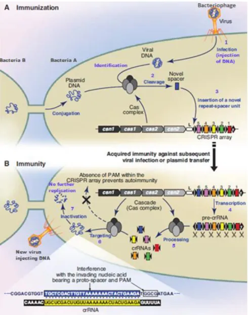
Most archaea and numerous bacteria have acquired immunity against plasmid transmission and infections caused by viruses that affect these prokaryotes called bacteriophages. This immunity is achieved by a highly developed adaptive immune system that is guided by RNA sequences which are encoded in CRISPR loci and the associated Cas genes (Figure 2A). CRISPR loci contain short, repetitive and partially palindromic base sequences of around 30 to 40 bp that are separated by similarly small and unique spacer sequences (Figure 2) obtained upon infection from plasmids or bacteriophages. Therefore, spacer sequences are responsible for yielding a genetic record of previous infections which in turn allows the prokaryote to avoid future infections from identical invaders (Figure 2B) 41,66.
Streptococcus pyogenes Cas9 is a large (1,368-amino-acid) multi-domain and multifunctional DNA endonuclease. It cuts double-strand DNA (dsDNA) 3 bp upstream of the PAM through its two different nuclease domains. The first one is an HNH-like nuclease domain that cleaves the DNA strand complementary to the guide RNA sequence, also known as the target strand, and an RuvC-like nuclease domain responsible for cleaving the DNA strand opposite to
the complementary strand, the nontarget strand27,28,67. Additionally, Cas9 also participates in crRNA maturation and spacer acquisition42.
The immunity provided by the CRISPR array can be divided into two phases, as shown in Figure 2. Initially during the immunization phase (Figure 2A), a Cas complex identifies sequences from plasmids or viral genome and integrates a newly acquired repeat-spacer section at the leader end of the CRISPR array, resulting in the immunization of the prokaryote. Subsequently, if another infection attempt of the same invader occurs, the immune response is activated (Figure 2B). During this stage of defence, the transcription of CRISPR sequence is set in motion resulting in a long pre-crRNA transcript that is processed into smaller and mature crRNAs that contains one spacer sequence at the 5’ end while the 3’ end contains a piece of CRISPR repeat sequence (Figure 2B). Afterwards, upon an infection of a familiar plasmid or bacteriophage, the mature crRNAs are used to guide the Cas endonucleases into recognizing and inactivating the foreign genetic material by cleaving plasmid or viral DNA (Figure 2B)13.
Figure 2-Crispr/Cas mechanism of action. A) Immunization phase: Upon insertion of foreign DNA into the affected
prokaryote, the genetic material is recognised by the Cas complex and, subsequently, cleaved and integrated as a newly acquired spacer into the leader end of the CRISPR array; B) Immunity phase: Transcription of the CRISPR array occurs and pre-crRNA is obtained which is further processed into short and mature crRNA. These mature crRNAs are then used as guides for Cas RNA-guided nucleases that typically form a complex to interfere with the invading DNA. The Cas complex then identifies and cleaves the corresponding target sequence of the foreign nucleic acid, thus inactivating it. The spacers are represented as coloured rectangles, the repeat sequences as black rhombus and the CRISPR leader sequence as a white box labelled L. Adapted from Horvath et al. 2010 (ref 1)
1.3.1 Mechanisms of different types of Crispr-Cas
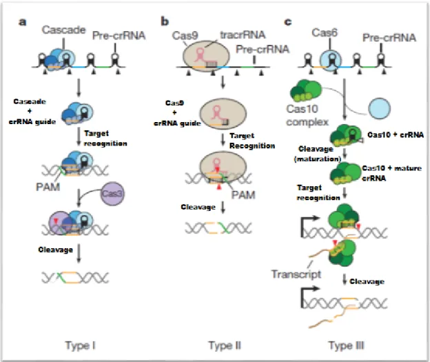
The cas1 and cas2 genes are present in all CRISPR/Cas systems and are known to be essential to the first phase of immunity provided by this defence mechanism. However, three distinct system types exist that can be distinguished based on the presence of accessory cas genes68. Despite the fact that the three types of CRISPR/Cas systems use the same method of DNA cleavage through crRNA-guided nucleases, they diverge in targeting requirements and the generation of crRNAs68.
The immunity provided by the type I CRISPR/Cas system is carried out by the Cas3 nuclease in combination with the Cascade complex (Figure 3A)20. After the pre-crRNA is produced by transcribing the CRISPR sequence, a Cascade subunit denominated Cas6e with endoribonuclease activity is responsible for the cleavage into small and mature crRNAs that stay linked to the complex and are, subsequently, employed by Cascade to find the target protospacer sequence20. Situated right upstream of the target sequence is a small sequence motif that is recognized by an additional subunit known as Cas869.
For the immunity mediated by the type I CRISPR/Cas system to occur, it is compulsory to recognize a PAM sequence. For this reason, an autoimmune response is avoided with the absence of a PAM sequence in the repeat sequences since crRNA is thus unable to target the spacers of the CRISPR sequence. When a matching sequence is flanked by a PAM, the Cascade complex is able to bind to the target DNA which results in an R-loop between the dsDNA and the crRNA. Finally, the target recognition by Cascade recruits and engages Cas3 that is responsible for introducing single-stranded DNA (ssDNA) breaks into the target plasmid or viral DNA70, consequently inactivating them71.
The immunity by the type II CRISPR/Cas system is achieved by using only a single cas gene called cas9 in combination with a crRNA guide for target recognition (Figure 3B). In contrast with the other types, the type II CRISPR/Cas system requires an additional short RNA known as the trans-activating crRNA (tracrRNA). This RNA is partially complementary to crRNA as it has an area that shares complementarity with the repeat sequences of CRISPR and additionally it establishes
a secondary structure facilitating its aggregation with the enzyme Cas9. The pre-crRNA is further processed into short crRNA guides by the cleavage mediated by ribonuclease (RNase) III that cleaves the double-strand RNA (dsRNA) formed between the pre-crRNA and the tracrRNA25.
Similarly, immunity mediated by the type II CRISPR/Cas system can only occur in the presence of a PAM sequence. This sequence is recognised by Cas9 through a PAM-binding domain but, differently from type I, in this immunity the PAM is located directly downstream of the sequence that is targeted. Cas9 has two nuclease domains known as HNH and RuvC that are responsible for the specific dsDNA breaks in both the DNA strands of the foreign DNA that is facilitated by the type II CRISPR/Cas system71. The target recognition is initiated by Cas9 transiently binding to PAM sequences of the foreign DNA, enabling the denaturation of the two DNA strands that are directly upstream of the PAM sequence. Targeted cleavage occurs after an R-loop forms due to a productive interaction of the denatured DNA with the crRNA guide in the aforementioned target area71.
In the immunity provided by the type III CRISPR/Cas system, a repeat-specific endoribonuclease that does not belong to a complex, called Cas6, is responsible for cleaving the pre-crRNA (Figure 3C). A sequence at the 5’ end of the spacer denominated crRNA tag contains 8 nucleotides remaining from the repeat sequence due to the CRISPR/Cas type processing 72. Afterwards, the short crRNAs produced through the cleavage mediated by Cas6 are relocated, by an unidentified mechanism, to a Cas10–Csm complex in case of a type III-A system or to Cas10– Cmr complex in case of a type III-B system73. These complexes are responsible for trimming the 3’ end of the crRNAs at intervals of 6 nucleotides, resulting in a mature crRNAs74. While the other system types depended solely on recognizing DNA sequences, the immunity provided by the type III CRISPR/Cas system additionally necessitates the transcription of the target sequence and complementarity to the transcript and the target non-template DNA strand by the crRNA for cleavage to occur75. Therefore, type III Crispr/Cas is able to target both the foreign DNA and its transcript, leading to the crRNA-guided cleavage by the Cas10 complex76. The cleavage of the non-template strand occurs in the palm domain of Cas10, while the cleavage of the RNA transcripts for the type III-A is mediated by the backbone subunit Csm3 and for the type III-B system by the backbone subunit Cmr476.
Figure 3- Mechanisms of different Crispr/Cas types. A) Type I systems possess a Cas protein complex (Cascade) that
is responsible for cleaving each repeat at the base of the stem–loop structure in the pre-crRNA, which results in the formation of short crRNA guides. Subsequently, the Cascade in combination with crRNA examines the target DNA to find a protospacer that is flanked by a PAM sequence. Finally, the crRNA anneals to the target strand forming an R-loop and, afterwards. the Cas3 nuclease is recruited and activated into cleaving the target sequence downstream of the PAM, resulting in the destruction of the opposite strand; B) Type II system encode a small tracrRNA that is aggregated to Cas9 and is partially complementary to repeat sequences of CRISPR. RNase III is responsible for cleaving the repeat/tracrRNA to obtain crRNA guides for the Cas9 nuclease. This enzyme after recognizing a target sequence, mediates the cleavage of both strands of the protospacer/crRNA R-loop; C) Type III systems encode a repeat-specific endoribonuclease that does not belong to a complex called Cas6, which is responsible for cleaving the pre-crRNA into crRNA that is subsequently relocated into the Cas10 complex where it is cleaved to produce a mature crRNA. The complex additionally necessitates transcription of the target sequence and complementarity to the transcript and the target DNA non-template strand by the crRNA for cleavage of both sequences to take place. Adapted from Marrafini et al. 2015 (ref 22)
The type III CRISPR/Cas system depends on the disparity in base pairing of the crRNA and the sequences that are adjacent to the protospacer to avoid targeting the CRISPR array72. Therefore, no autoimmunity is caused by this system because the DNA targeting is prevented when the crRNA tag is totally complementary to the DNA repeats within the CRISPR locus.
Contrarily, DNA targeting is permitted in case of incomplete complementarity between the sequences flanking the protospacer and the crRNA tag72.
1.4 Delivery Methods of CRISPR/Cas9
The sgRNAs and Cas9 can be delivered by different methods depending on the application including DNA constructs, mRNA constructs or Cas9 ribonucleoproteins77. While both crRNA and tracrRNA can be delivered separately, the combination into a single chimeric gRNA simplifies both design and delivery. The gRNA can either be expressed from a plasmid inside the cell or generated via in vitro transcription77. One way of delivery is through PCR amplicons containing an expression cassette2. PCR-based sgRNA delivery attaches the custom sgRNA sequence onto the reverse PCR primer used to amplify a U6 promoter template and the resulting amplicon could be co-transfected with a Cas9 expression plasmid. Since this method removes the need for plasmid-based cloning and sequence verification, it is suitable for testing or co-transfecting numerous sgRNAs for generating large knockout libraries or other scale-sensitive applications2.
Early efforts in mammalian cells focused on encoding Cas9 in the form of DNA plasmids63,78. In this method, a single plasmid can be used to encode both the chimeric gRNA and the Cas9 under separate promoters. Typically, the Cas9 is encoded as a fusion to a nuclear localization signal that mediates intranuclear transduction upon expression, thus allowing access to the genetic material of the cell63,78,79. The delivery of the plasmid DNA into the cells is attained either via standard chemical transfection or electroporation methods77.
A more complex delivery method is packaging the Cas9 DNA in a single-stranded form within a non-integrating virus such as adeno-associated virus (AAV)80. However, the maximum packaging capacity for AAV is approximately 4.5 kb81, which makes combination of both the Cas9 and cognate gRNA into a single capsid challenging. As such, smaller Cas9 variants82 and several systems to create split Cas9 enzymes83–85, allowing division between two AAV vectors, have been developed but both suffer from reduced efficacy82–85. Another path for AAV-based delivery lies in the use of significantly smaller Cas9 orthologs from other species as reported by Ran et al.86.
Cas9 isolated from S. aureus can edit mammalian genomes with similar efficiencies to S. Pyogenes Cas9 while being more than 1 kb shorter when encoded in DNA form, allowing packaging within a single AAV capsid86.
Another way of reducing the size of Cas9 delivered to cells is to generate RNA transcripts of the gene. Cas9 mRNA delivery has been extensively adopted for the ex vivo modification of mammalian embryos by microinjection87–89, and more recently in human somatic cell lines90, or primary cells91 via electroporation. RNA constructs can also be delivered in viral form, usually in the form of integrase defective lentiviral particles. The lentivirus capsid has a substantially larger capacity for nucleic acid (approximately 8 kb)92 compared to AAV, and so it can be engineered to express both Cas9 and up to four gRNAs simultaneously93,94.
A more recent line of investigation has been to deliver ribonucleoproteins (RNPs), which are composed of Cas9 protein precomplexed with gRNA90,95,96. Delivering Cas9 in protein form leads to fewer observed off-target mutations than delivery via plasmid DNA90,95,96. Non-standard techniques for the delivery of Cas9 RNPs are also being explored. These include lipid-based transfection reagents97, nanoparticle carriers of Cas998 and non-lipid carriers that mimic vesicle-like structures99.
The range of delivery options for Cas9 RNPs is further expanded upon modification of the Cas9 protein termini to allow for covalent functionalization such as conjugation to cell penetrating peptides (CPPs)100,101.
2. APPLICATIONS
Engineered nucleases have quickly become a frequently used method by researchers for targeted genome editing, especially because of the recent arrival of CRISPR with the combination of the highly customizable Cas9102.
Genome editing with these engineered nucleases has been used in a quick, easy and efficient manner to modify endogenous genes in a diverse set of biomedical relevant cell types and in organisms that have previously been difficult to modify genetically 32. It has quickly overcome TALENs103 and ZFNs104 where editing was excessively complex and arduous.
In the research arena, versatile CRISPR-enabled genome editing has been used in various fields, such as medicine65, biotechnology105 and biology106 with diverse applications such as: controlling transcription, modifying epigenomes, conducting genome-wide screens, imaging chromosomes,manipulating biological circuits facilitating the generation of synthetic materials, correcting gene mutations, control the expression of entire genes offering longer term expression alteration compared to other methods such as RNAi102,107. CRISPR systems are already being used to improve genetic disorders in animals and are likely to be employed soon to treat human diseases of the eye and blood108. Beyond biomedical applications, this system is already being used to expedite crop109,110 and livestock breeding111, develop new antimicrobials112 and control insects that carry diseases with gene drives113. The CRISPR technology is already providing a new class of genetic models that are more suited for diverse applications such as rapid diagnostics, fundamental disease research, drug screening and therapy development, in vivo editing and correction of inherited conditions114.
Even though the genome-wide specificities of the CRISPR/Cas9 technology remain to be fully understood, the ability of these systems to execute targeted alterations of genome sequences and gene expressions with high efficiency will unquestionably alter biological research and encourage the development of new molecular therapeutics for diseases affecting mankind.114
The applications of the CRISPR/Cas9 system in human pathologies will be further explored in the following chapters.
2.1 Antiviral Therapy
Antiviral therapy through designer nuclease CRISPR/Cas9 has become a promising new tool against human infecting viruses. This method has been used successfully to target viral genes or host genes that encode vital receptors to inhibit infection and replication of viruses115.
2.1.1 HIV
The human immunodeficiency virus (HIV) is a member of the genus Lentivirus, part of the family Retroviridae116 that causes a viral infection and progresses to an acquired immunodeficiency syndrome (AIDS). AIDS is responsible for a progressive deterioration of the human immune system, allowing lethal opportunistic infections and cancers to flourish117. Transmission of HIV occurs by blood transfer, pre-ejaculate, semen, vaginal fluids, and from mother to child during pregnancy, delivery or breastfeeding118.
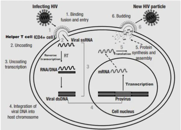
HIV infects essentially the human immune system cells including the vital helper T cells (CD4+ T cells) (Figure 4), dendritic cells and macrophages. The infection, consequently, leads to low levels of helper T cells through numerous mechanisms, including apoptosis of uninfected bystander cells, pyroptosis of abortively infected T cells, direct viral killing of infected cells, and by CD8 cytotoxic lymphocytes killing the infected cells. Cell-mediated immunity can be lost if helper T cell numbers drop below a set level, which leads to increasing susceptibility to opportunistic infections119.
Therapeutic interventions mainly target two key retroviral enzymes: reverse transcriptase and protease. In combination, these antiviral drugs have greatly diminished both mortality and morbidity in HIV infected patients. However, when administered over prolonged periods, these drugs induce considerable toxicity, and their effectiveness is undermined by the emergence of drug-resistant strains of HIV120.
Although current anti-HIV therapies can inhibit HIV-1 replication, the viruses that have Figure 4- HIV replication cycle. The main steps in HIV replication are sequentially numbered from 1 to 6. (1) Virus
binds to CD4 and the appropriate coreceptor resulting in fusion of the viral envelope and the cellular membrane, leading to the viral nucleocapsid being released into the cytoplasm. (2) The viral RNA is uncoated. (3) subsequently, reverse transcription mediated by the Reverse Transcriptase (RT) occurs. (4) The obtained viral dsDNA then migrates into nucleus of the affected CD4+ cell and is posteriorly integrated into the cellular DNA by the enzyme Integrase. (5) Afterwards, transcription of the proviral DNA mediated by the cellular RNA polymerase II occurs producing mRNAs. Then, translation of these RNA molecules is facilitated by the cellular polyribosomes. (6) Genomic RNA and proteins of viral origin are transported to the cellular membrane, where assembly occurs. Immature virions are released. The viral protease is then responsible for processing the polypeptide precursors to generate mature viral particles. After migrating to the cell’s plasma membrane, the virus particles suffer a budding process, resulting in the release of the new HIV particle. Adapted from Fanales-Belasio et al. 2010 (ref 122)
The CRISPR/Cas9 system has proved to be useful for eliminating latent HIV-1 by targeting its genomic DNA. Several groups have reported that they successfully disrupted the expression of HIV-1 provirus in infected cells utilizing the CRISPR/Cas9 system121–124. More recently a team successfully excised the HIV-1 provirus in three different animal models using an all-in-one adeno-associated virus vector to deliver multiplex sgRNAs and Staphylococcus aureus Cas9 (saCas9)108. The published results demonstrate the plausibility of engineering Cas9/gRNA to precise and efficiently obtain a prophylactic and therapeutic method against AIDS.
2.1.2 Epstein-Barr Virus
Epstein-Barr virus (EBV) or human herpesvirus 4, belongs to the herpes virus family and is one of the most common human viruses125. The virus usually spreads through bodily fluids, mostly saliva125. EBV can cause several health issues such as infectious mononucleosis, some forms of cancer and can also affect the nervous and autoimmune systems125.
Most infections occur during infancy and early childhood and present either no symptoms or nonspecific symptoms. However, in adolescents and young adults, EBV infection commonly results in infectious mononucleosis with symptoms including fever, sore throat, lymphadenopathy and splenomegaly. Additionally, other symptoms and signs may be present such as headache, fatigue, rash and hepatomegaly125.
EBV infects B cells of the immune system and epithelial cells. After controlling the initial lytic infection, EBV persists latently in B cells for the rest of the affected person’s life. In some cases, the virus may reactivate in asymptomatic form and people with weakened immune systems can even develop symptoms. If EBV reactivates it can infect others125.
Currently there is no treatment for Epstein-Barr Virus, however some measures can be taken to help relieve the symptoms including fluid intake, resting and using medication for pain and fever125.
In 2014, Wang et al.126 reported to have successfully applied CRISPR/Cas9 as an antiviral treatment in human cells by specifically targeting the genomes of latent Epstein-Barr virus. There
was a halt in proliferation and an associated reduction in viral load in cells from a Burkitt’s lymphoma with latent EBV after being exposed to the CRISPR/Cas9 system that targeted the viral genome.
In 2015 Yuen et al.127 used two gRNAs to introduce a 558bp deletion in the promoter region of BamHI-A rightward transcript (BART) which encodes viral microRNAs (miRNAs) on the EBV genome. Numerous latently infected human epithelial cell lines such as nasopharyngeal carcinoma C666-1128 cells were successfully edited. They observed efficient editing of the EBV genome by the CRISPR/cas9 system as the whole pBART region was eliminated which resulted in the loss of miR-BART expression and activity. Their results represented the first genetic evidence that the major promoter for the expression of BART is pBART. Finally, after cells expressing Cas9 and gRNAs were selected with puromycin, a recombinant virus with the intended deletion was obtained and deep sequencing revealed no off-target cleavage127.
Van Diemen and his team129 showed that the CRISPR/Cas9 system is able to directly edit the genome of latent EBV in EBV-positive tumor cells and that targeting vital areas of the virus dsDNA efficiently reduces the content of viral genome in latently infected cells. They designed gRNAs aiming for the viral EBV nuclear antigen 1 (EBNA1) and numerous parts of the origin of replication (OriP) of the Epstein-Barr virus that are involved in its replication and episome maintenance130,131.
Their team used Burkitt’s lymphoma Akata-Bx1 cells as model system. These cells carry a recombinant EBV expressing green fluorescence protein (eGFP) under control of the cytomegalovirus (CMV) promoter132. Therefore, the expression of the eGFP is a signal of EBV presence in these cells129. After the transduction of the cells with their corresponding gRNAs, a loss of eGFP expression was observed. A near full loss of eGFP from most of the cells was observed after introducing a combination of these active gRNAs. Finally, they showed that the most efficient method was targeting EBNA1 with two different gRNAs, which induced over 95% loss of EBV genomes129.
The results obtained by Van Diemen’s team demonstrate that viral genome content in latently EBV infected cells can by efficiently reduced by the CRISPR/Cas9 system when it is
targeted at vital areas of the EBV dsDNA and that this reduction could culminate on a termination of the tumorigenic, cell cycle-promoting roles carried out by EBV and the loss of anti-inflammatory functions and counter-apoptotic features of gene products encoded by the virus. Therefore, this approach might be a new therapeutic strategy to fight malignancies associated with the Epstein-Barr virus129.
In early 2017, Ma and colleagues133 used the CRISPR/Cas9 system to perform genome-wide loss-of-function screens to identify host dependency factors that are critical for the EBV infected lymphoblastoid and Burkitt lymphoma B cell growth and survival. They managed to identify multiple non-redundant mechanisms by which EBV prevents apoptotic responses to oncogene stress in transformed B cells and identified key EBV-induced synthetic lethal targets for therapeutic intervention.
Yuen et al.134 reported suppression of EBV in latently infected nasopharyngeal carcinoma cells when using the CRISPR/Cas9 system. They studied the possibility of the CRISPR/Cas9 system to induce a reduction of EBV levels by targeting the genome of the virus in infected nasopharyngeal carcinoma cells. They engineered several gRNAs aimed towards different areas of the viral genome and transfected them into C666-1 cells128. The team observed a reduction by half of the viral DNA levels in C666-1 cells and even though this effect lasted for weeks134. Lastly, they observed that the survival of C666-1 cells did not change. However, these cells became sensitized to the chemotherapeutic effect of 5-fluorouracil and cisplatin134. The authors consider that this work provides the proof-of-concept for suppressing EBV DNA load with CRISPR/Cas9 and that it may lead to a potential new strategy to sensitize EBV infected nasopharyngeal carcinoma cells to chemotherapy which in turn might reduce the necessary dose of these drugs, thereby alleviating the side effects. However, the team has concerns about possible off-target effects and the adequate delivery vehicle. All these issues need to be addressed before CRISPR/Cas9 technology can enter the next phase of medical applications134.
2.1.3 Hepatitis B virus
Hepatitis B is a viral infection affecting the liver caused by the Hepatitis B virus (HBV)135. The virus is transmitted from person to person through contact with the infected blood, semen or other infected body fluids136. Normally, it is an acute infection with a short duration, but it can also become a lasting, chronic infection. The risk of developing a chronic infection is associated with the age of the infected person. Most (90%) infected infants become chronically infected, while only a small fraction of infected adults (2% to 6%) become chronically ill135. It is best to prevent Hepatitis B infection by getting vaccinated as the chronic illness can lead to severe health problems, including hepatocellular carcinoma or cirrhosis136.
It may take approximately 90 days for symptoms to appear. The initial infection may present no symptoms in some people, while others may develop a rapid onset of sickness. The symptoms commonly last some weeks and this initial stage of the disease rarely results in death of the infected person135. The presence of symptoms differs depending on the age. Most children under age 5 and newly infected immunosuppressed adults are asymptomatic, whereas 30% to 50% of persons over 5 years old have initial symptoms136. The symptoms are mainly non-specific and can include: Fatigue, fever, nausea, loss of appetite, vomiting, abdominal pain, joint pain, jaundice, dark urine and clay-coloured bowel movements135.
Since 1982, there has been an effective vaccine against hepatitis B. The vaccine is 95% effective in preventing early viral infection and the progress into chronic illness and liver cancer136. There is no specific treatment for acute hepatitis B and the only therapeutic option available is to treat the patient’s symptoms relieving their discomfort. For example, lost fluids from diarrhea and vomiting are replenished to guarantee appropriate nutritional balance136. On the other hand, chronic hepatitis B infection can be treated with antiviral medication. The treatment can slow down the development of cirrhosis, decrease occurrence of liver cancer and ultimately improve long-term survival of those affected by the virus. However, the abovementioned antiviral therapy only suppresses the viral replication and therefore does not provide a cure for hepatitis B136.
Several in vitro and in vivo studies demonstrated that using the CRISPR/Cas9 system to target the HBV cccDNA is successful and efficiently inhibits HBV replication137–142. Seeger et al.137 showed that their team could inhibit HBV infections up to eightfold by testing several HBV-specific gRNAs. They demonstrated that Cas9 could certainly cleave the virus-derived cccDNA and that the resulting cleaved cccDNA is swiftly repaired, possibly by the NHEJ pathway. Even though the authors of this study did not show the experimental feasibility, they consider that this method can be applied to future investigations that study the role of host genes in the viral life cycle by inactivating relevant cellular genes.
Lin et al.138 designed eight sgRNAs that target the P1 and XCp genes on HBV. In this study, they found a significant reduction in the production of core and HBsAg proteins in Huh-7 hepatocytes derived from cellular carcinoma cells. In a mouse model, it was shown that this method could cleave the intrahepatic plasmid that contained the viral genome and enable its clearance, which resulted in a decrease of serum surface antigen levels138.
Furthermore, Zhen et al.139 used CRISPR-Cas9 to target the Hepatitis B antigen (HBsAg) on HBV in vitro culture and within in vivo systems that were confirmed by quantitative enzyme-linked immunosorbent assay (ELISA) and quantitative polymerase chain reaction (qPCR). The total amount of HBsAg secreted into the cell culture and in the mouse serum was reduced after editing the gene with CRISPR-Cas9. In the same study, they also found that no HBsAg-positive cells persisted in the liver tissue of CRISPR-Cas9 treated mice, and this was confirmed by immunohistochemistry139.
CRISPR/Cas9 system has also been used by Dong and colleagues140 to target the HBV cccDNA, which is highly stable, and a prime target for the inhibition of HBV infection. This work showed a reduction in the generation of the virus in Huh7 cells and in HepG2.2.15 HBV-replication cells140. Similarly, Kennedy et al.141 used Cas9 and HBV-specific sgRNAs. The total viral DNA load was reduced by up to 1000-fold and cccDNA was reduced by up to 10-fold, and outstandingly remaining viral DNA was mutated141.
These studies can be further expanded and explored with the hope of developing a novel therapeutic strategy, not only against chronic HBV infection, but also against the wider family of other hepatitis viruses in the future.
2.1.4 Human papillomavirus
Human papillomavirus (HPV) is a DNA virus belonging to the papillomavirus family and can cause infection in humans affecting the skin and the moist membranes lining of the body (cervix, anus, mouth and throat)143,144.
HPV is the most common sexually transmitted infection (STI) worldwide and there are more than 170 types of HPV, of these at least 13 are known to cause cancer143,144. The virus is primarily transmitted by sexual interaction and, generally, people are infected with it soon after the beginning of sexual activity144. Most HPV infections do not cause symptoms but some HPV types (primarily types 6 and 11) can cause warts143. A small proportion of infections with some types of HPV can persist and progress to various types of cancer such as: cervical cancer (approximately 70% of cervical cancer cases can be attributable to HPV infection), cancers of the anus, vulva, vagina, and penis144.
Moreover, persistent infection with specific types of HPV, most frequently types 16 and 18, have the highest risk for development of genital cancers143. Symptoms of cervical cancer normally only appear after it has gotten to a more advanced stage and may include non-specific symptoms: loss of appetite and weight loss; fatigue; leg, back or pelvic pain; a single swollen leg; vaginal odorous discharge or discomfort; and irregular, abnormal or intermenstrual vaginal bleeding after sexual intercourse143.
There are currently 3 vaccines that protect against some cancer causing HPV types: Cervarix® protects against HPV types 16 and 18145, Gardasil® protects against HPV types 6, 11, 16, 18146 and Gardasil 9® which in addition to protecting against infection by strains covered by the previous generation of Gardasil, also protects against five other HPV strains (31, 33, 45, 52, 58) that are the cause for one fifth of cervical cancers147.