diagnosis might be presumptive, if there is a typical cu-taneous presentation with unknown etiology.5
We present a review of 8 cases diagnosed between January 2008 and December 2016 in the Neonatal Unit or followed in outpatient departments in Centro Hos-pitalar de São João (CHSJ), Porto (Table I).
cAse reports
cAse 1
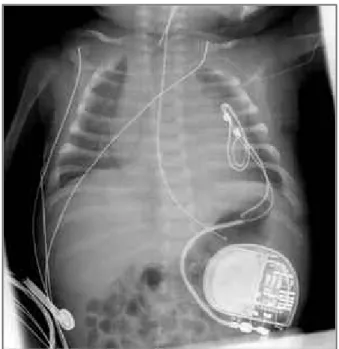
Female newborn (NB) with prenatal diagnosis (PND) of complete heart block (CHB). Her mother had ANA 1/1000 speckled pattern, Anti-dsDNA <10.0 UI/mL, anti--Ro/SSA positive and was assumed as having a probably Sjögren syndrome (SS), despite the absence of symp-toms. In the past, this woman underwent to a salivary gland biopsy, with inconclusive result, and to a salivary gland scintigraphy, which showed a decreased salivary secretion. She was treated with salbutamol, dexametha-sone, hydroxychloroquine (HCQ) 200 mg and intra-venous immunoglobulin (IVIG) during pregnancy. After birth, the child was admitted to the Neonatal Intensive Care Unit (NICU), where a pacemaker was implanted (Figure 1). Almost 2 years later, the child has ascending aortic dilatation (18mm) without other structural cardiac abnormalities and developed no further complications. cAse 2
Female NB with PND of CHB (Figure 2) and suspe cted pulmonary valve stenosis. Consequently, her mother was diagnosed with Systemic Lupus Erythematosus (SLE) in the last week of gestation, not receiving any treatment during pregnancy. She had ANA 1/320 nu-cleolar pattern, anti-dsDNA 0.5 UI/mL, anti-Ro/SSA and anti-La/SSB positive and iron deficiency anaemia unresponsive to oral iron. The NB was admitted to NICU, where she received a definitive pacemaker and pulmonary valve stenosis was excluded. On day 2 she had thrombocytopenia, abnormal liver function tests and positivity for anti-Ro/SSA and La/SSB. At the age of 1. Faculdade Medicina Universidade do Porto
2. Serviço de Pediatria, Centro Hospitalar de São João, Porto 3. Serviço de Neonatologia, Centro Hospitalar de São João, Porto 4. Serviço de Cardiologia Pediátrica, Centro Hospitalar de São João, Porto
5. Serviço de Reumatologia, Centro Hospitalar de São João, Porto
Neonatal lupus – case series of a tertiary hospital
Teixeira AR1, Rodrigues M2, Guimarães H3, Moura C4, Brito I5 ACTA REUMATOL PORT. 2017;42:318-323
AbstrAct
Neonatal lupus (NL) is a very rare condition with an es-timated incidence of 1 in 20.000 pregnancies. It is caused by the transplacental passage of autoantibodies anti-Ro/SSA, anti-La/SSB antibodies and/or anti-U1 RNP antibodies into the fetal circulation. The mother may be completely asymptomatic or have a known in-flammatory rheumatic disease, such as Sjögren syn-drome (SS) or Systemic Lupus Erythematosus (SLE). Clinical mani festations are diverse, being the most com-mon cutaneous and cardiac. The authors present a case series of eight cases diagnosed with NL between Jan-uary 2008 and December 2016 in a tertiary hospital and a brief revision of the literature.
Keywords: Anti-U1 RNP antibodies; Neonatal lupus;
Anti-La/SSB antibodies; Anti-Ro/SSA antibodies.
IntroductIon
Neonatal lupus (NL) is an autoimmune disorder with different manifestations. Cutaneous and cardiac mani-festations are the most common and may coexist in 10% of cases, being the first one more harmless1-3.
Usu-ally, NL is observed at birth but the skin lesions may appe ar only during the first weeks of life. Their mot hers have positive autoantibodies anti-Ro/SSA (90% of the cases), anti-La/SSB and/or anti-U1 RNP4.
Diagnosis of NL requires a high level of suspicion. It is made by clinical examination or complementary diagnos tic tests (for instance, echocardiography or elec-trocardiography) and laboratory demonstration of NL--associated antibodies in the mother and/or child.4The
8 months, dilated cardiomyopathy with an ejection fraction of 21%, pericardial effusion and a lupus myo -carditis was diagnosed. Despite medical treatment, she died at age of 2 from cardiac heart failure.
cAse 3
Male NB with PND of CHB and mild to moderate car-diomegaly. This was the brother of case 2 and their mother maintained antiRo/SSA and antiLa/SSB posi -ti vity. She was treated with prednisolone and HCQ during pregnancy, and she started salbutamol 32mg/d and dexamethasone 8 mg/d on week 26. NB was also
admitted to NICU where he underwent pacemaker placement. Echocardiogram didn’t show heart abnor-malities. At day 23, he developed hypoxemic respira-tory failure and required mechanical ventilation. He didn t respond to antibiotics and no infectious agents were found. The chest radiograph showed bilateral in-terstitial infiltrates (Figure 3), characterized as ground glass opacities in chest CT scan. A diagnosis of lupus pneumonitis was presumed and he received IV methylprednisolone with clinical improvement. He was positive for anti-Ro/SSA. He is now 14 months and has had a favourable evolution.
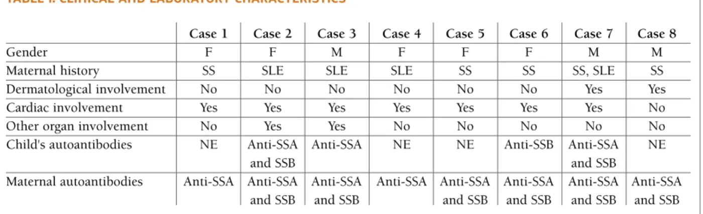
FIGure 1.Case 1; Electrocardiogram – Evidence of ventricular pacing (spike) previous to each QRS complex tAbLe I. cLInIcAL And LAborAtory chArActerIstIcs
Case 1 Case 2 Case 3 Case 4 Case 5 Case 6 Case 7 Case 8
Gender F F M F F F M M
Maternal history SS SLE SLE SLE SS SS SS, SLE SS
Dermatological involvement No No No No No No Yes Yes
Cardiac involvement Yes Yes Yes Yes Yes Yes Yes No
Other organ involvement No Yes Yes No No No No No
Child's autoantibodies NE Anti-SSA Anti-SSA NE NE Anti-SSB Anti-SSA NE
and SSB and SSB
Maternal autoantibodies Anti-SSA Anti-SSA Anti-SSA Anti-SSA Anti-SSA Anti-SSA Anti-SSA Anti-SSA and SSB and SSB and SSB and SSB and SSB and SSB F: Female; M: Male; SS: Sjögren Syndrome; SLE: Systemic Lupus Erythematosus; NE: Not Evaluated
cAse 4
Female NB with PND of bradyarrhythmia. The mo ther had anti-Ro/SSA positive and ANA 1/1000 speckled pattern but was asymptomatic, and only developed SLE four years later. The NB is one of two twins. In the NICU, she was diagnosed with first-degree AV block with episodes of second-degree AV block. Her sister didn’t have any NL manifestations. Currently, this girl is 5 years old, asymptomatic and is followed by Pedia -tric Cardiology. She never required pacing and main-tains first-degree AV block.
cAse 5
Female NB with a PND of CHB. The mother has SS with ANA 1/320 speckled pattern, anti-Ro/SSA and anti-La/SSB positive. After she was born, she was ad-mitted to NICU. She is currently 8 years old and she was always asymptomatic, never requiring pacing. She presents left auricular and ventricular dilatation, mild mitral and aortic insufficiency with a normal ejection fraction and an enlargement of the pulmonary trunk. Five years later, her mother remained antiRo/SSA posi -tive and delivered a girl without any NL manifestations. cAse 6
Female NB with a PND of CHB. The mother had ANA 1/1000 speckled pattern, anti-Ro/SSA and anti-La/SSB positive. She was treated with salbutamol and dexame thasone but just diagnosed with SS months after pre gnancy, when symptoms appeared. The NB was admi -tted to NICU and required pacemaker placement. The NB had ANA 1/320 speckled pattern and positive anti--La/SSB. She currently has mild tricuspid insufficiency
and is followed by Pediatric Cardiology. Two years la ter, this mother had a girl without any NL manifestation. cAse 7
Male NB born at term by elective caesarean section. Due to bradycardia he was admitted to NICU, where a mild facial rash was also identified. He was dis-charged at day 3 after spontaneous resolution of brady-cardia, echocardiogram did not show any abnormali-ties. His mother has SS/SLE overlap, with ANA 1/1000 homogenous pattern, dsDNA 162.6 UI/mL, anti--Ro/SSA and anti-La/SSB positive. She was treated with HCQ and aspirin during pregnancy. The child had ANA 1/320 speckled pattern, Ro/SSA and anti--La/SSB positivity. The skin lesions worsened progres-sively, becoming a malar rash and a maculopapular rash on scalp, neck, trunk and abdomen (Figure 4). Skin biopsy showed vacuolar degeneration of basal layer, perivascular inflammatory lymphocytic infiltra-tion of the superficial dermis and dermal mucinous deposits with normal direct immunofluorescence, which corresponds to NL diagnosis. He was treated with 1% topical hydrocortisone and the lesions disap-peared without sequel.
cAse 8
Male NB had a malar rash at birth. His mother has SS, FIGure 2. Case 2; Color M-mode fetal echocardiography with
evidence of complete heart block (CHB); 1 – Atrial Heart Rate (HR), 2 – Ventricular Heart Rate
FIGure 3. Case 3; Chest radiograph at day 24 with bilateral interstitial infiltrates
with ANA 1/1000 speckled pattern, anti-dsDNA <10.0 UI/mL, anti-Ro/SSA and anti-La/SSB positive. She was treated with HCQ and aspirin. During the pregnancy, she also developed hypothyroidism and received levothyroxine from week 27. At 1 month of age, he was evaluated by Dermatology due to worsened malar rash, which became annular and erythematodesqua-mative. After treatment with 1% topical hydrocorti-sone, the lesions resolved completely within 5 days. Given the favourable outcome, no further tests were performed.
dIscussIon
NL is a rare disease with an estimated incidence of 1 in 20.000 pregnancies, but it seems be underdiagno -sed. On one hand, it is possible that mothers don’t have an inflammatory rheumatic disease. On the other hand, cutaneous manifestations may mimic rash from other causes and some other manifestations are mild, transient and might go unnoticed. Even if noticed, the
attribution of manifestations to NL may be difficult5,6.
Cutaneous NL has a female predominance, with a fe-male-male ratio of 2:1 to 3:1.7Cardiac NL has an equal
female-male distribution4.
It is accepted that the transplacental passage of these IgG autoantibodies from the second trimester is res -ponsible for the appearance of NL8.
Approximately 40-60% of mothers are asymp-tomatic or without known autoimmune diseases when the infants are diagnosed with NL, as we verified in cases 1, 2, 4 and 6. 50% of them will later develop symptoms related to autoimmunity, like in case 44,5,9.
Mothers may have SLE, SS, rheumatoid arthritis, mixed connective tissue disease and undifferentiated connective tissue disease4,5. In our series, mothers had
as much SLE as SS (Table I).
Although 98% of NL cases are positive for anti--Ro/SSA, La/SSB and U1RNP, just 1-2% of the women with these antibodies have neonates with the disease4.
In subsequent pregnancies, this incidence is higher, around 25%. In women with Ro/SSA or anti--La/SSB antibodies, recurrence of CHB in subsequent pregnancies is 15%.5In this case series, three women
had subsequent pregnancies: two women with anti--Ro/SSA and anti-La/SSB positivity, but just one had a child with NL, and another woman with anti-Ro/SSA positivity, who had a healthy child.
In fact, only some neonates exposed to the antibo -dies developed NL. Perhaps other factors may predis-pose for the disease, like the titers of maternal anti-bodies, genetic predisposition and environmental fac-tors. In case 4, a dizygotic twin pregnancy resulted in just one affected fetus, which might be genetically sus-ceptible4.
Cutaneous lesions usually appear between the 4th--6th week of life, but may be present at birth10. They
consist on multiple red macules that evolve.by clea ring
the center and developing an annular configuration2.
Typically, they are localized in photoexposed areas, like the head or neck, but may also occur on the trunk or the extremities2,3. If the periorbital area is affected, a
typical signal named “raccoon eyes” or “owl eyes” is originated10. They resolve within 4-6 months, when
maternal autoantibodies disappear from the circula-tion. Very rarely, it remains as permanent telangiecta-sia.3Despite the spontaneous resolution, treatment
may include sun avoidance, sunscreen and mild topi-cal steroids, as in cases 7 and 82,3. It is important to
es-tablish the differential diagnosis with other patholo-gies like atopic dermatitis and neonatal acne3. Skin FIGure 4. Case 7; Malar rash and maculopapular rash on
biopsy is only necessary when there is doubt about the NL diagnosis5.
Heart involvement is mostly diagnosed between 1824th weeks of gestation. It is a potentially lifethreate -ning complication, with a mortality rate of 15-30%, especially during fetal, neonatal and infant periods11.
The CHB is most commonly associated with maternal anti-Ro/SSA antibodies with or without anti-La/SSB, as in our 6 cases of cardiac involvement11,12. The
autoantibodies induce inflammation and fibrosis of atrio -ventricular node12. Depending on the degree of
scarring, the AV block severity is different. First and se -cond-degree AV blocks may reduce its degree or even resolve completely, but third-degree is irreversible11.
Some individuals may develop other severe com-plications such as valvular insufficiency or endocar-dial fibroelastosis that may progress to end-stage heart failure and death. Recently, an association between CHB in NL and aortic dilatation has been reported, as in case 112. Furthermore, some authors described a
dif-fuse myocardial disease, before or after birth, and iso-lated cardiomyopathy, like in case 211.
After the PND of AV block, it is advisable to start dexamethasone/betamethasone and/or IVIG therapy, due to their potential benefit in reversing first or se -cond degree block and preventing the development of CHB in foetuses, respectively. In our case report, 3 ca -ses were treated with dexamethasone and 1 with also IVIG but they maintained the CHB since the block was already established. Several studies had found that, if foetal ventricular rate <5055 beats/min, there is pro -fit in adding a sympathomimetic11.
Recently, has been suggested a role of Toll-like Re-ceptors (TLR) in the pathogenesis of cardiac-NL. Con-sequently, HCQ, an inhibitor of TLR ligation, has been studied in women with SLE. Results demonstrated that HCQ prevents disease flare during pregnancy and, in patients with anti-Ro/SSA antibodies and previous child with cardiac NL, HCQ may reduce the recur-rence of cardiac NL in subsequent pregnancies8,13. In
case 1 and 3, HCQ couldn’t prevent the appearance of CHB but, in case 7 may played a role in reversing bradycardia. Case 8 only had cutaneous manifesta-tions.
Newborns with cardiac involvement should be ad-mitted to NICU, as we verified in this case series. It is known that 63% of all recognized cases of AV block re-quire pacemaker implantation, which happened in 4 of our 6 cases of cardiac involvement11.
The hepatobiliary system may also be affected, with
only asymptomatic and transient elevation of liver function tests, such as in case 2, or with severe liver failure4,10.
Hematological abnormalities occur in 10-35% of the NL cases. It may include thrombocytopenia (like in case 2), neutropenia and anemia, isolated or in any combination, which resolve within days to months6.
The central nervous system (CNS) is only affected in 1.4% of patients with NL. It is most commonly asym ptomatic and neuroimaging-determined14.
Very rarely, the respiratory system may be affected with a pneumonitis-like presentation, like in case 3.4
There is no doubt that early detection of this con-dition is paramount, especially when there is cardiac involvement, since adequate treatment may reverse lower grade HB. Pregnant woman with anti-Ro/SSA and anti-La/SSB should be referred to experienced cen-tres early on, since surveillance with serial echocar-diography and ultrasonography is fundamental, as is adequate treatment of the maternal underlying condi-tion.
correspondence to
Ana Raquel Martins Teixeira
Rua Dr. Eduardo Santos Silva, 400, 3B E-mail: [email protected]
reFerences
1. Porcel Chacon R, Tapia Ceballos L, Diaz Cabrera R, Gutierrez Perandones MT. Neonatal lupus erythematosis: a five-year case review. Reumatol Clin, 2014; 10: 170-173.
2. Moretti D, Cimaz R, Vannucci G, Marino A, De Martino M, Gre-co A. Cutaneous neonatal lupus: a case report and review of the literature. Int J Dermatol, 2014; 53: 1508-1512.
3. Barcelos A, Fernandes B. Cutaneous manifestations of neona-tal lupus: a case report. Acta Reumatol Port, 2012;37:352-354. 4. Hon KL, Leung AK. Neonatal lupus erythematosus.
Autoim-mune Dis, 2012; 2012: 301274.
5. Johnson B, Overview of neonatal lupus. J Pediatr Health Care, 2014; 28: 331-341.
6. Chao MM, Luchtman-Jones L, Silverman RA. Hematological complications of neonatal lupus: case report and review of the literature. J Pediatr Hematol Oncol, 2013; 35: e344-346. 7. Silverman E, Jaeggi E. Non-cardiac manifestations of neonatal
lupus erythematosus. Scand J Immunol, 2010; 72: 223-225. 8. Lazzaroni MG, Dall’Ara F, Fredi M, et al. A comprehensive
re-view of the clinical approach to pregnancy and systemic lupus erythematosus. J Autoimmun, 2016.
9. Klein-Gitelman MS. Neonatal Lupus: What We Have Learned and Current Approaches to Care. Curr Rheumatol Rep, 2016; 18: 60.
10. Inzinger M, Salmhofer W, Binder B. Neonatal lupus erythema-tosus and its clinical variability. J Dtsch Dermatol Ges, 2012; 10: 407-411.
11. Yildirim A, Tunaoolu FS, Karaaoac AT. Neonatal congenital heart block. Indian Pediatr, 2013; 50: 483-488.
12. Brito-Zeron P, Izmirly PM, Ramos-Casals M, Buyon JP, Kha-mashta MA. Autoimmune congenital heart block: complex and unusual situations. Lupus, 2016; 25: 116-128.
13. Izmirly PM, Pisoni CN, Khamashta MA, et al. Maternal use of hydroxychloroquine is associated with a reduced risk of re-current antiSSA/Ro-antibody-associated cardiac manifestations of neonatal lupus. Circulation, 2012; 126: 76-82.
14. Chen CC, Lin KL, Chen CL, Wong AM, Huang JL. Central ner-vous system manifestations of neonatal lupus: a systematic re-view. Lupus, 2013; 22: 1484-1488.