www.reumatologia.com.br
REVISTA BRASILEIRA DE
REUMATOLOGIA
Study conducted at the Unit of Rheumatology and Clinical Pathology of Hospital Geral de Fortaleza, Fortaleza, CE, Brazil. * Corresponding author.
E-mail: [email protected] (M.R.M. Callado)
0482-5004/$ - see front matter. © 2013 Elsevier Editora Ltda. All rights reserved.
Original article
Usefulness of anti-dsDNA antibody screening with
chemiluminescence followed by coni rmation by indirect
immunofl uorescence
Maria Roseli Monteiro Callado
a,*, José Rubens Costa Lima
b,
Maria Nancy de Alencar Barroso
c, Antonio Tiago Mota Pinheiro
d,
Moisés Francisco da Cruz Neto
d, Maria Arenilda de Lima Abreu
c, Walber Pinto Vieira
e,fa School of Medicine, Universidade de São Paulo, São Paulo, SP, Brazil
b Epidemiological Surveillance Cell, Municipal Department of Health of Fortaleza, Fortaleza, CE, Brazil c Laboratory of Clinical Pathology, Hospital Geral de Fortaleza, Fortaleza, CE, Brazil
d Universidade Federal do Ceará, Fortaleza, CE, Brazil
e Rheumatology Unit, Hospital Geral de Fortaleza, Fortaleza, CE, Brazil f School of Medicine, Universidade Estadual do Ceará, Fortaleza, CE, Brazil
a r t i c l e i n f o
Article history:
Received 28 May 2012 Accepted 23 April 2013
Keywords:
Anti-dsDNA
Autoimmune diseases Systemic lupus erythematosus Chemiluminescence
Indirect immunol uorescence
a b s t r a c t
Objective: The purpose of this study was to evaluate the performance of a
chemilumines-cent immunoassay (CLIA) to detect anti-dsDNA antibodies, using the indirect immunol uo-rescence test (IIF) on Crithidia luciliae as a reference.
Methods: The automation system demonstrated 81% efi ciency, 100% sensitivity and 82%
specii city according to the intrinsic validation process performed using 179 consecutive samples from 169 patients in the beginning of 2011. These patients were subsequently di-vided into 3 groups according to the co-reactivity of anti-dsDNA results using the 2 meth-ods (reactive, non-reactive and discrepant results).
Results: Upon data analysis, 77% (129/169) of the tests were requested by rheumatologists,
and 57% (97/169) of the samples were from lupus patients. Both the reactive and non-reac-tive results of the CLIA were well dei ned and standardised, and automation reduced the manual labor required by 70% in a safe and high-quality manner. Furthermore, the high prevalence of patients with lupus and nephritis among the CLIA false-positive results cor-roborates the hypothesis that the actual index of CLIA false positivity is lower than that initially found in this study.
Utilidade da triagem dos anticorpos anti-dsDNA por
quimioluminescência, seguida de coni rmação por imunofl uorescência indireta
Palavras-chave:
Anti-dsDNA Doenças autoimunes Lúpus eritematoso sistêmico Quimioluminescência Imunol uorescência indireta
r e s u m o
Objetivo: Avaliar o desempenho de um imunoensaio quimioluminescente (CLIA) para os
anticorpos anti-dsDNA, utilizando como referência o teste de imunol uorescência indireta (IFI) sobre Crithidia luciliae.
Métodos: O sistema de automação foi previamente aprovado com 81% de ei ciência, 100%
de sensibilidade e 82% de especii cidade, por processo de validação intrínseca em 179 amostras consecutivas de 169 pacientes no início de 2011. A seguir, esses pacientes foram subdivididos em três grupos de acordo com os resultados da pesquisa dos anticorpos anti--dsDNA nas duas metodologias (reagentes, não reagentes e resultados discrepantes).
Resultados: Na análise dos dados: 1) 77% (129/169) dos exames haviam sido solicitados por
médicos reumatologistas; 2) 57% (97/169) das amostras eram de pacientes lúpicos; 3) Os resultados de CLIA, reagentes e não reagentes, estavam bem dei nidos e padronizados; 4) A automação reduziu em 70% as passagens pela técnica manual com segurança e quali-dade; 5) A alta prevalência de pacientes lúpicos e com nefrite entre os resultados de CLIA falso-positivos corrobora a hipótese de que o índice real de falsa positividade do CLIA seja menor que o encontrado inicialmente neste estudo.
© 2013 Elsevier Editora Ltda. Todos os direitos reservados.
Introduction
The study of anti-double-stranded DNA (anti-dsDNA) auto-antibodies is useful for the diagnosis and management of systemic lupus erythematosus (SLE),1 especially in patients
with lupus nephritis.2 Automated assays have been
intro-duced as a more rapid alternative for anti-dsDNA antibody screening in the major laboratories.3 Although
radioimmu-noassay tests are recognised as a more specii c method, such tests are less commonly used because they require the use of radioactive material.4 Automated assays process large
vol-umes of clinical samples quickly and at lower cost than tra-ditional methods.5,6
The implementation of serological testing in a clinical pa-thology laboratory requires an intrinsic validation process to evaluate test performance by comparison to a reference method, according to sensitivity, specii city and efi ciency parameters. This validation process evaluates features of the new test rather than those of the population to which it is being applied, which enables the collection of consistent re-sults that are independent of disease prevalence.7 These
val-idation methods may be approved for replacing techniques (change of reactive supplier), improving quality (an addition to the technique in use) and/or reducing laboratory operat-ing costs. This type of analysis does not require approval of an ethics committee because the origin of the biological sample should not be disclosed.
From 2002 to 2006, the prevalence of positive antinucle-ar antibody (ANA)-Hep-2 test results at the Hospital Geral de Fortaleza (HGF) was studied. Among the 6,000 samples analysed, negative results were obtained in 84% of cases,8
which justii ed the performance of autoimmune screening tests using an automated method to reduce the test time
and chance of human error resulting from the interface with the equipment.
The objective of this study was to analyse the perfor-mance of a chemiluminescent immunoassay (CLIA) for the detection of anti-dsDNA antibodies, using the indirect im-munol uorescence assay (IFA) on Crithidia luciliae as a refer-ence. Upon approval of an internal protocol of the Clinical Pathology Laboratory of HGF for the intrinsic validation of an automation system for screening ANA and anti-dsDNA antibodies, this project was developed to analyse medical records of the clinical samples studied. This study was ap-proved by the Research Ethics Committee of HGF under pro-tocol number 060705/11; all authors signed the trustee state-ment and declared no conl icts of interest.
Materials and methods
Sample
Anti-dsDNA antibody tests were requested in subsequent consultations for patients with discrepant results between the two methods during the CLIA validation period and were monitored for one year (March/11 to March/12), without in-volvement of the study authors.
Laboratory analysis
Chemiluminescence assay (CLIA): LIAISON_dsDNA (DiaSo-rin, Saluggia, Italy) is a CLIA that uses magnetic particles coated with a synthetic dsDNA oligonucleotide, which en-sures the absence of contamination with histones and other nuclear proteins. A monoclonal antibody labelled with an isoluminol derivative is used as a conjugated antibody to detect IgG anti-dsDNA antibodies.5 All test procedures were
performed automatically in a primary sample using the LI-AISON® system. The reactivity pattern was dei ned by the
manufacturer as non-reactive (< 20 IU/mL), in the grey zone (20-25 IU/mL) or reactive (> 25 IU/mL).
Indirect immunol uorescence assay (IFA): These tests were performed using the commercially available method (Euroimmun, Lubeck, Germany) according to the manufac-turer’s technical recommendations. The sera were 1/10 di-luted in phosphate-buffered saline solution and incubated on glass slides with the antigen substrate (Crithidia luciliae), where the anti-dsDNA antibodies present bind to the ki-netoplast and are revealed by a specii c l uorescein isothio-cyanate-labelled anti-gamma-globulin. Internal positive and negative controls were conducted in each test routine. Cell staining was examined using a l uorescence micro-scope (model Nikon YS2H) under 400x magnii cation. Sera with positive results in the 1/10 screening were expressed in semiquantitative titres.
Statistical analysis
The data were collected in a Microsoft Excel® spreadsheet. Sensitivity, specii city and efi ciency tests were carried out for validation of the serological test using the analysis of anti-dsDNA antibodies by IFA as a reference test.
Results
The intrinsic evaluation was performed with 179 serum samples, which were analysed using both techniques. The CLIA was positive in 41 (23%) serum samples, negative in 132 (74%) and indeterminate (grey area) in six samples (3%). The six indeterminate sera samples were grouped with the positive samples for comparing sensitivity, specii city and efi ciency of the method compared to IFA. The comparison between the two methods revealed that 15 samples (8.4%) were positive in both techniques, 132 (73.7%) were double negative, 32 (17.9%) were false positive in CLIA, and none were false negative in CLIA, revealing a sensitivity of 100%, specii city of 82% and an efi ciency of 81% for CLIA. After this analysis, the laboratory implemented screening of anti-dsD-NA antibodies by automation, in which positive results were re-evaluated by IFA for coni rmation. In this new screening process, the manual phase was reduced by 74% (132/179) of
the previous total test-bench effort, limiting the need for manual testing in each of the 4 tests previously performed using the IFA method.
The intrinsic evaluation (179 serum samples) comparing the CLIA and IFA methods involved 169 patients with eight duplicate sera and one triplicate sample. The CLIA results of multiple samples were negative in seven patients and dis-crepant in one patient (32 and 13.8 IU/mL), with double serum samples positive in the patient with three dsDNA anti-body test requests over a 2-month period (154.6, 46 and 37.5 IU/mL). All these sera samples were negative using the IFA method. An analysis of these results will be presented later.
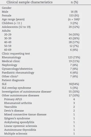
The epidemiological and demographic characteristics of this patient sample are shown in Table 1. Patients were clas-sii ed according to the diagnoses in their medical records. One third of the sample (55 patients) comprised patients under diagnostic investigation due to clinical suspicion of SLE, where the test requests were made due to the presence
Table 1 – Epidemiological characteristics of the sample (n = 169).
Clinical sample characteristics n (%)
Gender
Male 16 (9)
Female 153 (91)
Age range (years) (n = 166)a
Children (< 11 ) 3 (2%) Adolescents (12 to 19) 20 (12%) Adults
20-29 54 (33%)
30-39 43 (26%)
40-49 28 (17%)
50-59 12 (7%)
60-69 6 (4%)
Clinic requesting test
Rheumatology 123 (73%)
Medical clinic 19 (11%)
Nephrology 7 (4%)
Gynaecology/obstetrics 7 (4%) Paediatric rheumatology 6 (4%)
Other clinicb 7 (4%)
Patient diagnosis
SLE 92(54%)
SLE overlap syndrome 5 (3%) Investigation of autoimmune diseasec 55 (33%)
Other autoimmune diseases 17 (10%)
Primary APLS 4
Rheumatoid arthritis 3
Vasculitis 2
Devic’s disease 2
Mixed connective tissue disease 1
Sjögren’s syndrome 1
Ankylosing spondylitis 1 Linear systemic sclerosis 1 Autoimmune thyroiditis 1
Multiple sclerosis 1
SLE, systemic lupus erythematosus; APLS, antiphospholipid antibody syndrome.
a Three patients did not have records and their ages were not
mentioned in the laboratory records.
b Emergency, endocrinology, neurology and ICU.
c The patients without medical records (n = 3) were included in this
of several signs or symptoms (e.g., arthralgia, arthritis, kid-ney failure, haemolytic anaemia, purpura, Raynaud’s disease and paresthesias) related to or present in SLE.
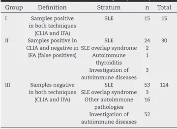
Patients were divided into three groups as dei ned in Table 2, according to the results obtained in the anti-dsDNA antibody testing using the two methodologies. The sera from Group I belonged to 15 lupus patients. Of these, 12 patients had a previous diagnosis of lupus nephritis, one demonstrat-ed serositis, and another had sufferdemonstrat-ed from SLE for seven months, presenting with evidence of positive inl ammatory activity, lymphopenia and consumption of the complement components C3 and C4. The last patient, who had juvenile rheumatoid arthritis (JRA) and had been in treatment for two years, tested positive for ANA and anti-dsDNA antibodies in this laboratory revaluation nine months before meeting the criteria for a diagnosis of SLE. The results of the screening for anti-dsDNA antibodies using the CLIA technique in the 15 sera samples from Group I remained in the 240 IU/mL to 32.6 IU/mL range with a mean of 167 IU/mL, median of 198 IU/mL and mode of 240 IU/mL, and the IFA titres ranged from 1/640 to 1/20. Eleven sera samples demonstrated readings greater than 125 IU/mL in the CLIA and IFA titres of 1/320 or 1/640. The values for the four remaining sera samples (63, 39, 36 and 32.6 IU/mL) showed titres of 1/320, 1/20 1/160 and 1/320, respectively.
The relevant information recorded in the medical records of each patient in Group II (clinical condition and laboratory changes, with reasons for requesting the anti-dsDNA tests) is listed in Table 3. These data showed that 87% (26/30) of the cases labelled as false-positive in CLIA had SLE, and 65% (17/26) of these patients had a previous diagnosis of lupus ne-phritis, with a description of signs and/or symptoms of clini-cal progression of disease in 50% (13) of cases. In addition, iso-lated laboratory changes compatible with active disease were present in 23% (6/26) of patients (cases 5, 8, 11, 19, 25 and 29). The results of the anti-dsDNA antibody testing using CLIA in Group II (Table 4) was in the range of 184-20 IU/mL, with a mean of 59 IU/mL and median 45 IU/mL. Four sera sam-ples (cases 1 to 4) were positive with values 5 times greater
than the cut-off point indicated by the manufacturer. Sera classii ed in the ‘grey zone’ accounted for 3.5% (6/169) of the sample studied.
The clinical condition of each patient in Group II (Table 4) was paired with the historic presence of autoantibodies and the progression of detection of anti-dsDNA antibodies in sera since disease onset. The ANA results were available and positive in 97% (29/30) of patients. Anti-Sm antibodies were detected in 38% (10/26) of SLE patients in this group. A previous history of reactivity to dsDNA (anti-dsDNA by IFA) occurred in 50% (13/26) of lupus patients; however, this information was not available for two patients from other units (cases 25 and 26), and four patients under diagnostic investigation were undergoing tests for the i rst time (cases 1, 4, 12 and 21).
Further evaluations of anti-dsDNA antibodies were re-quested in 77% (23/30) of patients within one year. Among the seven remaining patients, three did not have SLE (cases 1, 12 and 21), three had lupus in clinical and laboratory remission (3, 10 and 18) and case number 5, with clinical remission, had haematuria at the time of intrinsic validation.
Two patients (cases 2 and 17) were studied using multi-ple sera sammulti-ples. Case 2, which demonstrated a trimulti-ple posi-tive evaluation in CLIA (154.6, 46 and 37.5 IU/mL) had been diagnosed with SLE and lupus nephritis for four years. This patient showed a positive result for anti-dsDNA antibodies by enzyme-linked immunosorbent assay (ELISA) (80 U) at the onset of the disease (2007), which was not coni rmed by IFA in three tests conducted in the 2009 to 2010 period, although the IFA test became positive after this patient experienced convulsive symptoms for one month. Case 17, which demon-strated discrepant results in the CLIA (32 and 13.8 IU/mL), had been diagnosed with SLE and lupus nephritis 2 months prior, although anti-dsDNA results using both CLIA and IFA for this patient remained negative after six months.
Ten patients became reactive by IFA within one year of their i rst evaluation (cases 2, 9, 23 and 26 up to 3 months; case 29 after i ve months; cases 14 and 15 after 9 months and cases 7, 20 and 22 after an interval of 12 months).
The remainder of the sample (Group III) consisted of 58% (56/97) of the total number of patients with SLE, 95% (52/55) of the patients being tested for autoimmune diseases and the majority (94%, 16/17) of patients affected by other autoim-mune diseases. There were seven duplicate samples in the in-trinsic evaluation of this group that belonged to i ve patients with SLE, 1 under diagnostic investigation and one with rheu-matoid disease. The CLIA results in Group III showed values in the range of 19 to 0.5 IU/mL (Fig. 1), with a mean of 5.5 IU/mL, median of 4 IU/mL and mode of 0.5 IU/mL.
A prior history of reactivity to dsDNA using IFA in all SLE patients who participated in the study (n = 97) was investigat-ed using the minvestigat-edical and/or laboratory records (Table 5), and we found that 48% (47/97) of the samples were reactive, with i ve patients in Group I (positive in both methodologies) un-dergoing anti-dsDNA testing for the i rst time. Clinical activity measured using the Systemic Lupus Erythematosus Disease Activity Index (SLEDAI) was not available in all records during the test request period, which prevented the study of clinical progression (periods of disease activity or remission) related to the presence of anti-dsDNA antibodies.
Table 2 – Dei nition of strata for analysis (n = 169).
Group Dei nition Stratum n Total
I Samples positive in both techniques
(CLIA and IFA)
SLE 15 15
II Samples positive in CLIA and negative in
IFA (false positives)
SLE SLE overlap syndrome
Autoimmune thyroiditis Investigation of autoimmune diseases
24 2 1
3 30
III Samples negative in both techniques
(CLIA and IFA)
SLE SLE overlap syndrome
Other autoimmune pathologies Investigation of autoimmune diseases
53 3 16
52 124
Discussion
Currently, the most commonly used techniques for detecting anti-dsDNA antibodies are immunoenzymatic assay and IFA, the latter being more specii c and capable of detecting anti-bodies with moderate and high afi nity related to SLE activity.9
ELISA-based methods, although quantitative, reproducible and automated, exhibit lower precision in terms of clinical performance because they detect low-avidity anti-dsDNA autoantibodies, which generally have little clinical relevance and may be present in other connective tissue diseases, in-l ammatory or infectious diseases and in normain-l subjects.10
However, in recent years, a new generation of ELISAs for the detection of anti-dsDNA antibodies has been introduced into the market, and these new reagents provide greater antigen purii cation, making them more selective for the detection of intermediate- and high-avidity antibodies.5 The CLIA method
evaluated in this study is included in this group.
The performance of the CLIA-LIAISON assay in this in-trinsic evaluation was satisfactory and produced 100%
sen-sitivity, 82% specii city and 81% efi ciency when compared to IFA. This same CLIA reagent has been tested by the Italian Society of Laboratory Medicine Study Group on Autoimmune Diseases5 in an extrinsic evauation7 with a clinical samples
from 52 patients with SLE, 28 patients with other connective tissue diseases, 36 patients with hepatitis C virus (HCV) and 24 patients with other acute viral diseases. These authors re-ported 84.6% sensitivity, 82.9% specii city and 83.6% efi ciency of the method, which is similar to the results obtained in the present study, although the difference in sensitivity may be attributed to the clinical samples examined. This study also analysed the performance of the automated test for the de-tection of anti-dsDNA antibodies, according to the reality ex-perienced by the local population, where the majority (57%) of patients who undergo this exam have SLE. The Italian study also included patients with HCV who eventually had positive CLIA testing for anti-dsDNA antibodies.5
The performance of the CLIA test in identifying a negative reaction was adequate in this study, with measures of central tendency in Group III convergent with values less than three times the maximum negativity suggested by the manufac-Table 3 – Relevant information for Group II patients (n = 30)
Pat. G Age Reasons for requesting anti-dsDNA test
Time Current condition
Clinical Laboratory changes
1 F 35 Undei ned arthralgia 1 y under investigation ANA-reactive 2 F 25 SLE + nephritis 4 y activity (convulsion) lymphopenia, Ļ C ', proteinuria
3 F 60 SLE 15 y remission no change
4 F 34 Additive polyarthritis 2 y under investigation ANA-reactive
5 F 33 SLE + nephritis 11 y remission haematuria ++
6 F 21 SLE + nephritis 7 m activity ĻC', haematuria, proteinuria
7 F 24 Mucocutaneous SLE 2 m remission no change
8 F 57 SLE 22 y remission lymphopenia
9 F 28 SLE + nephritis 5 y activity anaemia, ĻC', proteinuria
10 F 18 SLE + nephritis 2 y remissiona no change
11 F 49 SLE 5 y remission CRP+, ĻC3
12 F 49 Autoimmune thyroiditis 3 y arthralgia haematuria + 13 F 33 SLE + nephritis 8 y activity lymphopenia, Ļ C ', haematuria
14 F 22 SLE 10 m remission no change
15 M 13 SLE 2 m activity ĻC'
16 F 43 SLE + nephritis 5 y activity lymphopenia, haematuria, proteinuria 17 F 34 SLE + nephritis 2 m activity lymphopenia, ĻC', ESR and CRP,
haematuria, proteinuria
18 F 27 SLE + DM/DP 4 y remission CRP
19 F 14 Mucocutaneous SLE 5 m remission ĻC'
20 F 27 SLE + nephritis 6 y activity ĻC', proteinuria 21 F 49 Undei ned kidney failure 1 m under investigation proteinuria 22 F 22 SLE + nephritis 4 y activitya haematuria, proteinuria
23 F 23 SLE + nephritis 7 y activitya proteinuria
24 F 28 SLE + nephritis 3 y activitya haematuria, proteinuria
25 F 19 SLE + nephritis 5 y remission lymphopenia
26 M 34 SLE + nephritis 3 y activitya, haemodialysis ĻC', haematuria, proteinuria, creatinine
27 F 46 SLE + nephritis 5 y activitya leukocyturia
28 F 29 SLE + nephritis 18 m activity, haemodialysisb ĻC3, proteinuria, leukocyturia
29 F 61 SLE + SS 10 y remission lymphopenia, ĻC',
30 F 22 SLE + nephritis 3 y evaluation after pregnancy
no change
Pat, patient; G, gender; F, female; y, year; ANA, antinuclear autoantibody; SLE, Systemic lupus erythematosus; C’, complement; m, month; CRP, C-reactive protein; M, male; ESR, erythrocyte sedimentation rate; DM/DP, dermatomyositis/dermatopolymyositis; SS, systemic sclerosis
Table 4 – Presence of autoantibodies in the serum of Group II patients (n = 30). Pat. Current clinical
condition
Previous history of other autoantibodies
Anti-dsDNA history
Previous Current Later evaluations
(IFA) CLIA (IU/mL) CLIA (IU/mL) IFA (titre) (interval)
1 Investigation ANA tnc 183.5 tnc tnc
-2 Activity ANA NR 154.6 49.0 1:80 1 m
3 Remission ANA R 134.1 tnc tnc
-4 Investigation ANA, Cardio G and M tnc 125.5 tnc NR 5 m
5 Remission ANA, Sm, Cardio G and M R 111.1 tnc tnc
-6 Activity ANA, Sm, RNP NR 92.5 199.9 NR 10 m
7 Remission ANA NR 92.1 54.1 1:80 12 m
8 Remission ANA R 85.6 19.0 tnc 5 m
9 Activity ANA, Ro, La NR 84.6 189.3 1:320 3 m
10 Remissiona ANA R 68.6 tnc tnc
-11 Remission ANA, Sm R 51.5 96 e 15.2 NR 2 e 6 m
12 Investigation ANA, Ro tnc 50.9 tnc tnc
-13 Activity ANA, Sm NR 49.5 38. 2 e 52 NR 3 e 12 m
14 Remission ANA NR 45.2 45.1 1:160 9 m
15 Activity ANA, Sm, Ro NR 44.9 26.7 1:80 9 m
16 Activity ANA, Ro, La R 36.1 4.75 tnc 12 m
17 Activity ANA NR 32.0 12.5 NR 6 m
18 Remission ANA R 31.4 tnc tnc
-19 Remission ANA, Sm, Cardio G tnc 31.1 28.4 NR 3 m
20 Activity ANA, Sm, RNP R 30.2 45.7 1:80 12 m
21 Investigation tnc tnc 28.7 tnc tnc
-22 Activitya ANA R 26.6 33.6 1:40 12 m
23 Activitya ANA R 26.5 34.4 1:160 1 m
24 Activitya ANA, Sm, RNP, anti-p NR 25.4 tnc NR 2 m
25 Remission un un 22.7 22.2 NR 10 m
26 Activitya ANA, Ro, Cardio G un 22.1 56.3 1:40 2 m
27 Activitya ANA, RNP R 22.0 45.6 NR 2 m
28 Activity ANA, Sm, Cardio G NR 20.9 8.33 tnc 10 m
29 Remission ANA, Sm, RNP R 20.6 32.5 1:40 5 m
30 Remission ANA R 20.3 18.3 tnc 11 m
Pat, patient; IFA, indirect immunol uorescence assay; CLIA, chemiluminescent immunoassay; ANA, antinuclear autoantibody; tnc, test not conducted; NR, non-reactive; m, month; R, reactive; Cardio G, anticardiolipin G; Cardio M, anticardiolipin M; Sm, anti-Sm; RNP, anti-RNP; Ro, anti-SSA(Ro); La, anti-SSB(La); un, evaluation unknown; anti-p, anti-ribossomal p.
a In the presence of pulse therapy with methylprednisolone and cyclophosphamide, anti-ribosomal p.
Table 5 – Previous history of anti-dsDNA (IFA) in lupus patients (n = 97).
Group Reactive
Non-reactive
1st time UN Total
I 10 0 5 0 15
II 13 10 1 2 26
III 19 32 5 0 56
Total 42 42 11 2 97
un, evaluation unknown.
Fig. 1 – Frequency of CLIA results (IU/mL) in Group III (n = 124).
0 5 10 15 20 25
0 2 4 6 8 10 12 14 16 18 IU/mL
n turer (up to 19 IU/mL). In addition, the low frequency (3.5%) of grey zone results enabled a clear dei nition of the positiv-ity of the method. The identii cation of patients who consti-tuted Group III demonstrated the specii city for anti-dsDNA antibodies used in the test; of the 45 CLIA-reactive sera, 91% (41/45) were from SLE patients.
The availability of the clinical samples also facilitated qualitative analysis of the type of patient who receives anti-dsDNA testing in the hospital, and these results re-vealed a clinical and epidemiological profile similar to that found in the pathology of lupus where this autoantibody is prevalent.11 The vast majority of patients were female
After the establishment of automated screening for anti-dsDNA antibodies, positive samples and those with results in the grey zone in CLIA were tested by IFA, using Crithidia luciliae as a substrate. The implementation of this routine led to the optimisation of time and laboratory personnel,6 reducing the
requirement for manual procedures by more than 70% and also reducing the likelihood of procedural and random errors that could compromise the quality and accuracy of the re-leased tests. The potential for cost reduction with this new detection approach will be analysed in a subsequent study.
International trials recommend the use of automated reagents for the detection of anti-dsDNA antibodies,3,12-15
al-though the gold standard method in clinical and laboratory research remains IFA.12,16 Because the array of laboratory
methods for the detection of anti-dsDNA antibodies is con-tinuously increasing, tests traditionally used in routine work are still far from becoming standardised and widely accepted. Moreover, physicians should be aware that the agreement rates between laboratories, the interpretation of results and the diagnostic accuracy are dependent on the analytical vari-ability and the population of patients being studied.17 In the
present study, the technical laboratory conditions and the re-ferral of patients’ serum samples were maintained within the normal working routine of the institution.
This study demonstrated that screening of anti-dsDNA au-toantibodies using CLIA is a safe (100% sensitivity) and rapid method that could improve the quality of tests available to pa-tients. Among the study i ndings, it should be noted that most of the CLIA results labelled as false positives belonged to lupus patients with clinical and/or laboratory disease activity, some of whom were coni rmed months later as positive by IFA.
Confl icts of interest
The authors declare no conl icts of interest.
R E F E R Ê N C I A S
1. Ghirardello A, Villalta D, Morozzi G, Afeltra A, Galeazzi M, Gerli R, et al. Diagnostic accuracy of currently available anti-double-stranded DNA antibody assays. An Italian multicentre study. Clin Exp Rheumatol. 2011;29(1):50-6. 2. Heidenreich U, Mayer G, Herold M, Klotz W, Al-Jazrawi SK,
Lhotta K. Sensitivity and specii city of autoantibody tests in the differential diagnosis of lupus nephritis. Lupus. 2009;18(14):1276-80.
3. Lemarié R, Jacomet F, Goutte B, Bonnafoux C, Tridon A, Evrard B. The anti-dsDNA antibodies: validation of an original two step strategy of detection. Ann Biol Clin (Paris). 2011;69(1):47-53.
4. Launay D, Schmidt J, Lepers S, Mirault T, Lambert M, Kyndt X, et al. Comparison of the Farr radioimmunoassay, 3 commercial enzyme immunoassays and Crithidia luciliae immunol uorescence test for diagnosis and activity assessment of systemic lupus erythematosus. Clin Chim Acta. 2010;411(13-14):959-64.
5. Antico A, Platzgummer S, Bassetti D, Bizzaro N, Tozzoli R, Villalta D. Diagnosing systemic lupus erythematosus: new-generation immunoassays for measurement of anti-dsDNA antibodies are an effective alternative to the Farr technique and the Crithidia luciliae immunol uorescence test. Lupus. 2010;19(8):906-12.
6. Meroni PL, Schur, PH. ANA screening: an old test with new recommendations. Ann Rheum Dis. 2010;69(8):1420-2. 7. Ferreira AW, Ávila SLM. Diagnóstico Laboratorial das
principais doenças infecciosas e autoimunes. 2.ed. Rio de Janeiro: Guanabara Koogan; 2001.
8. Callado MRM, Vieira RMRA, Araújo VMA, Callado CM, Costa Lima JR, Rodrigues JNA, et al. Prevalência dos anticorpos antinucleares (ANA) no Hospital Geral de Fortaleza no período de jan/2002 a dez/2006. Jornal da Liga dos Reumatologistas do Norte-Nordeste (LIRNNE). 2007;3:118-22. 9. Kim KH, Han JY, Kim JM, Lee SW, Chung WT. Clinical
signii cance of ELISA positive and immunol uorescence negative anti-dsDNA antibody. Clin Chim Acta. 2007;380:182–5.
10. Smeenk RJT. Detection of autoantibodies to dsDNA: Current insights into its relevance. Clin Exp Rheumatol. 2002;20:294-300.
11. Pisetsky DS. In: JH, Stone JH, Crofford LJ, White PH (eds.). Primer on the Rheumatic Diseases. 13.ed. Springer/ Arthritis Foundation; 2008.
12. Yang JY, Oh EJ, Kim Y, Park YJ. Evaluation of Anti-dsDNA antibody tests: Crithidia luciliae immunol uorescence test, immunoblot, enzyme-linked immunosorbent assay, chemiluminescence immunoassay. Korean J Lab Med. 2010;30(6):675-84.
13. Fiegel F, Buhl A, Jaekel HP, Werle E, Schmolke M, Ollert M, et al. Autoantibodies to double-stranded DNA--intermethod comparison between four commercial immunoassays and a research biosensor-based device. Lupus. 2010;19(8): 957-64.
14. El-Chennawi FA, Mosaad YM, Habib HM, El-Degheidi T. Comparative study of antinuclear antibody detection by indirect immunol uorescence and enzyme immunoassay in lupus patients. Immunol Invest. 2009;38(8):839-50. 15. Suh-Lailam BB, Chiaro TR, Davis KW, Wilson AR, Tebo AE.
Evaluation of a high avidity anti-dsDNA IgG enzyme-linked immunosorbent assay for the diagnosis of systemic lupus erythematosus. Int J Clin Exp Pathol. 2011;4(8):748-54. 16. Chiaro TR, Davis KW, Wilson A, Suh-Lailam B, Tebo AE.
Signii cant differences in the analytic concordance between anti-dsDNA IgG antibody assays for the diagnosis of systemic lupus erythematosus-Implications for inter-laboratory testing. Clin Chim Acta. 2011;412(11-12):1076-80. 17. Ghirardello A, Villalta D, Morozzi G, Afeltra A, Galeazzi