UNIVERSIDADE DE LISBOA
FACULDADE DE CIÊNCIAS
Departamento de Química e Bioquímica
Development of nanosystems for HIV vaccine delivery
Ana Isabel Neves de Matos
Dissertation
Master in Biochemistry
Specialization: Biochemistry
UNIVERSIDADE DE LISBOA
FACULDADE DE CIÊNCIAS
Departamento de Química e Bioquímica
Development of nanosystems for HIV vaccine delivery
Ana Isabel Neves de Matos
Dissertation
Master in Biochemistry
Specialization: Biochemistry
Supervisors: Carla Real Afonso, PhD, FCUL
Helena Florindo, PhD, FFUL
i
Acknowledgements
The progress of this master thesis was only possible with teamwork. For this reason, I want to thank all people that worked with me this year.
I express my deepest gratitude to my supervisor, Prof. Helena Florindo, not only for the opportunity to develop this project in her laboratory, but also for the unconditional help, support and friendship throughout this year.
I am grateful to my supervisor, Prof. Carla Afonso, for accepting embarks on this adventure and for helping me with confocal microscopy, as well as Filipe.
I thank to all my group colleagues by teaching, advice and guidance, especially Carina Peres, Eva Zupančič and Joana Silva. Your friendship has made everything much easier.
A special thanks to Melissa Sirage by strong friendship attained this year that will remain for live.
I am grateful to Prof. Liana Silva for all the advice given.
I am very thankful to Ana Salgado and Prof. Ana Viana for helping me with HPLC and AFM analysis, respectively.
I would like to thank Fundação para a Ciência e a Tecnologia (FCT) for the financial support: PTDC/SAU-FAR/119389/2010 and Pest- OE/SAU/UI4013/2011.
Finally, my sincere gratitude to my family and friends for their patience, strength and care at all times. A special thank you to my parents for the unconditional love. An extra special thanks to João, my safe haven.
ii
Abstract
Purpose: Despite the improvements achieved in the current therapy regimens, the development of prophylactic, safe and highly immunogenic vaccines to control and eradicate the Human Immunodeficiency Virus (HIV) remains one of the most exigent and challenging tasks. To prevent HIV infection, highly specialized antigen presenting cells, as dendritic cells (DCs), have to elicit effective humoral and cellular immune responses. In fact, the induction of both virus-specific neutralizing antibodies (IgG and IgA), at the site of viral entry, and cytotoxic T lymphocyte along with a Th1 response seem to be crucial to impair the dissemination of the virus. As a promising strategy for HIV delivery systems, polymeric nanoparticles (NPs) can protect HIV-1 antigens from unfavorable conditions after administration, in addition to acting as adjuvants by enhancing the antigen uptake by DCs. T20 and T1249 antigenic peptides are HIV-1 fusion inhibitors derived from the gp41 helical region adjacent to the C-terminal sequence that have never been associated to a particulate delivery system. This project aims to design a HIV-1 vaccine throught the entrappement of HIV-1 antigenic peptides (T20 and T1249) into polymeric NPs.
Materials and Methods: Polymeric NPs were prepared by the double emulsion solvent evaporation method and characterized. Polyvinyl alcohol, block co-polymer Pluronic® F127 and glycol chitosan were included in vaccine formulation to promote their potential adjuvant effect and/or improve NP stability. Model antigen ovalbumin and HIV-1 gp41 peptides, T20 and T1249, were entrapped or co-entrapped into NPs. Characterization of NPs was performed in terms of size, zeta potential and surface morphology by Dynamic Light Scattering, Laser Doppler Electrophoresis and Atomic Force Microscopy, respectively. Entrapment efficiency (EE, % (w/w)) and loading capacity (LC, µg/mg) of the entrapped agents were quantified by MicroBCA®, High Performance Liquid Chromatography and fluorescence. DCs were used to evaluate the
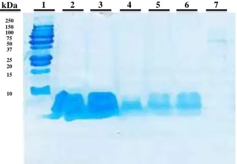
in vitro cytotoxicity of NPs by the AlamarBlue® assay and the cellular uptake and intracellular trafficking of rhodamine-labeled NPs by confocal microscopy. The peptide integrity was also assessed by sodium dodecyl sulphate polyacrylamide gel electrophoresis.
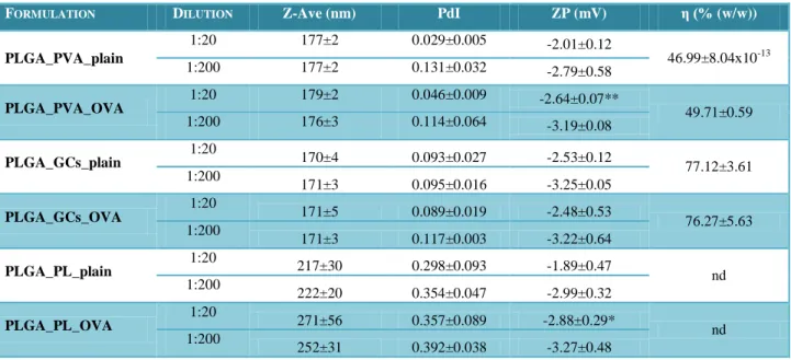
Results: NPs presented mean diameters in the range of 170-180 nm, with a
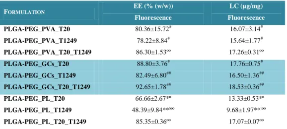
narrow size distribution (PdI < 0.15), surface charge close to neutrality and spherical shape. These antigen delivery systems presented high EE and LC, and no cytotoxic
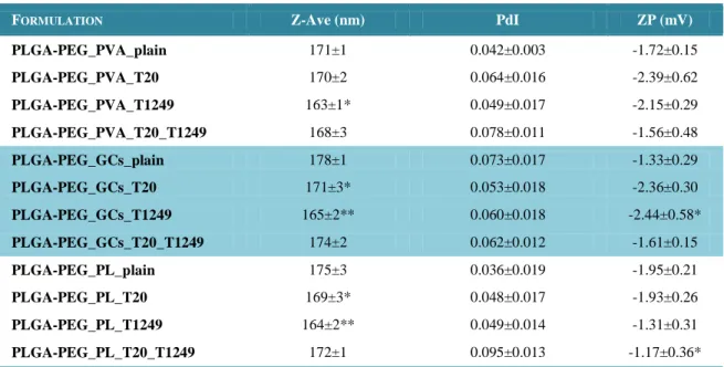
iii effect on DCs, in the range of tested concentrations (0.125-1 mg/mL), after 24, 48 and 72 h of incubation. According to NP physicochemical characteristics, PLGA-PEG_GCs formulation was selected for further studies. Peptide structure was maintained intact, not being affected by the double emulsion procedure. Confocal microscopy confirmed the internalization of NPs, demonstrating their endosomal localization and a tendency to accumulate close to the endoplasmic reticulum.
Conclusions: The results herein described show that stable and reproducible
PLGA-PEG_GCs NPs produced using a modified double emulsion solvent evaporation method constitute a promising platform for the successful delivery of HIV antigens to DCs, in order to develop a HIV-1 vaccine.
Keywords
HIV-1 vaccine, PLGA-PEG nanoparticles, HIV-1 gp41 peptides, targeted delivery systems, dendritic cells, intracellular trafficking
iv
Resumo
Objetivo: Apesar dos avanços alcançados nas terapêuticas atuais, o desenvolvimento de vacinas profiláticas, seguras e altamente imunogénicas para controlar e erradicar o vírus da imunodeficiência humana (HIV) continua a ser uma das tarefas mais exigentes e desafiantes. Para prevenir a infeção por HIV, as células apresentadoras de antigénio (APCs) altamente especializadas, têm de induzir respostas imunitárias humorais e celulares eficazes. Mais especificamente, a indução de anticorpos neutralizantes específicos contra o vírus (nomadamente as imunoglobulinas (Ig)G, IgA e IgE) secretados pelos linfócitos B, especialmente no local de entrada do vírus (resposta tipo Th2), e a estimulação de linfócitos T citotóxicos juntamente com uma resposta do tipo Th1 parecem ser cruciais para travar a disseminação deste vírus. Deste modo, a persistência da infeção por HIV parece estar intimamente associada à inadequada estimulação de uma resposta imunológica eficaz e específica capaz de destruir o vírus. Esta resposta é bastante complexa, não existindo uma correlação directa entre a infeção por este agente e eventuais mecanismos de proteção ou controlo da disseminação da doença.
O HIV liga-se ao receptor CD4 via glicoproteína gp120 do invólucro viral, a qual sofre uma alteração conformacional, facilitando a sua ligação a um dos dois co-recetores de referência, CCR5 ou CXCR4. A interação da gp120 com o recetor CD4 e os co-recetores é vital para que o vírus se ligue à célula. Contudo, para finalizar o processo de infeção vírus-célula, a glicoproteína gp41 do invólucro viral terá posteriormente de interagir com um recetor de fusão na célula. Após a fusão, o conteúdo viral é libertado para o interior da célula com a subsequente transcrição do RNA viral em DNA, que será transportado até ao núcleo e integrado no genoma celular, sendo posteriormente traduzido em proteínas. Estas proteínas virais, juntamente com o RNA viral, são transportados para a superfície celular iniciando a formação de novos viriões maduros. Os linfócitos T e B, os macrófagos e as células dendríticas (DCs) podem expressar os co-recetores CCR5 ou CXCR4, estando portanto suscetíveis à infeção por HIV. No entanto, apenas as DCs podem ligar-se à glicoproteína gp120 sem que haja fusão membranar.
As DCs são atualmente reconhecidas como entidades fundamentais para a estimulação de uma resposta imunológica robusta e específica contra o HIV, a qual se deve à sua capacidade de migração para os órgãos linfáticos, zonas ricas em linfócitos
v T, onde apresentarão os antigénios fagocitados e consequentemente levarão à estimulação dos linfócitos T CD4+ e CD8+, promovendo um eficiente e prolongado controlo da replicação do vírus no hospedeiro. No entanto, o papel destas APCs na imunologia do HIV está longe de ser um tema consensual. Estas células podem capturar, processar e apresentar o HIV desencadeando uma resposta eficaz e específica. Contudo, uma vez infetadas, as DCs podem infetar os linfócitos T, promovendo a disseminação do vírus e a progressão da doença. A fase crónica e assintomática da doença é caracterizada por um decréscimo da carga viral, persistindo o vírus latente em tecidos extravasculares, nódulos linfáticos, DCs e nos linfócitos T CD4+. Embora seja reconhecido o papel dos linfócitos T no controlo da infeção crónica por HIV-1, a imunidade especificamente induzida contra este microrganismo decresce após períodos prolongados da supressão do vírus, não conseguindo controlar a replicação do vírus e consequentemente originando a progressão da doença. Apesar destes doentes serem tratados com medicamentos antiretrovirais, reconhecidos como a melhor opção terapêutica atualmente disponível, estes tratamentos são dispendiosos, apresentam efeitos secundários significativos inerentes à sua toxicidade, dificultando a adesão à terapêutica por parte do doente por longos períodos. Mesmo na presença de medicamentos antiretrovirais, o vírus permanece latente no organismo em níveis não detetáveis, nomeadamente em linfócitos T CD4+ memória. Estes estão inativos, constituindo reservatórios que protegem o vírus da resposta imunológica e da terapêutica antiretroviral, tornando praticamente impossível a eliminação total do vírus com as abordagens terapêuticas actualmente disponíveis.
O desenvolvimento de vacinas preventivas e de abordagens imunoterapêuticas contra a infeção por HIV tem sido bastante dificultado pela complexidade dos mecanismos imunológicos associados. De fato, a mutação do vírus, a capacidade deste ultrapassar a resposta imunológica humoral, bem como a manipulação das APCs evitando a maturação destas e portanto a eficaz apresentação dos antigénio aos linfócitos T, têm contribuído para a falta de eficácia obtida por inúmeros candidatos. Deste modo, torna-se desejável o desenvolvimento de novas estratégias capazes de modelar a resposta imunológica contra a infeção por HIV. As nanopartículas (NPs) poliméricas constituem sistemas promissores para o transporte de antigénios HIV-1. De fato, estes nanosistemas protegem os antigénios de HIV-1 das condições desfavoráveis após a sua administração e podem atuar como adjuvantes, aumentando o reconhecimento e a internalização de antigénios pelas DCs. Assim, este projeto único e
vi inovador tem como objetivo criar uma vacina contra o HIV-1 através da encapsulação dos péptidos antigénicos inibidores de fusão do HIV-1, derivados da sequência da região helicoidal adjacente ao C-terminal da gp41, T20 e T1249 em NPs poliméricas. É importante referir que estes péptidos de HIV-1 nunca foram associados a partículas poliméricas.
Materiais e Métodos: As NPs poliméricas foram preparadas utilizando o método
de dupla emulsão com a evaporação do solvente. O álcool polivinílico, o co-polímero Pluronic® F127 e o glicol quitosano foram incluídos na formulação da vacina para promover o seu potencial efeito adjuvante e/ou aumentar a estabilidade das NPs. O antigénio modelo ovalbumina e os péptidos de HIV-1 gp41, T20 e T1249, foram incorporados ou co-incorporados nas NPs. A caracterização da NPs foi realizada tendo em conta o tamanho, a carga e morfologia da sua superfície por Dispersão Dinâmica de Luz, Dispersão Electroforética da Luz e Microscopia de Força Atómica, respetivamente. A eficiência de incorporação (EE, % (w/w)) e capacidade de carga (LC, µg/mg) dos agentes incorporados foram quantificados por MicroBCA®, Cromatografia Líquida de Alta Eficiência e fluorescência. As DCs foram usadas para avaliar a citotoxicidade in
vitro das NPs através do ensaio AlamarBlue® e a captura e tráfego intracelular das NPs marcadas com rodamina foram estudados por microscopia confocal. A integridade da estrutura dos péptidos foi ainda confirmada por electroforese em gel de poliacrilamida em condições desnaturantes, utilizando o dodecil sulfato de sódio.
Resultados: As NPs apresentaram diâmetros médios compreendidos entre
170-180 nm, com uma distribuição de tamanhos restrita (PdI < 0,15), carga superficial próxima da neutralidade e forma esférica. Estes sistemas de transporte de antigénios apresentaram elevados EE e LC, e nenhum efeito citotóxico em DCs, na gama de concentrações testadas (0,125-1 mg/mL), após 24, 48 e 72 h de incubação. Tendo em consideração os resultados previamente descritos, nomeadamente as características físico-químicas das NPs, a formulação PLGA-PEG_GCs foi selecionada para os restantes estudos. A estrutura dos péptidos foi mantida intacta, não sendo afetada pelo processo de dupla emulsão. A microscopia confocal confirmou a internalização das NPs, demonstrando a sua localização endosomal e uma tendência para se acumularem perto do retículo endoplasmático.
Conclusões: Os resultados aqui descritos mostram que NPs estáveis e
reprodutíveis foram produzidas utilizando um método de dupla emulsão com evaporação de solvente modificado. Importa no entanto realçar que as NPs
PLGA-vii PEG_GCs constituem uma plataforma promissora para o transporte e eficaz apresentação de antigénios de HIV, tendo em vista o desenvolvimento de uma vacina eficaz contra a infeção por HIV-1.
Palavras-chave
Vacina contra a infeção por HIV-1, nanopartículas PLGA-PEG, péptidos de HIV-1 gp41, sistemas de transporte, células dendríticas, tráfego intracelular
viii
List of Contents
1. State of art………1
1.1. Incidence and mortality………...1
1.2. Biology of HIV………..1
1.3. HIV infection process………2
1.4. HIV transmission………...4
1.5. HIV pathogenesis and immune response………...6
1.5.1. HIV reservoir sites………...8
1.5.2. Humoral immune response against HIV………..8
1.5.3. Cellular immune response against HIV………...9
1.6. Antiretroviral therapy………...10
1.7. HIV vaccine development………11
1.7.1. Antigens, adjuvants and delivery systems……….12
1.7.1.1. Polymeric nanoparticles as HIV vaccine delivery system………14
1.7.1.2. HIV nanovaccine based on dendritic cells targeting……….16
1.7.1.3. HIV nanovaccine based on peptide HIV-1 fusion inhibitors……21
1.7.2. PLGA nanoparticles as promising HIV vaccine delivery systems………23
Goals………...28
3. Materials and Methods……….29
3.1. Materials………..29
3.2. Methods………30
3.2.1. Preparation of nanoparticles………..30
3.2.2. Physicochemical characterization of nanoparticles………...31
3.2.2.1. Size and zeta potential analysis……….31
3.2.2.2. Surface morphology analysis………32
3.2.3. Antigen loading analysis………32
3.2.4. Cell culture conditions………...34
3.2.5. In vitro cell viability assay……….34
3.2.6. Peptide integrity assessment ……….35
3.2.7. Cell uptake and intracellular trafficking studies………36
3.2.8. Statistical analysis………..37
4. Results……….38
ix
4.1.1. Ovalbumin entrapped-nanoparticles………...38
4.1.2. HIV-1 peptides entrapped-nanoparticles………...43
4.2. Cell viability assay………...46
4.3. Evaluation of peptide integrity……….48
4.4. Uptake and intracellular trafficking of nanoparticles………..49
5. Discussion………...52
5.1. Composition of nanoparticles………..52
5.2. Physicochemical characterization of nanoparticles……….55
5.3. Antigen loading analysis………..59
5.4. Cytotoxic effect of nanoparticles……….62
5.5. Peptide integrity evaluation……….64
5.6. Uptake and intracellular trafficking of nanoparticles………..65
6. Conclusions and Future Prospects………...68
6.1. Evaluation of peptide antigenicity by Imunoblotting………..69
6.2. Cellular uptake and intracellular trafficking studies of peptide-loaded NPs…...69
6.2.1. Flow cytometry analysis………70
6.2.2. Confocal microscopy imaging analysis……….70
6.3. Study of endocytic pathways involved in NPs internalization………70
6.4. In vivo assay testing HIV-1 prophylactic effect………...71
6.4.1. Serology……….71
6.4.2. Cytokines assays………71
6.5. Antibody reactivity against HIV-1 peptides………71
6.6. Neutralizing assay………72
6.7. Lyophilization process optimization and NPs stability studies………...72
x
List of Figures
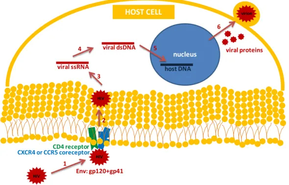
Figure 1 – HIV infection process………..4
Figure 2 – The desired immune response elicited by a HIV vaccine………...…..19
Figure 3 – Analysis of size, shape and surface morphology of NPs by AFM……...40/41 Figure 4 – Effect of NPs on cell viability………...………...46/47 Figure 5 – Evaluation of the integrity of HIV-1 peptides, T20 and T1249, entrapped in
PLGA-PEG_GCs NPs……….48
Figure 6 – Uptake and intracellular trafficking of NPs by BMDCs, and colocalization
analysis………...50/51
List of Tables
Table 1 – Composition of plain and antigen-loaded PLGA or PLGA-PEG NPs……...31 Table 2 – Physicochemical properties of plain and ovalbumin(OVA)-entrapped PLGA
NPs………...38
Table 3 – Physicochemical properties of plain and ovalbumin(OVA)-entrapped
PLGA-PEG NPs………..39
Table 4 – Entrapment efficiency (EE, % (w/w)) and loading capacity (LC, µg/mg) of
the ovalbumin (OVA) entrapped in PLGA and PLGA-PEG NPs………...42
Table 5 – Physicochemical properties of plain or HIV-1 peptides (T20 and
T1249)-entrapped PLGA-PEG NPs……….43
Table 6 – Physicochemical properties of HIV-1 peptide-loaded PLGA-PEG NPs, one
month after preparation………...44
Table 7 – Entrapment efficiency (EE, % (w/w)) and loading capacity (LC, µg/mg) of
xi
List of Abbreviations
η: Yield
λmax: Maximum wavelength
Abs: Antibodies
AFM: Atomic Force Microscopy
AIDS: Acquired Immune Deficiency Syndrome AP: Alkaline phosphatase
APs: Ammonium persulfate APCs: Antigen presenting cells ART: Antiretroviral therapy
ATCC: American Type Culture Collection BCA: bicinchoninic acid
BMDCs: Bone marrow dendritic cells bNAbs: Broadly neutralizing antibodies BSA: Bovine serum albumin
BSP: Betamethasone sodium phosphate CaCl2: Calcium chloride
CD40L: CD40 ligand
CHR: Helical region adjacent to the C-terminal CLRs: C-type lectin receptors
CNS: Central nervous system
CpG: Unmethylated cytosine-phosphate-guanine oligodeoxynucleotides Cs: Chitosan
CTL: Cytotoxic T lymphocytes DCM: Dichloromethane DCs: Dendritic cells
DC-SIGN: Dendritic cell specific intercellular adhesion molecule-grabbing
non-integrin
DLS: Dynamic Light Scattering DNA: deoxyribonucleic acid DPBS: Dulbecco’s PBS dsRNA: Double stranded RNA EE: Entrapment efficiency EFV: Efavirenz
xii EMA: European Medicine Agency
EP: External aqueous phase ER: Endoplasmic reticulum
FBS: Heat inactivated fetal bovine serum FDA: Food and Drug Administration FGF: Fibroblast growth factor
GALT: Gut-associated lymphoid tissues GCs: Glycol chitosan
GM-CSF: Granulocyte-macrophage colony stimulating factor gp: Glycoprotein
6HB: Six-helix bundle HAART: Highly active ART HCl: Hydrochloric acid
HIV: Human Immunodeficiency Virus
HIV-1: Human Immunodeficiency Virus type 1 HIV-2: Human Immunodeficiency Virus type 2 HPLC: High Performance Liquid Chromatography HR: Helical region
HSV-2: Herpes Simplex Virus type 2 ICH: International Conference Guidelines ID: Intradermal IFN: Interferon Ig: Immunoglobulin IL: Interleukin IM: Intramuscular IN: Intranasal
IP: Internal aqueous phase IS: Infectious synapse
ISCOMs: Immunostimulating complexes LB: Loading buffer
LC: Loading capacity LCs: Langerhans cells
LDE: Laser Doppler Electrophoresis LPS: Lipopolysaccharide
xiii MALP: Macrophage-activating lipopeptide
MBD: Methyl-CpG binding domain protein MgCl2: Magnesium chloride
MHC: Major histocompatibility complex MIP: Macrophage inflammatory protein MPLA: Monophosphoryl lipid A MRI: Magnetic Resonance Imaging mRNA: Messenger RNA
MTT: Thiazolyl blue tetrazolium bromide Mw: Molecular weight
Na2HPO4: sodium phosphate dibasic
NaCl: sodium chloride
NaH2PO4: sodium phosphate monobasic
NHR: Helical region adjacent to the N-terminal NK: Natural killer
NPs: Nanoparticles
NPs-Rho: Rhodamine-labeled NPs
OVA: Ovalbumin, albumin from chicken egg white PALS: Phase Analysis Light Scattering
PAMP: Pathogen associated molecular patterns PBS: Phosphate buffered saline
PCL: Poly(caprolactone) PdI: Polydispersity index pDNA: Plasmid DNA PEG: Poly(ethylene glycol) PEI: Poly(ethyleneimine) PEST: Penicillin/streptomycin PG: Poly(γ-glutamic acid) PGA: Poly(glycolic acid) PI: Protease inhibitors PL: Pluronic® F127
PLA: Poly(lactic acid)
PLGA: Poly(lactic-co-glycolic) PLL: Poly-L-lysine
xiv pNPP: p-nitrophenylphosphate
poly(I:C): Poly(inosinic-cytidylic) acid PRR: Pattern recognition receptors PVA: Poly(vinyl alcohol)
RAL: Raltegravir
RES: reticuloendothelial system
Rho-grafted PLGA: Rhodamine 6G derivative-grafted PLGA RNA: Ribonucleic acid
Rr: Pearson’s correlation coefficient RTI: Reverse transcriptase inhibitors SC: Subcutaneous
SD: Standard deviation SDS: Sodium dodecyl sulfate
SDS-PAGE: SDS-polyacrylamide gel electrophoresis SHIV: Simian and human immunodeficiency chimeric virus Siglec-1: Sialic acid-binding immunoglobulin-type lectin-1 siRNA: Small interfering RNA
SIV: Simian immunodeficiency virus
STAT: Signal transducer and activator of transcription TAP: Transporter associated with antigen processing Tc: T cytotoxic
TC: Transcutaneous
TEMED: N,N,N',N'-tetramethylethylenediamine TGF: Transforming growth factor
Th: T helper
TLR(s): Toll-like receptor(s) TNF: Tumor necrosis factor Treg: T regulatory
UNAIDS: Joint United Nations Program on AIDS VS: Virological synapse
w/o/w: water-in-oil-in-water Z-Ave: Z-Average (size)
1
1. State of art
1.1. Incidence and mortality
The Human Immunodeficiency Virus (HIV), the causative agent of Acquired Immune Deficiency Syndrome (AIDS) was identified in the early 1980s and became an increasing global health, social, and economical concern (1). HIV/AIDS has high prevalence rates in developing nations, such as sub-Saharan African countries and other important regions including the Caribbean, Latin America, and South and Southeast Asia, with important socioeconomic, family, and public health burdens (2).
In 2007, it was estimated that 33 million of people were infected by HIV and 25 million have died since the first reported cases. Moreover, two million of new infections as well as deaths per year are annually reported. Despite the increased number of new infectious per year, Joint United Nations Program on AIDS (UNAIDS) reported a slight decrease in HIV pandemic since the beginning of the 21st century (2.7 million in 2007 vs. 3.0 million in 2001), justified by the expanding access to antiretroviral drugs that increased lifespan and the quality life of HIV infected people (3).
1.2. Biology of HIV
HIV belongs to the genus Lentivirus and the family of Retroviridae (4). The virus is a spherical nanostructure (around 100-150 nm) composed by an outer viral envelope and an inner nucleocapsid enclosing the genetic material in the form of two copies of single stranded ribonucleic acid (RNA). The RNA genome (9.5 kB) contains nine genes that code for structural (Gag, Pol and Env), regulatory (Tat and Rev) and accessory (Nef, Vif, Vpr and Vpu) proteins (5). Gag protein is cleaved into matrix (p17), capsid and nucleocapsid (p24) proteins. Env is a homotrimeric type I integral membrane protein cleaved into glycoprotein 160 (gp160) that forms two noncovalently attached envelope glycoproteins embedded in the lipid bilayer of outer viral membrane, gp120 surface protein and gp41 transmembrane protein, responsible for the virus/cell fusion subsequent to the recognition of CD4 receptor and CCR5 or CXCR4 coreceptors present on host cell membrane (6). The Pol protein encodes essential viral enzymes such as reverse transcriptase, integrase and protease that are enclosed within a cage of capsid and matrix (4). Regarding accessory proteins, Vif can infect otherwise non-permissive cell types inducing productive infections and allowing the increase of viral
2
infectivity; Vpr transport the viral genome to the nucleus in the viral life cycle; and Vpu and Nef degrade cellular CD4 receptor and increase virus budding/spreading (7). Summarizing, HIV retrovirus contains the RNA genome into the nucleocapsid, which is converted to deoxyribonucleic acid (DNA) by reverse transcriptase and inserted into host cell genome, causing an extended and lethal disease.
Two distinct types of HIV (HIV-1 and HIV-2) were identified as causative agents for HIV infection and disease in humans (8). Apart from other differences, such as their geographical origin and the organization of their genome, HIV-1 is the most prevalent and efficiently transmitted, associated with a faster progression to immunodeficiency (9). HIV-1 is divided into three groups: the prevalent M, N and O. The M group is responsible for most of the epidemic and contains eleven subtypes termed A-K, where B causes the majority of infections in America, Europe, Asia and Australia, whereas subtypes A, C, D and E are widely founded in Africa (10). HIV-2 mostly prevalent in West Africa, India, Portugal and Portuguese African colonies is diversified into eight virus groups termed A–H, being group A the most prevalent (11).
1.3. HIV infection process
HIV infects preferably cell types that express cell-surface protein CD4 such as macrophages, lymphocyte T-cells and dendritic cells (DCs) (12). HIV entry into the target cell can be divided into three steps: attachment, coreceptor interaction and fusion. Attachment of HIV-1 to host cell membrane is not an easy task and is mediated by the Env protein (13). Env is synthesized as gp160, a complex of noncovalently associated glycoproteins that is cleaved during the transport to the cell surface into two vital glycoproteins for viral binding to human cells, gp120 and gp41 (120 and 41 kDa, respectively). At the beginning of HIV entry process, the viral envelope gp120 interact with the host cell-surface receptor CD4. Consequently, conformational changes and structural rearrangements are induced in gp120 exposing the coreceptor-binding site and allowing its binding to a chemokine coreceptor (CXCR4 or CCR5) on the host cell surface (14). Together, CD4 and coreceptor binding trigger additional fusogenic conformational changes in gp41 that cause its release and insertion into the host cell membrane, completing the fusion process between virus and target cell (15, 16).
Specifically, HIV-1 gp120 contains an inner core constituted by five conserved protein domains (C1-C5) that interact noncovalently with gp41 transmembrane protein, forming
3
a i) trimeric envelope spikes; ii) an outer structure composed by five variable protein domains (V1-V5); and iii) numerous N-linked glycosylation sites that forms the most part of the spike-exposed surface. This surface protein mediates virus binding to cellular receptors. Particularly, the interaction between gp120 and CD4 receptor induces a conformational change leading to the formation of a third domain or bridging sheet, responsible for the interaction of inner and outer domains with viral coreceptors (14). HIV-1 gp41 is composed of three domains (an extracellular termed ectodomain, a transmembrane and an intracellular named endodomain) that constitute the transmembrane subunit of Env, embedded in the viral membrane by their transmembrane domains. Through the ectodomains, gp41 and the Env surface subunits constituted by gp120 molecules remains noncovalently associated. Immediately after gp120-coreceptor binding, the gp120-gp41 complex is dissociated and the unstable gp41 structure is released, exposing a fusion domain. Subsequently, the fusion peptide present on gp41 ectodomain is inserted into the lipid cell membrane, eliciting the virus-cell anchoring. Following the insertion, two heptad-repeated helical regions (HR) adjacent to the N- and C-terminal (NHR and CHR, respectively) of the ectodomain are energetically reorganized under conformational changes, creating a thermostable trimer of heterodimers termed six-helix bundle (6HB) structure. This 6HB consists of three NHR domains in the inner core and three CHR domains packed on the outside and associated with NHR domains in an antiparallel manner, representing the fusion-active core of gp41 (17). As a result, the fusion peptide and transmembrane domain of gp41, along with their associated membranes, are brought into close proximity producing a juxtaposition of viral and host cell membranes and their complete fusion (18, 19).
Following the fusion process, the capsid core is disrupted and the viral content containing RNA, reverse transcriptase and integrase, is released inside the host cell cytoplasm. After the successful entry of the content, the viral RNA template is degraded and reverse transcribed into its complementary DNA strand by the viral enzyme reverse transcriptase associated with other viral proteins, producing a double-stranded viral DNA. The newly synthesized viral DNA is then transported to the host cell nucleus where is processed and inserted into the host cell genome by viral enzyme integrase (20). Once transferred viral DNA, the target cell is permanently infected. Without cell stimulation, the HIV can persist as a provirus in a latent state for several years (21). However, host cell activation will elicit the transcription of proviral DNA into viral RNA by RNA polymerase, the viral RNA transport outside the nucleus and its
4
translation into viral proteins by the ribosome. The newly produced viral proteins (capsid, envelope and auxiliary proteins) together with two copies of the viral genomic RNA are transported to the cell surface beginning the assembly of new virus particles (22). This assembly induces a curvature in plasma membrane allowing that non-infectious and immature virions sprouting from host cell, with its consequent explosion. The immature virion is then reorganized into its mature form with the help of HIV protease that cleaves the Gag and Pol polyproteins into their structural proteins and functional enzymes (10). The HIV entry process described above is illustrated on Figure 1.
Figure 1 – HIV infection process.
1.4. HIV transmission
HIV-1 is transmitted through the horizontal route, that includes hetero- or homosexual contact (vaginal or anal sexual intercourse), or contaminated blood contact (blood transfusions, drug addiction: sharing of contaminated needles among injected drug users, accidents); or vertical route from mother to child during pregnancy, at childbirth or during breastfeeding (23). Overall, the efficiency of HIV-1 transmission is dependent on the concentration of virus in body fluids (blood, semen, vaginal secretions
1 3 4 6 Env: gp120+gp41 CD4 receptor HIV HIV CXCR4 or CCR5 coreceptor HIV viral ssRNA viral dsDNA host DNA nucleus virion HOST CELL viral proteins
1. Entry 2. Fusion 3. Core release 4. Reverse transcription 5. Viral DNA insertion 6. Virions production
2
5 and breast milk contain higher virus load) and the immunological and cellular host vulnerability (24).
Regarding the sexual contact, HIV can penetrate in vaginal and cervical mucosa either as free virions or cell associated, being macrophages the primary transmission carriers (25). In the genital mucosa, the presence of several immune cells mainly DCs, macrophages, T and B lymphocytes that express either CXCR4 or CCR5 coreceptors, enhance the spread of the virus (26). In addition, the unique capacity of DCs to binding HIV gp120 without the membrane fusion process due to the presence of a dendritic cell-specific HIV-1 binding protein, DC-SIGN (dendritic cell cell-specific intercellular adhesion molecule-grabbing non-integrin), facilitates the viral transport to secondary lymphoid organs increasing the infectivity of HIV in T cells (27). Moreover, epidermal immature DCs, named Langerhans cells (LCs), are abundant in the cervico vaginal epithelium and can extend the initial HIV replication (local amplification) through the uptake of HIV antigens, mediating its transmission across intact genital epithelium and presenting them to specific T cells in the surrounding tissues (28). The capacity of all DCs, including infected/HIV-bearing LCs, to carry virions across the epithelium due to their ability to migrate to T cell-rich lymph nodes, has highlighted their crucial role in vaginal HIV transmission (29).
Rectal transmission is also a straightforward route for viral infection enhanced by the presence of a single columnar epithelial lining in rectum and terminal colon. Mainly, M cells abundant in these tissues can present antigens to sub-epithelial lymphocytes and macrophages ensuring its constant lumen sampling (30).
It has been known that HIV-1 replicates more promptly in target cells co-cultured with infected cells, unlike cell-free virus inoculation (31). HIV-1 cell-to-cell transmission requires transient adhesive junctions between infected and target cells (32). Two types can be differentiated by the infection status of the donor cell. Virological synapse (VS), also known as cis-infection, is formed by HIV-infected cells. The binding of Env expressed by the infected donor cell to CD4 receptor on the target cell is a characteristic of transmission via VS. VS assembly and target cell infection is also co-receptor and fusion-dependent (33). In contrast, infectious synapse (IS) is formed by uninfected cells that have captured free HIV-1. This process also known as trans-infection is mediated by a variety of DC receptors (DC-SIGN, sialic acid-binding immunoglobulin-type lectin (Siglec)-1 and heparin sulphate proteoglycans) used to capture HIV-1 virions and to maintain them in an infectious state adsorbed within
6
complex surface-connected plasma membrane invaginations. The surface-bound virus will be released after the contact between DCs and uninfected target cell, such as a CD4+ T helper (Th) lymphocyte, interacting with receptors on the target cell (34). In addition, the transfer either via VS (infected immature DCs) or IS (uninfected mature DCs) can be directed by DC maturation cycle, where DC phenotype varies from an immature state with higher phagocytic ability in the mucosal tissues to a mature state with higher ability to present antigen in lymph nodes. It has also been reported that mature DCs are resistant to HIV-1 infection but also much more efficient at HIV-1 trans-infection, whilst immature DCs are capable of limited HIV-1 replication (35).
1.5. HIV pathogenesis and immune response
Clinically, HIV-1 infected patients may undergo three stages: acute phase, chronic phase (first asymptomatic and later non-AIDS defining symptoms) and terminal or illness phase, defined by AIDS.
During the following days post initial infection, the virus readily infects CD4+ Th cells to produce new virions (up to 1010 new virus particles/day) being locally amplified, at the mucosal site. Accordingly, virus spreads as free or infected cell-associated form, via regional lymph nodes from the local entry to lymphoid organs, principally the gut-associated lymphoid tissues (GALT), spleen and bone marrow, being accompanied by a burst in viral replication (acute infection) (36). Until the first six weeks, a high viral load expressed as several million of viral genomic RNA copies/mL plasma and the fast depletion of CD4+
Th and CD8+ T cytotoxic (Tc) cells are particularly observed in the peripheral blood and gastrointestinal tract, where there is a massive viral production (37). During this acute phase exists an increased risk for sexual HIV transmission consequence of high blood and genital viral load (38). Moreover, a proportion of the newly infected patients have experienced the clinical “acute phase syndrome” described by fever, fatigue, sore throat, skin rash, enlarged lymph nodes, diarrhea, nausea and general malaise symptoms. Within six months post infection, the immune system reacts with specific cellular responses and HIV-1 specific antibodies, which recovers immediately and gradually CD4+ Th and CD8+ Tc cell levels, respectively. Never completely depleted, viral load is reduced attaining a steady state viremia (viral setpoint) defined by
7
the equilibrium between viral replication (fitness) and viral suppression by the intense immune response (latent phase) (39).
During the chronic phase, characterized initially by the absence or presence of residual clinical symptoms, in addition to the high viral mutation rate and virus persistence in several reservoir sites, is observed an exhaustion of the immune system accompanied by T-cell depletion. The last, especially the continuous decrease of CD4+ Th cells is a result of the i) limited regeneration capacity of the immune system (including thymic atrophy in adults), ii) direct morphologic alterations of infected cells (cytopathic effects), iii) loss of CD8+ Tc cell response efficacy, iv) apoptosis of uninfected CD4+ Th and CD8+ Tc lymphocytes and v) lymphoid organs degeneration. Moreover, the depletion of T cells, especially the massive loss of CD4+ Th cells in gastrointestinal tract, may affect the protective barrier of the intestinal mucosa, allowing bacteria and bacterial toxins such as lipopolysaccharide (LPS, a cell-wall component of Gram-negative bacteria) and possibly gastrointestinal viruses to cross into blood circulation, inducing a pathological over-activation of the immune system (40).
Furthermore, T regulatory (Treg) lymphocytes, a subset of T cells with suppressor activity capable of blocking the activation (proliferation and cytokine production) of T cells (41), have appeared increased in the GALT during HIV infection. Treg cells were reported as protectors from productive infection and pathogenic disease (42); but, on the other hand, Treg cells were associated with the establishment of HIV infection in humanized mice (43). Therefore, whether this accumulation prevents disease progression by inhibition of immune activation or increases opportunistic infections probability in gastrointestinal tract remains unclear. Consequently, immune response influenced by Treg cells against HIV infection may be destructive, resulting in diseases such as lymphoid interstitial pneumonitis and autoimmunity (e.g. immune thrombocytopenic purpura), or protective. (44) A possible explanation for such diverging results is arguably the heterogeneity of this population, not exclusive to HIV-infection. Treg cells are mostly characterized based in the expression of surface markers (CD4+, CD25+, CD127-) and the transcription factor FoxP3. However, the phenotypic characterization of HIV, wherein different CD4 subsets become infected with different kinetics, can affect the expression of Treg surface markers, CD4+ Th cell plasticity and FoxP3 stability (45).
Finally, the loss of immune surveillance characterized by decreased CD4+ Th cell counts and rising viral load, causing the appearance of opportunistic infections normally
8
prevented by cell-mediated immunity (i.e. infections with mycobacteria, fungi, protozoa and viruses), leads to a symptomatic clinical stage, designated by AIDS, within about ten years after primary infection for the majority of rapid progressors (46). The progression of infection is also encouraged by the increase of HIV genetic diversity due to intense error-prone reverse transcription and evolutionary pressure to evade the immune system, generating viruses resistant to cellular and humoral immune responses. Nevertheless, a curious small number of individuals named long term survivors or non progressors remain without symptoms, with stable CD4+ Th cells number and low to intermediate viral loads, for more than ten years (47). The rapid progressors and long term non-progressors differ in the immune system ability to control virus infection. More curiously, in normal survey people it was discovered an apparently harmless mutation (the D32 mutation), which results in CD4+ Th cells not presenting CCR5, capable to confer a natural resistance to HIV infection (48).
1.5.1. HIV reservoir sites
Reservoir sites are cellular or anatomical locations able to protect the virus from biological elimination ensuring a persistent and undetected replication. Overall, cellular reservoirs are particularly composed by macrophages, lymph node DCs and memory CD4+ Th cells and allow the residence and surviving of HIV infection for long periods (49). Mainly, macrophages are important viral reservoirs outside the bloodstream able to transport HIV and infect the central nervous system (CNS), causing neurocognitive disorders, namely HIV-associated dementia. This cell population is also the key in maintaining the HIV replication cycle when CD4+ Th cells are largely depleted, at later stages of infection (50). Anatomical reservoir sites of HIV include mainly lymphoid organs (in particular the spleen, lymph nodes, and GALT) and CNS, but also testicles and female genital tract (51). As discussed above, lymphoid organs are directly implicit in the circulation and production of HIV-susceptible lymphocytes, being its destruction well correlated with progressive HIV disease.
1.5.2. Humoral immune response against HIV
In order to control viral transmission preventing infection of host cells, the adaptative immune system induces a humoral immune response mediated through the production of virus-specific antibodies by B lymphocytes (52). Post infection, extracellular viral proteins are taken up by antigen presenting cells (APCs), essentially
9
DCs, macrophages and B cells; and processed into small peptides that will be presented to CD4+ Th cells once complexed with major histocompatibility complex (MHC) class II molecules at cell surface. In particular, the activation of Th2 cells will stimulate naïve B cells through the production of cytokines, including interleukin (IL)-4, 5, 6, IL-10 and transforming growth factor (TGF)-β. Therefore, specific epitopes or intact virus recognize the surface of immunoglobulin (Ig)M on naïve B cells inducing their differentiation into both antibodies-producing plasma cells (IgG, IgA, IgE) and memory B cells. Within the antibodies produced a few weeks to several months after HIV-1 infection against gp120, gp41, the nucleocapsid (p24) and the matrix (p17), mainly anti-HIV Env antibodies predominantly target one of the three crucial regions for viral entry into CD4+ Th cells (gp120, gp41 or the binding sites for CD4 and CXCR4 or CCR5 co-receptors), presenting a neutralizing capacity able to prevent virus attachment or inhibit the viral entry. Moreover, antibodies can attach and kill HIV infected cells via antibody dependent cellular cytotoxicity mediated by their Fc moiety and natural killer (NK) cells (53, 54). However, the virus easily mutates and escapes from immune pressure exerted by autologous neutralization, which uses a minority of anti-HIV Env antibodies (55). In addition, the ability to neutralize heterologous viruses using generated broadly neutralizing antibodies (bNAbs) is achieved by only 20% of infected individuals during the chronic infection, being insufficient to control or eliminate established HIV-1 infection (56). As expected, long-term non-progressor individuals present a strong neutralizing antibody response.
1.5.3. Cellular immune response against HIV
When the viremia peak attain the maximum level within 1-2 weeks, specific cellular immune responses mediated by CD8+ Tc lymphocytes are triggered as other arm of the adaptative immune system to combat viral infection. MHC class I molecules at cell membrane display peptide fragments resulting from intracellular processing of viral proteins, by the proteasome, for recognition by CD8+ Tc lymphocytes. In this case, the engagement of T cell receptors with MHC-peptide complexes activates Th1 cells, producing IL-2, interferon (IFN)-γ and tumor necrosis factor (TNF)-α, which induce CD8+ Tc lymphocytes maturation and differentiation into memory or effector cells. CD4+ Th cells induced by APCs are a crucial help in an optimal CD8+ Tc cell response through the secretion of cytokines and the modulation of cellular functions. Specifically,
10
after massive depletion during acute phase, the remaining CD4+ Th cells need to produce at least IL-2 and IFN-γ to proliferate after antigen stimulation and provide help to CD8+ Tc lymphocytes (57). Polyfunctional CD8+ Tc cells producing IFN-γ, TNF-α, IL-2, macrophage inflammatory protein (MIP)-1β, perforines and granzymes display the capacity to exert different effector functions such as: i) directly eliminate HIV-infected cells; ii) induce apoptosis of infected cell through the interaction between the Fas ligand on CD8+ Tc cells and Fas receptor on infected T cells; and iii) suppress virus replication and block viral entry into CD4+ Th cells, controlling partially the viremia (58). To promote polyfunctional CD8+ Tc cells and initiate target cell lysis more rapidly, a strong avidity of T cell receptor for the epitope-MHC class I-complex is necessary (59). However, the introduction of escape mutations in specific CD8+ Tc cell epitopes (60), the Nef-mediated down-regulation of MHC class I, the rate of cytokine production and T-cell signaling, and the CD4+ Th cell function weakness (61) become responsible for virus escape from CD8+ Tc mediated cell response, resulting in a reduced control of the infection and an increased viral load.
1.6. Antiretroviral therapy
Despite the HIV ability to persist in cellular and anatomical reservoir sites (latent state), currently the best option to preserve the immune system after HIV infection, prolonging and maximizing viral suppression, is the antiretroviral therapy (ART) (62). The first antiretroviral drug, a nucleotide mimic that targets HIV-1 reverse transcriptase, was introduced in the mid-1980s (63). Over the years, more than 30 individual antiretroviral drugs to treat HIV infection were developed, being inserted into different classes such as reverse transcriptase inhibitors (RTI), protease inhibitors (PI), entry inhibitors (CCR5 antagonists and fusion inhibitors), and integrase inhibitors.
Usually, antiretroviral treatment leads to increase levels of CD4+ Th cell, improving their function and immune competency (64). Additionally, the application of highly active antiretroviral therapy (HAART), that involves the use of agents from at least two distinct classes of antiretrovirals, can suppresses viremia and partially restores CD4+ Th cell number, improving the lifespan expectancy of patients infected with HIV-1 (65). However, most of these antiretroviral drugs displays inadequate physicochemical properties (e.g. poor solubility, permeability and stability) which affect the optimal absorption, biodistribution and sustained antiretroviral effect. Furthermore,
11
these therapies presents several disadvantages such as the inherent toxicity caused by the lifelong treatment with important adverse effects (cardiovascular complications (66), renal and hepatic diseases, lipodystrophy and diabetes mellitus (67)), insufficient efficacy, drug resistance, drug interactions, poor bioavailability, increase in viral load post therapy cessation and high costs (~US$10 000 per patient per year), particularly for drugs recently approved (68). Predominantly, drug resistance has been reported for each antiretroviral drug currently used in HIV therapy, being the most common cause of antiretroviral treatment failure (69). As a result, it is necessary the continual development of new inhibitors that can be used against resistant strains of HIV-1 virus, as well as, new classes of drugs against unexploited targets. Also, cheaper and more widely available therapies to suppress and/or eliminate the viral reservoirs, even if the treatment is stopped or interrupted, are a clearly need.
Taking into account the lifelong treatment exposure, costs, the risk of antiretroviral drugs and the incapacity to restore effective HIV-specific T cell responses, vaccination is a generally considered approach to tackle all these problems.
1.7. HIV vaccine development
The future control of the HIV-1 pandemic is based on the development of a safe, effective and inexpensive HIV vaccine for therapeutic and/or prophylactic use. HIV vaccine development is perhaps the most important and challenging goal remaining in HIV-AIDS research and its discovery will contribute to the solution of one of the major global health problem of our time. The RV144 trial (Thailand) recently reported the successful use of a prime-boost combination of two vaccines: a canarypox-HIV recombinant vector encoding Gag, Pro and gp120 followed by a recombinant gp120 protein, which prevented HIV infection (31.2% efficacy) (70). In fact, a HIV vaccine development is a major research challenge due to the numerous infection-related hurdles including the virus latency, the high genetic variability, the potential cell-to-cell transmission and the down-regulating MHC class I in infected cells (71). Accordingly, a HIV vaccine must prevent the acquisition of infection, inducing a durable and robust protective immunity (prophylactic effect) and/or stimulate sufficiently potent immune responses that limits virus replication in infected persons in order to diminish disease progression and viral transmission (therapeutic effect) (72). To prevent or control HIV infection, the new vaccine have to induce the production of bNAbs, activate
HIV-12
specific CD4+ and CD8+ T cells, polyfunctional T cell responses and induce long-term memory cells (73).
In fact, prophylactic vaccines focus on limiting viral entry into host cells through the production of broadly reactive anti-HIV neutralizing antibodies (mainly IgG and IgA) that bind to free virus particles, crucially on mucosal tissues. Despite the extreme variability of HIV Env antigenic epitopes complicating the induction of bNAbs, HIV-neutralizing IgA antibodies have been isolated in uninfected individuals frequently exposed to the virus (74). In addition, the passive transfer of human neutralizing antibodies in animal models can protect against viral challenge (75). However, an enormous quantity of antibodies is needed for the protection of viral transmission. The strategy is also based on high quality CD8+ Tc memory and effector cells to eliminate infected cells if cellular infection prevented by antibodies fails, stimulating polyfunctional and sustained CD4+ Th responses (76). Moreover, such vaccine should induce HIV specific CD8+ Tc cell to eliminate infected cells and CD4+ Th cells to help in the induction and maintenance of B and CD8+ Tc cell responses (77).
Therapeutic vaccine have to enhance HIV-1 specific CD4+ Th and CD8+ Tc cell-mediated immune responses in infected individuals to control virus replication and eliminate infected cells (78).
1.7.1. Antigens, adjuvants and delivery systems
Vaccine strategies use antigens to be recognized by the immune system promoting the induction of an adaptive immune response. Traditional HIV vaccines designed to mimic natural infection are mainly composed of live attenuated or inactivated virus, which often cause many undesired side effects for human use (79). Unsafety of these vaccines and advances in biotechnology encouraged the development of new vaccine strategies using part of the pathogen, such as recombinant or synthetic proteins or peptides, or nucleic acids such as plasmid DNA (pDNA) or messenger RNA (mRNA) encoding viral proteins. As compared to pDNA, mRNA is easier to engineer, effective in protein production not needing to cross the nuclear membrane (80), and eliminates the insertion of mutations on cell genome (81). Despite the greater safety of these antigens comparing with live attenuated or killed pathogens, they are typically insufficiently immunogenic to initiate adaptative immune responses when administered
13
alone; therefore, adjuvants or vaccine delivery systems are necessary to induce an adequate immunity (82).
The development of long-lasting antigen-specific immune responses implies an adjuvant which stimulates the innate and adaptative immune system, more specifically by activating APCs. Therefore, an effective adjuvant will help the long-term efficacy of antigen vaccines (83). Furthermore, delivery systems are also required to insure an optimal, protector and modulator delivery of single or multiple vaccine antigens and/or adjuvants to APCs, in a targeted and prolonged manner (84). In fact, when administered alone, antigens tend to elicit a Th2-type immune response or even tolerance to the antigen, while when co-administered with adjuvants tend to induce Th1-type immune response. Aluminium-based adjuvants (generally referred to as alum) were the first vaccine adjuvants to be widely used worldwide, being the only ones approved by the Food and Drug Administration (FDA) for human use more than 60 years (85). Although alum adjuvants are generally well tolerated, they have important side effects and safety concerns such as the formation of granulomas after injection, increase of vascular permeability, toxic effects to macrophages, and if renal function is poor, their higher toxicity can be linked to neurodegenerative disorders, including Alzheimer’s disease (86, 87). Alum is not suitable for all antigens and is a weak adjuvant for producing antibodies and initiating cell-mediated immunity (88). Consequently, a great need exists for new, safe and potent immunostimulatory adjuvants and antigen delivery systems able to stimulate both Th1 and Th2-type immune responses. There are several other experimental adjuvants that have been shown to activate APCs more directly, including LPS and unmethylated cytosine-phosphate-guanine oligodeoxynucleotides (CpG). However, the administration of high doses of these adjuvants seems to cause toxic effects. Particularly, repeated daily injections of CpG in mice for 14 days of treatment damaged lymphoid tissues and caused hepatic toxicity (89). Other types of adjuvants, including microemulsions (MF59 or Freund incomplete adjuvant) and saponins (QS-21) were approved for human use in Europe (90). However, adjuvant safety and efficacy on a wide scale have yet to be confirmed. Thus, improved adjuvants such as particulate systems have demonstrated both non-toxic and strongly immunogenic characteristics required for developing new vaccine technology. The latter also act as a delivery system carrying immunostimulatory molecules and promoting the presentation of vaccine antigens to the immune system. HIV vaccine delivery systems based on particulate adjuvants, such as liposomes, virosomes, immunostimulating complexes (ISCOMs),
14
emulsions and biocompatible polymeric nano- and microparticles have been widely investigated due to their safety profile, ability to protect and enhance immunogenicity of antigens, when delivered by diverse administration routes (e.g. intramuscular (IM), subcutaneous (SC), intradermal (ID), and intranasal (IN)) (91).
1.7.1.1. Polymeric nanoparticles as HIV vaccine delivery system
Biodegradable polymeric nanoparticles (NPs) or nanospheres are colloidal nanosystems composed of natural or synthetic macromolecular substances, having a size range between 10 and 1000 nm (92). Hydrophobic or hydrophilic HIV drugs, as well as biomolecule-like proteins, peptides, pDNA and small interfering RNA (siRNA) can be loaded into polymeric NPs through dissolution, encapsulation, adsorption or conjugation (93). These nano-scaled polymeric particles have shown a great potential to become the next generation vaccine delivery systems/adjuvants due to the protection of antigens from proteolytic degradation in vivo after systemic or mucosal immunization and the enhancement of antigen uptake by APCs in a targeted manner, facilitating the induction of potent immune responses comparing to soluble molecules (94). Furthermore, polymeric NPs facilitate co-delivery of other immunomodulators or adjuvants. Moreover, these nanocarriers are a sustainable source of retained antigens releasing them in a sustained manner for a long period to APCs (depot effect), avoiding the need for repeated administrations (95). In addition, formulation process and scaling-up of polymeric NPs are much easier and less expensive than liposomal formulations, especially in terms of ability to manipulate several physicochemical properties (96). In summary, polymeric nanosystems present good toxicity profile, possibility of drug-release modulation, high drug payloads, relative low cost, easiness to produce and possible scaling up. Using polymeric nanocarriers, the drug uptake is increased and cell toxicity is diminished, due to the slow-release properties of these systems. Particularly, synthetic polymers such as poly(lactic acid) (PLA), poly(γ-glutamic acid) (PG), poly(lactic-co-glycolic) acid (PLGA), poly(caprolactone) (PCL), poly(methyl methacrylates) (PMMA) and natural polymers like chitosan (Cs) are widely used for the formulation of nanoparticulate delivery systems for HIV vaccines.
Liard et al. (2011) investigated the cellular and humoral immune responses in mice after injection of HIV-1 p24 antigen-coated PLA NPs by three different skin layers, transcutaneous (TC), ID and SC application. Regarding the cellular response, TC
15
and ID routes induced significant levels of p24-specific CD8+ effector T cells producing IL-2, TNF-α and IFN-γ cytokines, as well as p24-specific CD4+ Th cells. Higher levels of systemic p24-specific IgG1 and IgG2a antibodies were produced by ID and SC immunization (humoral immunity). As a primary line of protection, a higher number of vaginal mucosa CD8+ Tc cells was found after TC and ID skin immunizations. Significant levels of vaginal p24-specific IgA were generated choosing ID and TC skin routes for priming. Thus, systemic and mucosal cellular and humoral responses are influenced by the skin route of p24-coated PLA NPs vaccine administration (97).
Caputo and colleagues (2009) developed anionic surfactant-free nanospheres as HIV-1 vaccine carriers composed of a PMMA polymeric-inner core with Tat antigen electrostatically linked onto hydrophilic anionic Eudragit L 100-55 outer surface. HIV-1 vaccines administered by IM, SC or IN routes revealed efficient and long-lasting cellular (Tat-specific cytotoxic T lymphocytes) and humoral (IgG) immune responses in mice elicited by particulate systems as opposed to Tat alone or Tat delivered with alum adjuvant (98).
Another example was shown by Himeno et al. (2010) evaluated immune responses and protective efficacy against an inoculation of simian and human immunodeficiency chimeric virus (SHIV) after rhesus macaques’ immunization with HIV gp120-carrying PG NPs. Despite PG NPs did not offer protection against SHIV in macaques, maintaining higher values of viral load, they induced stronger responses for both gp120-specific cellular and humoral immunity than gp120 in solution (99).
Zhu et al. (2012) developed PLGA NPs encapsulating a CD4+ Th cell epitope fused with an HIV Env CD8+ Tc lymphocyte epitope and toll-like receptor (TLR) ligands (macrophage-activating lipopeptide (MALP)-2 + Poly(inosinic-cytidylic) acid (poly(I:C)) + CpG). These NPs were subsequently encapsulated in Eudragit FS-30D microspheres for oral administration to mice. These microparticles induced colorectal immunity and protection of animals against rectal and vaginal viral challenge. The induction of mucosal immunity and protection of antigens from gastrointestinal environment by PLGA NPs demonstrate their great potential for delivery of HIV vaccines (100). Furthermore, as mentioned before, nanosystems are able to develop multivalent vaccines carrying several antigenic substances simultaneously. Accordingly, Lamalle-Bernard et al. (2006) developed surfactant-free anionic PLA NPs acting as delivery system coadsorbing two vaccine antigens, HIV-1 p24 and gp120 proteins. Divalent PLA-p24/gp120 induced high antibody-mediated responses in mouse after SC
16
administration, suggesting its potential application as multivalent vaccine delivery systems (101).
DermaVir (Genetic Immunity LLC, United States/Hungary) is a topical therapeutic vaccine in development (human phase II clinical trials) for patients being treated with antiretroviral drugs composed of HIV pDNA material encapsulated in mannosylated poly(ethyleneimine) (PEI) NPs. Dermavir is able to target mice epidermal LCs upon topical administration (102) allowing the induction of CD8+ Tc response and efficiently inhibiting and controlling Simian immunodeficiency virus (SIV) replication in infected rhesus macaques (103).
1.7.1.2. HIV nanovaccine based on dendritic cells targeting
Next generation of vaccines and immunotherapy technology is based on the ability to deliver antigens and immune stimuli to the most effective APCs, DCs, in a targeted and prolonged manner to develop a protective immune response against infection with subsequent activation of T-cell mediated immunity (104). DCs represent the sentinels of the immune system, residing in most peripheral tissues and bridging innate and adaptative immunity. Known as the most potent activators of naïve T cells, DCs are able to modulate the adaptative immunity by antigenic peptide presentation to CD4+ and CD8+ T lymphocytes through MHC class I and class II pathways, respectively (105). However, several complex processes including antigen transport to DC-rich areas across physiological barriers, DC binding, and antigen uptake, processing and presentation, as well as the immunogenicity and integrity of antigens influence immune responses.
Biodegradable polymeric NPs have been designed to target DCs in new nanovaccine strategies, enhancing the antigen uptake process compared with soluble antigen alone. First of all, nanovaccine (foreign substance) induces an inflammatory or innate immune response, involving inflammatory mediator secretion. In particular, the secretion of MIP-1α in the peripheral tissues activates and recruits immature DCs, through their exclusive chemokine receptors, CCR1 and CCR5 (95, 106). Subsequently, immature DCs localized strategically in peripheral tissues (skin and mucosal surfaces) capture efficiently the nanovaccine and are activated to migrate into local secondary lymphoid tissues (spleen, lymph nodes and mucosal lymphoid system, mucosa’s and gut) to interact with naïve T cells for an adaptative response. This DC-maturation
17
process, induced by exposure to “danger signals”, is accompanied by a reduction of uptake capacity, acquisition of cellular motility with the development of characteristic dendrites, and migration to lymph nodes to efficiently present the antigen and further activate naïve T cells (107, 108). These issues are also accompanied by the down regulation of immature receptors and up regulation of CCR7, which directs DCs into the T cell area of lymph nodes through the production of CCR7 ligand, MIP-3β, by the resident DCs in T cell areas (109). In particular, DC maturation is induced by the recognition and interaction between highly conserved portions of molecular structures produced by pathogens, termed “danger signals” or pathogen associated molecular patterns (PAMP), and specific pattern recognition receptors (PRR), such as TLRs and C-type lectin receptors (CLRs) (110). Particularly, the interaction between TLRs and hydrophobic portions of ligands is also coupled to the expression of host-derived inflammatory molecules such as CD40 ligand (CD40L), TNF-α, IL-1, IL-6, and IFN-α initiating the adaptive immune response (111, 112). In addition, the contact with TLRs also induces cytokines and chemokines (IL-12 and IFN-γ) and co-stimulatory molecules (CD80 and CD86) mediators of innate immunity that are essential for the induction of adaptative response (113). In contrast, CLRs that recognizes various saccharides, such as D-mannose, L-fucose, and N-acetylglucosamine help in internalization for further processing and presentation by DCs (114).
Following uptake process, antigens are released intracellularly and fragmented into peptides presenting at least 12 amino-acid by proteases in endosomes or phagosomes, which also contain high concentrations of MHC class II molecules (endolysosomal pathway). Before exported to the cell surface to bind specifically the receptors of CD4+ Th lymphocytes, antigenic peptides are loaded onto MHC class II molecules (115). On the other hand, antigenic peptides encapsulated into NPs can be processed into normally 8-9 amino-acid peptides and presented on cell surface by MHC class I molecules to activate CD8+ Tc lymphocytes, in a mechanism termed cross-presentation only operated by DCs. The exact mechanism is poorly understood; however, two distinct routes have been proposed: i) in a transporter associated with antigen processing (TAP)-dependent pathway, antigen escapes from the endosomes into the cytosol, before lysosomal fusion, being degraded by cytosolic proteasomes into small peptides, which are then transported via TAP into the endoplasmic reticulum (ER) and loaded onto MHC class I molecules; ii) in a TAP independent manner favored by the low pH values in endosomal vesicles, the antigen is processed by endosomal acid