Cytopathologic evaluation of patients submitted to radiotherapy
for uterine cervix cancer
CÁTIA MARTINS LEITE PADILHA1*, MÁRIO LÚCIO CORDEIRO ARAÚJO JUNIOR2, SERGIO AUGUSTO LOPESDE SOUZA3
1MSc in Pathology from Universidade Federal Fluminense (UFF). Staff (Cytopathology), Instituto Nacional de Câncer (Inca), Rio de Janeiro, RJ, Brazil
2PhD in Medical Sciences from Universidade do Estado do Rio de Janeiro (Uerj). MD, Anatomic Pathologist, and Vice-director (HC2) of INCA, Rio de Janeiro, RJ, Brazil
3Postdoctoral Fellowship from Universidade Federal do Rio de Janeiro (UFRJ). Adjunct Professor, Faculdade de Medicina da Universidade Federal do Rio de Janeiro (FM-UFRJ), Rio de Janeiro, RJ, Brazil
S
UMMARYStudy conducted at the Department of Radiology, School of Medicine, Universidade Federal do Rio de Janeiro, Rio de Janeiro, RJ, Brazil
Article received: 8/23/2016
Accepted for publication: 10/19/2016
*Correspondence:
HUCFF, UFRJ Address: Rua Prof. Rodolpho
Paulo Rocco, 255 Rio de Janeiro, RJ – Brazil Postal code: 21941-913 [email protected]
http://dx.doi.org/10.1590/1806-9282.63.04.379
Cervical cancer is an important public health problem. Pap smear is the leading strategy of screening programs for cervical cancer worldwide. However, delayed diagnosis leads to more aggressive and less effective treatments. Patients with uterine cervix malignancies who are referred for radiotherapy have advanced-stage disease, which results in high rates of locoregional recurrence. The use of radio-therapy as a treatment for cervical cancer causes morphological changes in neo-plastic and non-neoneo-plastic epithelial cells, as well as in stromal cells, which make it difficult to diagnose the residual lesion, resulting in a dilemma in cytopatho-logical routine. Based on the difficulties of cytopathologic evaluation for the follow-up of patients treated with radiotherapy for cervical cancer, our objective was to describe the actinic cytopathic effects. Our paper was based on a structured review including the period from June 2015 to April 2016, aiming at an explor-atory-descriptive study. Bibliographic investigations were carried out through selection and analysis of articles, list of authors and keywords, selection of new articles focused on the analysis of bibliographic references to previously selected documents, as well as textbooks of recognized merit. The most incident actinic cytopathological alterations as described in the literature are: cellular gigantism, nuclear and cytoplasmic vacuolization, dyskeratosis, bi- and multinucleated (B/M) cells, macro and multiple nucleoli, anisokaryosis, anisonucleolosis and nuclear pyknosis. To date, a protocol has not been established that can precisely differ-entiate the morphological characteristics between benign cells with actinic effects from recurrent malignant cells on post-radiotherapy smears.
Keywords: radiotherapy, uterine cervix neoplasms, actinic effects, cytopathology.
INTRODUCTION
Cervical cancer is an important public health problem worldwide. Its incidence is higher in less developed coun-tries, compared to the more developed ones.1 The disease
usually begins after the age of 30 years, and its risk in-creases quickly until it reaches a peak between the ages of 50 and 60 years. According to Instituto Nacional de Câncer (Inca, in the Portuguese acronym), 16,340 new cases of cervical cancer were expected in Brazil in 2016, with an estimated risk of 15.85 cases per 100,000 women. In the Northern Region, for example, this malignant tu-mor is the most incident among the female population.1
Pap smear (Papanicolaou) is the leading strategy of screening programs for cervical cancer worldwide. In Brazil,
the strategy recommended by the Ministry of Health is cytopathological examination in women aged 25 to 64 years. For an effective cervical cancer control program, the orga-nization, integrity and quality of services and actions in the care chain must be guaranteed, as much as patient treatment and follow-up.2,3 Pap smears are considered highly effective,
low-cost, and are well accepted by the population.4
Patients with uterine cervix malignancies who are referred for radiotherapy have advanced-stage disease, which results in high rates of locoregional recurrence.5 In
cases of cervical cancer, cytopathological examination should be performed to control possible residual neoplasm or recurrence of neoplasm after radiotherapy.2,6,7
Follow-up of cervical cancer patients treated with curative intent is based on the premise that early detec-tion of a recurrence would result in decreased morbidity and mortality from this disease. Currently, follow-up protocols vary widely, especially in relation to the num-ber of tests and intervals. There are no formal recom-mendations for an ideal program to monitor these pa-tients. However, the importance of performing periodic exams (physical, cytopathological, colposcopic and im-aging) is a consensus.2,3,8
According to the handbook of gynecologic oncology practice by Hospital A.C. Camargo (Manual de condutas em ginecologia oncológica, 2010), clinical and colpocytological reevaluations every 3-4 months in the first 2 years, with intervals of 6 months from the third to the fifth year of follow-up and annual return after 5 years, are recom-mended for follow-up of patients irradiated due to cervi-cal cancer, in addition to individualized imaging tests.8
The use of radiotherapy as a treatment for cervical cancer causes morphological changes in neoplastic and nonneoplastic epithelial cells, as well as in stromal cells. These alterations make it difficult to diagnose the
re-sidual lesion, resulting in a dilemma in cytopathological routine.9 Actinic cellular atypia may produce false-positive
results, but also false-negatives, given the difficulty in collecting adequate samples due to changes in the anat-omy of the cervix and vaginal canal, mainly caused by brachytherapy.10 Subjectivity in the interpretation of
changes is also a limitation of the method.2
Based on the difficulties of cytopathologic evaluation for the follow-up of patients treated with radiotherapy for cervical cancer, our objective was to describe actinic cytopathic effects in the follow-up of patients with cervi-cal cancer after radiotherapy.
METHOD
This paper was based on a structured review that includ-ed the interval from June 2015 to April 2016, and followinclud-ed the methods proposed by Villas et al.11 and Mendes et
al.,12 aiming at an exploratory-descriptive study.
Biblio-graphic investigations were carried out through selection and analysis of articles, list of authors and keywords, se-lection of new articles focused on the analysis of biblio-graphic references to previously selected documents, as
well as textbooks of recognized merit. The main purpose of exploratory-descriptive studies is to characterize aspects of a given research object compared to previously accu-mulated knowledge. They are particularly suitable because the object of study is not recurrent in the literature.13 Data
collection included journals indexed in the following databases: MedLine, LILACS, PsycINFO, SciELO Brasil, and the CAPES Portal: http://periodicos.capes.gov.br. There was no time limitation, but articles published be-tween 2005 and 2016 were prioritized.
THEORETICAL BASIS
Conceptually, ionizing radiation consists of electromag-netic waves with enough energy to cause electrons to detach from atoms and molecules, changing their struc-ture in a process known as ionization. As a result, they become electrically charged. There are several types of ionizing radiation and each has different penetration power, causing different degrees of ionization in matter.14,15
Ionizing radiation penetrates according to its type and energy. While alpha particles can be blocked by a sheet of paper, beta particles require a few millimeters of a mate-rial such as aluminum, to block them, while high-energy gamma radiation requires dense materials to block it, such as lead or concrete.14,15
Ionizing radiation can occur naturally, for example, by the decomposition of natural radioactive substances such as radon gas. The rate at which a radionuclide de-composes (becomes less radioactive) is called half-life, which is the time it takes for a radioactive material to reduce its activity by half. Depending on the radionuclide, this can range from fractions of a second to millions of years. It is possible to measure radiation in various mate-rials, even at very low levels, and the amount of measured radioactivity is expressed as a concentration.14,15
BIOLOGICAL EFFECTS OF RADIATION
Ionizing radiation interacts with living matter in a process that takes place at the atomic level. Biological molecules are mainly constituted by atoms of carbon, hydrogen, oxygen and nitrogen that can be ejected when irradiated. The transformation of a macromolecule by the action of radiation promotes harmful consequences that can be observed in the cells. Likewise, the generation of new chemical entities in the system also has an impact on the irradiated cell. On the other hand, water molecules are the
most abundant in the human body, with about 2 x 1025
ra-diolysis. After ionization, the water molecules undergo an electronic rearrangement with the possibility of pro-ducing free radicals due to the presence of atoms whose last layer does not have the number of electrons that would give stability to the structure.15-18
DNA is a macromolecule responsible for encoding the molecular structure of all cellular enzymes, and it is key to the process of establishing biological damage. By undergoing direct (ionizing) or indirect (through free radical attack) radiation action, DNA is exposed to basi-cally two types of damage: gene mutation and lysis.16-19
DNA lesions are the most biologically important because they can compromise vital processes such as cell replication and transcription.20 The different lesions
pro-duced by radiation, if left unrepaired, can compromise important biological functions such as DNA transcription and replication, leading to cell death. Failure to repair damage leads to mutagenesis when they are present in the DNA during replication.21
The distribution and repair of lesions caused in DNA depend on the nucleotide sequence, whether or not they are in transcribed regions, and the accessibility to DNA by its association with chromosomal proteins.22 Despite the
ability of human cells to remove nucleotides damaged by radiation by means of excision mechanisms, some lesions remain in the genome. Radiation-induced carcinogenesis involves the inactivation of one or more tumor suppressor genes or the activation of pro-oncogenes. The disease can also result from a gene product altered by a mutation.22
RADIOTHERAPY
Radiotherapy is a method capable of destroying tumor cells by employing a beam of ionizing radiation. A pre-calculated dose of radiation is applied at a given time to a volume of tissue encompassing the tumor, seeking to eradicate all tumor cells with the least possible damage to the surrounding normal cells, which play a vital role in the regeneration of the irradiated area. Ionizing radia-tion is electromagnetic or corpuscular in nature and car-ries energy. By interacting with the tissues, they produce fast electrons that ionize the medium and create chemical effects such as water hydrolysis and the breakdown of DNA strands. Cell death can then occur through a variety of mechanisms, from the inactivation of systems that are vital for the cell to its inability to reproduce. Tissue re-sponse to radiation depends on many factors, such as tumor sensitivity to radiation, location and oxygenation, as well as the quality and amount of radiation, and the total time it is administered. In order for the biological effect to reach a greater number of neoplastic cells and
tolerance of normal tissues to be respected, the total dose of radiation administered is usually fractionated in equal daily doses when external therapy is used.3,23
The rate of tumor regression represents the degree of sensitivity of the tumor to radiation. It depends funda-mentally on its cellular origin, its degree of differentiation, oxygenation and the clinical presentation. Most radio-sensitive tumors are radiocurable. However, some tumors spread despite local control and others have their sensitiv-ity so close to that of normal tissues that it is not possible to apply the eradication dose. Local curability is only achieved when the dose of radiation applied is lethal to all tumor cells, but does not exceed the tolerance of nor-mal tissues.3,23
Radiotherapy is used in approximately 60% of all di-agnosed cases of malignant tumors, including those most prevalent in Brazil, namely prostate, lung, breast and cer-vical cancers. This means that, out of every 100 patients, 60 will undergo radiotherapy in one of their evolutionary stages.19,23 In recent times, the most significant
develop-ment in the treatdevelop-ment of locally advanced cervical carci-noma has been the introduction of radiochemotherapy. However, there are some impediments to its administration, including elderly patients, patients with pre-existing dis-eases and patients who refuse chemotherapy. There are also financial issues, such as the cost of chemotherapy and the cost of managing toxicities.19
CERVICAL CANCER
Squamous or epidermoid carcinoma accounts for more than 80% of malignant cervical neoplasms. Adenocarci-noma (endocervical adenocarciAdenocarci-noma, endometrioid, clear cells, adenocystic, adenosquamous) is a less frequent type that affects the glandular epithelium and corre-sponds to about 10% of the cases.1,8 Other
histopatho-logical types that appear less frequently are sarcomas (embryonal rhabdomyosarcoma [children] and leiomyo-sarcoma), melanoma, small cell carcinoma (neuroendo-crine), and metastatic carcinoma.8,24
Microinvasive carcinoma represents the initial stage of stromal infiltration by neoplastic cells that ruptured the basement membrane, measuring up to 5 mm deep and 7 mm wide in the underlying cervical stroma. How-ever, it is only diagnosed microscopically.8,25-27 From the
practical point of view, it is impossible in smears to ac-curately ensure that a carcinoma lesion is microinvasive. The cytological pattern may resemble a high-grade squa-mous intraepithelial lesion or an invasive carcinoma. The category of high-grade squamous intraepithelial lesion with suspected invasion characteristics (Bethesda System) or high-grade intraepithelial lesion, which cannot rule out microinvasion (Brazilian Nomenclature for Cervical Reports), can be applied when neoplastic cells in syncytial clusters exhibit occasional nucleoli and parachromatin clearing. Frankly invasive squamous carcinoma shows in histopathological examination nests of neoplastic cells infiltrating the stroma beyond 5 mm depth from the
basement membrane.25-27
DIFFERENTIAL DIAGNOSES OF CERVICAL CANCER
In cervical neoplasms, differential diagnoses are essen-tially made by a process of elimination.26,27 The most
com-mon include:
• Squamous intraepithelial lesions, especially keratini-zing ones (absence of tumor diathesis, absence of “clear spaces” in nuclei characterizing the irregular distribution of chromatin, absence of nucleolus in other non-keratinized abnormal cells).
• Repair process (rare isolated cells, lower nuclear-cy-toplasmic ratio, less significant abnormalities in ch-romatin distribution, absence of tumor diathesis).
• Atrophy (absence of irregularity in nuclear margins,
absence of irregularities in chromatin distribution).
• Cytopathic changes by herpes virus
(multinuclea-tion, nuclear molding and chromatin rarefaction in other cells).
• Effect of radiotherapy/chemotherapy (macrocytosis,
polychromasia, cytoplasmic vacuolization, chroma-tin with “blurred” appearance).
• Poorly differentiated endocervical or endometrial
adenocarcinoma (glandular arrangements and sphe-rical arrangements, columnar cells in endocervical adenocarcinoma, frequent vacuolation and common neutrophil infiltration in endometrial adenocarcino-ma, sometimes eccentric nuclei, with less hyperchro-masia and more frequent and prominent nucleoli, absence of keratinized cells).
• Metastatic adenocarcinoma (characteristics similar
to those described above).26-29
POST-RADIOTHERAPY EFFECTS ON CELLS
Cellular and molecular changes induced by radiotherapy include: nuclear DNA destruction or damage, inhibition of protein synthesis, and denaturation/coagulation of proteins. Normal cells are also compromised, resulting in their death or in nuclear and cytoplasmic changes that may persist for many years.19,29,32,33 Cervical smear is
con-sidered an excellent method of investigation in the follow--up of patients undergoing radiation therapy for cervical cancer. The finding of malignant cells that persist after treatment or that reappear soon after allows immediate clinical and/or surgical intervention before the onset of any symptoms. It is important to note that after the begin-ning of radiotherapy, during a period between four and eight weeks, cervical smears will show abundant necrotic material, with many inflammatory cells and occasional malignant cells. Cytological examination, therefore, is not indicated at this stage to assess whether the neoplasm persists. After this period, all malignant cells disappear, and an atrophic cytological pattern is established.19,27,29,32,33
Cellular changes are related to both the acute and to the chronic phases following radiation therapy. It is not always easy to differentiate these changes from those of malignant cells. On the other hand, a cytopathologist with little ex-perience in this area may underestimate the changes, miss-ing the opportunity to detect early or recurrent cancer.19,29-31
Persistent or recurrent carcinoma is diagnosed in the cervicovaginal smear when malignant cells are identified in the course of radiotherapy or immediately after its completion. It indicates radioresistance of the neoplastic cells, being thus related to a poorer prognosis. Persistent malignant cells exhibit, in addition to irradiation-related alterations, others that are associated with malignancy. Thus, in addition to pleomorphism and hyperchromasia that are common in cells affected by irradiation, there is thickening of the nuclear margins and a well-preserved, coarse and irregularly distributed chromatin. Increased nuclear-cytoplasmic ratio is also observed. The presence of keratinized, pleomorphic cells may indicate malig-nancy. The most reliable criterion to establish the viabil-ity of neoplastic cells is the finding of mitoses.26,27,34
Since radiotherapy is associated with characteristic cytological changes, including nuclear activation, in-creased cytoplasm (with preservation of the nuclear-cy-toplasmic ratio), cynuclear-cy-toplasmic vacuolization, eosinophil-ia, polychromaseosinophil-ia, multinucleated giant cells, nuclear membrane blebbing and nuclear vacuolization, as well as repair cells, atypical stromal cells, endothelial cells and macrophages,29,31,35 cytological samples should be
true that radiotherapy is associated with unsatisfactory samples. According to Wright et al.,33 liquid-based
cytol-ogy considerably reduced the occurrence of unsatisfac-tory results in the studies performed by them, with only 2.7% of the cytopathological exams (8 of 294) being de-scribed as unsatisfactory. They concluded that the use of ThinPrep to perform post-irradiation Pap smears is associated with a high satisfactory cytology rate. Other studies using the SurePath method also corroborated these results.29,31,36-41 However, the method is still too
expensive to be used applied on a large scale.29
Cells with actinic effects may be confused with dyskary-otic atypia and produce false-positive results. False-negatives may also occur as a result of actinic changes, as well as dif-ficulty in collecting adequate samples because of changes in the anatomy of the cervix and vaginal canal, especially with brachytherapy, leading to inadequate treatment. It is a con-sensus among professionals who perform microscopic ana-lyzes (cytopathologists, cytologists and cytotechnicians)42
that it is often difficult to differentiate actinic alterations in normal cells from cellular atypia in recurrent tumors.2
RESULTS
Post-radiotherapy cytopathologic diagnosis
Several methods have already been used to test the radio-sensitivity of cervical cancer. Some measure the tumor response and others measure host response, such as the cytopathologic method.43 The quantitative and qualitative
analysis of SR-sensitive and RR-resistant cells, as described by Graham,44 served as a parameter for post-radiotherapy
diagnosis for a long time, but today it is no longer used in most services.2,9,34 Several authors have didactically
classified cytological changes caused by radiation as im-mediate or chronic, delimited in annual periods,34,45 or
acute, intermediate or chronic.9
Although the morphology generates difficulties to de-fine all the cytological changes induced by radiation, it is still considered by several authors an effective means of post-therapeutic control.45,46 In order to improve cytological
diagnosis, methods such as computerized cytometry, spe-cific immunoreactions, immunocytochemistry, and other techniques have been used. However, to date there is no effective protocol to predict the biological behavior of some cell types found in post-radiotherapy smears.45-48
Post-radiotherapy cytopathologic criteria
In general, almost all cells undergo radiation-induced changes.45 Cellular alterations, despite the previously
mentioned pattern, can display, depending on gravity, a wide and complex series of morphological modifications,
with the appearance of bizarre cytological formations that are difficult to interpret.49 Table 1 shows the main
cytological findings in post-radiotherapy smears.2,9,34,43-49
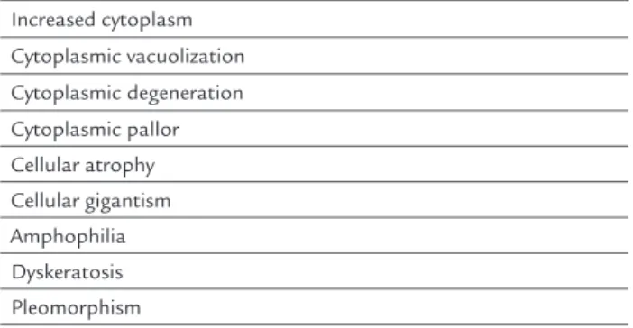
TABLE 1 Main cytological indings induced by radiation in cervicovaginal smears.
Increased cytoplasm Cytoplasmic vacuolization
Cytoplasmic degeneration Cytoplasmic pallor Cellular atrophy Cellular gigantism Amphophilia Dyskeratosis Pleomorphism
Nuclear increase (without compromising the nuclear-cytoplasmic ratio)
Nuclear vacuolization Nuclear degeneration Nuclear pallor Hyperchromasia
Dyskaryosis (present in malignant cells) Mitosis (typical or atypical)
Binucleation Multinucleation Karyorrhexis Nuclear pyknosis Anisokaryosis Necrosis
Leukocyte iniltrate Multinucleated giant cells Repair cells
Macro and multiple nucleoli Anisonucleolosis
CONCLUSION
To date, a protocol has not been established to precisely differentiate the morphological characteristics of benign cells with actinic effects from recurrent malignant cells on post-radiotherapy smears. However, there are several studies aimed at minimizing occasional diagnostic difficulties. The information presented here allows for a critical and reflexive analysis of the knowledge about the impact of radiotherapy on epithelial cells, allowing us to point out difficulties, limitations and potentialities that affect the medical prac-tice and the care provided during cytopathological follow--up of patients submitted to cervical cancer radiotherapy.
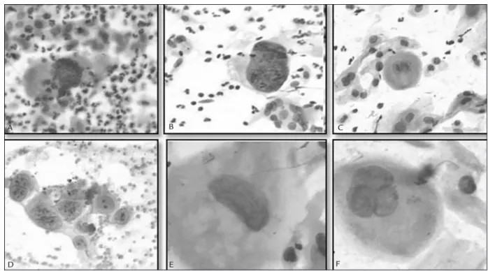
and cytoplasmic vacuolization, dyskeratosis, bi- and multi-nucleated (B/M) cells, macro and multiple nucleoli, aniso-karyosis, anisonucleolosis and nuclear pyknosis (Figure 1). The difficulties pointed out show the importance of considering in future studies the experience of the profes-sionals involved in the analysis of irradiated cells and a reflection on the subjectivity of the method. In general, our review provided insights to help coordinate further training for professionals dedicated to the analysis and diagnosis of cells under actinic effects, in addition to recommending complementary studies using techniques that contribute to the understanding of actinic alterations in order to increase prognostic acuity.
RESUMO
Avaliação citopatológica no seguimento de pacientes submetidas à radioterapia por câncer de colo uterino
O câncer de colo uterino configura-se como um importan-te problema de saúde pública. O importan-tesimportan-te citopatológico é a principal estratégia de programas de rastreamento dessa neoplasia maligna em todo o mundo. Entretanto, a demo-ra no diagnóstico ocasiona tdemo-ratamentos mais agressivos e menos efetivos. Pacientes com neoplasia maligna de colo uterino que são encaminhadas para radioterapia
apresen-tam doença em estádios avançados, e esse fato determina altos índices de recidiva locorregional. A utilização da ra-dioterapia como tratamento do câncer do colo uterino provoca alterações morfológicas não só nas células epiteliais neoplásicas e não neoplásicas como também nas células estromais, o que dificulta o diagnóstico da lesão residual e resulta em um dilema na rotina citopatológica. Com base nas dificuldades da avaliação citopatológica do seguimen-to das pacientes pós-radioterapia, o objetivo deste trabalho foi descrever os efeitos citopáticos actínicos. O trabalho teve como base uma revisão estruturada no período de junho de 2015 a abril de 2016, visando a um estudo explo-ratório-descritivo. As investigações bibliográficas foram realizadas por meio de seleção e análise dos artigos, lista de autores e palavras-chave; seleção de novos artigos foca-da na análise de referências bibliográficas dos documentos previamente selecionados e livros-texto de relevância con-ceitual. As alterações citopatológicas actínicas mais inci-dentes descritas na literatura são: gigantismo celular, va-cuolização nuclear e citoplasmática, disceratose, bi e multinucleações, macro e múltiplos nucléolos, anisocario-se, anisonucleolose e picnose nuclear. Até o momento, não se conseguiu estabelecer um protocolo que possa diferen-ciar precisamente as características morfológicas entre células benignas com efeitos actínicos das células malignas recidivantes em esfregaços pós-radioterapia.
FIGURE 1 Actinic cytopathological aspects.
Source: Padilha et al.2
A B C
Palavras-chave: radioterapia, neoplasias do colo do úte-ro, efeitos actínicos, citopatologia.
REFERENCES
1. Instituto Nacional de Câncer – INCA. Radioterapia [cited 2016 May]. Available from: http://www.inca.gov.br/conteudo_view.asp?ID=100.
2. Padilha CML, Feliciano GD, Padilha Filho LG. Analysis of actinic effect after radiotherapy in the uterine cervix carcinomas. J Am Sci. 2006; 2(3):23-28.
3. Instituto Nacional de Câncer José Alencar Gomes da Silva / INCA. Estimativa 2016: Incidência de Câncer no Brasil / Instituto Nacional de Câncer José Alencar Gomes da Silva. Coordenação de Prevenção e Vigilância. Rio de Janeiro: INCA; 2014.
4. Panobianco MS, Pimentel AV, Almeida AM, Oliveira ISB. Women diagnosed with advanced cancer of the cervix: coping with the disease and treatment. Rev Bras Cancerol. 2012; 58(3):517-23.
5. Yuan G, Wu L, Huang M, Li N, An J. A phase II study of concurrent chemo-radiotherapy with weekly nedaplatin in advanced squamous cell carcinoma of the uterine cervix. Radiation Oncol. 2014; 9:55.
6. Solomon D, Davey D, Kurman R, Moriarty A, O’Connor D, Prey M, et al.; Forum Group Members; Bethesda 2001 Workshop. The 2001 Bethesda Sys-tem: terminology for reporting results of cervical cytology. JAMA. 2002; 287(16):2114-9.
7. Ministério da Saúde. Instituto Nacional de Câncer José Alencar Gomes da Silva / INCA. Nomenclatura Brasileira para Laudos Citopatológicos Cervicais. Coordenação-Geral de Prevenção e Vigilância, Divisão de Detecção Precoce e Apoio à Organização de Rede. 3. ed. Rio de Janeiro: Inca; 2012. 8. Hospital AC Camargo. Manual de condutas em ginecologia oncológica. São
Paulo: FAP; 2010.
9. Powers CN. Radiation treatment effects in cervical cytology. Diagn Cytopathol. 1995; 13(1):75-80.
10. Zannoni GF, Velone GV, Carbone A. Morphological effects of radiochemo-therapy on cervical carcinoma: a morphological study of 50 cases of hyste-rectomy specimens after neoadjuvant treatment. Int J Gynecol Pathol. 2008; 27(2):274-81.
11. Villas MV, Macedo-Soares TDLVA, Russo GM. Bibliographical research method for business administration studies: a model based on scientific journal ranking. Braz Admin Rev. 2008; 5(2):139-59.
12. Mendes KDS, Silveira RCCP, Galvão CM. Revisão integrativa: método de pesquisa para a incorporação de evidências na saúde e na enfermagem. Texto & Contexto Enferm. 2008; 17(4):758-64.
13. Gil AC. Como delinear uma pesquisa bibliográfica. In: Gil AC, organizer. Como elaborar projeto de pesquisa. 4. ed. São Paulo: Atlas; 2002. 14. Okuno E. Efeitos biológicos das radiações ionizantes. Acidente radiológico
de Goiânia. Estud Av. 2013; 27(77):185-99.
15. Okuno E. Radiação: efeitos, riscos e benefícios. São Paulo: Harbra; 1988. 16. Vargas FG, Silva BS, Cardoso CO, Leguisamo N, Moraes CAR, Moraes CV,
et al. Impact of body weight on radiation exposure during invasive cardiac procedures. Rev Bras Cardiol Invasiva. 2012; 20(1):63-8.
17. Souza E, Soares JPM. Correlações técnicas e ocupacionais da radiologia intervencionista. J Vasc Bras (Porto Alegre). 2008; 7(4):341-50.
18. Ribeiro EL, Pessoa MB. The effects of electromagnetic radiation in the life of the human being: An analysis of the environmental paradigm. Rev Tecnologia e Sociedade. 2007. Available from: https://periodicos.utfpr.edu. br/rts/article/download/2502/1616.
19. Sharma M, Revannasiddaiah S, Gupta M, Seam RK, Gupta Mk, Rastogi M. Can pure accelerated radiotherapy given as six fractions weekly be an option in locally advanced carcinoma cervix: results of a prospective randomized phase III trial. J Can Res Ther. 2016; 12(1):103-8.
20. Friedberg EC, Walker GC, Siede W, Wood, RD, Schultz RA, Ellenberger T. DNA repair and mutagenesis. Washington: ASM Press; 2006.
21. Choi JH, Besaratina A, Lee DH, Lee CS, Pfeifer GP. The role of DNA polymerase iota in UV mutational spectra. Mutat Res. 2006; 599(1-2):58-65. 22. Matsumura Y, Ananthaswamy H. Toxic effects of ultraviolet radiation on
the skin. Toxicol Appl Pharmacol. 2004; 195(3):298-308.
23. Mundt AJ, Lujan AE, Rotmensch J, Waggoner SE, Yamada SD, Fleming G, et al. Intensity-modulated whole pelvic radiotherapy in women with gyne-cologic malignancies. Int J Radiat Oncol Biol Phys. 2002; 52(5):1330-7. 24. INCA/MS. Câncer do colo do útero. Rev Bras Cancerol. 2000; 46(4):351-4.
25. Castle PE, Schiffman M, Wheeler CM. Hybrid capture 2 viral load and the 2-year cumulative risk of cervical intraepithelial neoplasia grade 3 or cancer. Am J Obstet Gynecol. 2004; 191(5):1590-7.
26. Navarro C, Fonseca AJ, Sibajev A, Souza CIA, Araújo DS, Teles DA, et al. Cervical cancer screening coverage in a high-incidence region. Rev Saúde Pública. 2015; 49:17.
27. Solomon D, Nayar R. The Bethesda System for reporting cervical cytology: definitions, criteria, and explanatory notes. 2. ed. New York: Springer; 2004. 28. Pinto AP, Maia R. [Adenosquamous carcinoma of the cervix mimicking adenoid basal carcinoma: case report and review of the literature]. J Bras Patol Med Lab. 2007; 43(1):45-50.
29. Costa MOLP, Heráclio AS, Coelho AV, Acioly VL, Souza PR, Correia MT. Comparison of conventional Papanicolaou cytology samples with liquid--based cervical cytology samples from women in Pernambuco, Brazil. Braz
J Med Biol Res. 2015; 48(9):831-8.
30. Mcgoogan E, Colgan TJ, Ramzy I, Cochand-Priollet B, Davey DD, Grohs HK, et al. Cell preparation methods and criteria for sample adequacy. International Academy of Cytology Task Force summary. Diagnostic Cytology Towards the 21st Century: An International Expert Conference and Tutorial. Acta Cytol. 1998; 42(1):25-32.
31. Lu CH, Chang CC, Ho ESC, Chen SJ, Lin SJ, Fu TF, et al. Should adequacy criteria in cervicovaginal cytology be modified after radiotherapy, chemotherapy, or hysterectomy? Cancer Cytopathol. 2010; 118(6):474-81.
32. Zannoni GF, Velone GV. Accuracy of Papanicolaou smears in cervical cancer patients treated with radiochemotherapy followed by radical surgery. Am J Clin Pathol. 2008; 130(5):787-94.
33. Wright JD, Herzog TJ, Mutch DG, Gibb RK, Rader JS, Davila RM, et al. Liquid--based cytology for the postirradiation surveillance of women with gynecologic
malignancies. Gynecol Oncol. 2003; 91(1):134-8.
34. Gupta S, Mukherjee K, Gupta YN, Kumar M. Sequential radiation changes in cytology of vaginal smears in carcinoma of cervix uteri during radiotherapy. Int J Gynaecol Obstet. 1987; 25(4):303-8.
35. Shield PW. Chronic radiation effects: a correlative study of smears and biop-sies from the cervix and vagina. Diagn Cytophatol.1995; 13(2):107-19. 36. Alsharif M, McKeon DM, Gulbahce HE, Savik K, Pambuccian SE.
Unsatisfactory surepath liquid-based Papanicolaou tests: causes and significance. Cancer. 2009; 117(1):15-26.
37. Baker JJ. Conventional and liquid-based cervicovaginal cytology: a comparison study with clinical and histologic follow-up. Diagn Cytopathol. 2002; 27(3):185-8.
38. Bolick DR, Hellman DJ. Laboratory implementation and efficacy assessment of the ThinPrep cervical cancer screening system. Acta Cytol. 1998; 42(1):209-13.
39. Bur M, Knowles K, Pekow P, Corral O, Donovan J. Comparison of ThinPrep preparations with conventional cervicovaginal smears. Practical considerations. Acta Cytol. 1995; 39(4):631-42.
40. Carpenter AB, Davey DD. ThinPrep Pap Test: performance and biopsy follow--up in a university hospital. Cancer. 1999; 87(3):105-12.
41. Linder J, Zahniser D. The ThinPrep Pap Test. A review of clinical studies. Acta Cytol. 1997; 41(1):30-8.
42. Teixeira LA, Porto MAT, Souza LPA. A expansão do rastreio do câncer do colo do útero e a formação de citotécnicos no Brasil. Physis (Rio J). 2012; 22(2):713-31.
43. Gusberg SB. A consideration of the problem of radiosensitivity in cancer of the cervix. Am J Obstet Gynecol. 1956; 72(4):804-19.
44. Graham JB, Graham RM, Liu W. Prognosis in cancer of the uterine cervix based on the vaginal smear before treatment; SR: the sensitization response. Surg Gynecol Obstet. 1954; 99(5):11.
45. Murad TM, August C. Radiation-induced atypia. A rewiew. Diagn Cytopathol. 1985; 1(2):135-52.
46. Shield PW, Wrigth RG, Free K, Daunter B. The accuracy of cervicovaginal cytology in the detection of recurrent cervical carcinoma following radiotherapy. Gynecol Oncol. 1995; 41(3):223-9.
47. Davey DD, Gallion H, Jennings CD. DNA cytometry in postirradiation cervico-vaginal smear. Hum Pathol. 1992; 23(9):1027-31.
48. Wacthel MS, Thaler HT, Gangi MD, Hadju SI. Immunoperoxidase staining of cervicovaginal smears after radiotherapy. Acta Cytol. 1992; 36(3):305-9. 49. Masubuchi K, Kubo H, Tenjin Y, Ono M, Yamazaki M. Follow-up studies