Printed version ISSN 0001-3765 / Online version ISSN 1678-2690 http://dx.doi.org/10.1590/0001-3765201620150051
www.scielo.br/aabc
Production, purification and characterization of an
exo-polygalacturonase from
Penicillium janthinellum
sw09
Yuping Ma1
, Siwen Sun2
, Hui Hao1 and
CHunping Xu2
1
Technical Center of China Tobacco Henan Industrial Co. Ltd, Longhai road 72, Zhengzhou, Henan 450016, China 2
College of Food and Biology Engineering, Zhengzhou University of Light Industry, Dongfeng road 5, Zhengzhou 450002, China
Manuscript received on January 23, 2015; accepted for publication on March 3, 2015
aBSTRaCT
A soil isolate, Penicillium janthinellum sw09 has been found to produce significant amounts of an extracellular pectinase subsequently characterized as exo-polygalacturonase (exo-PG). By optimizing growth conditions, P. janthinellum sw09 produced high amount of exo-PG (16.54 units/mL). The crude enzyme was purified by gel filtration chromatography and two exo-PG activity peaks (designated as PGI and PGII) were revealed. On SDS–PAGE analysis, purified PGII using DEAE-Sepharose FFcolumn, was found to be a single band with a molecular mass of 66.2 kDa. The purified PGII exhibited maximal activity at the temperature of 45 oC and pH 5.0. The stability profiles show that PGII is more stable in the pH range of 4.0-8.0 and below 60 o
C. The Km and Vmax for the enzyme was 1.74 mg/mL and 18.08 μmol/ (mL•min), respectively. Due to this enzymatic characterization, this pectinase is an attractive candidate for applications in degradation of pectin.
Key words: Penicillium janthinellum, polygalacturonase, purification, characterization.
Correspondence to: Chunping Xu E-mail: [email protected]
inTRoDuCTion
Pectinases constitute a unique group of enzymes which catalyze the degradation of pectic polymers present in the plant cell walls (Torres-Favela et al.
2003). Polygalacturonases (PGs) (EC 3.2.1.67) are part of a group of pectinases that possess hydrolytic
functions and are able to cleave the α-1,4 glycosidic bonds between the two non-esterified galacturonic
acid residues in pectin and pectic substances (Mohamed et al. 2006, Niture 2008). PGs have been
classified according to their substrate specificity
and the position of the bonds which they hydrolyse.
Endo-PG was defined as hydrolysing the a-1,4- glycosidic bonds in the substrate at random, whilst
exo-PG hydrolyses the terminal link (Niture 2008).
These enzymes are used in different sectors, such as paper, textile, cellulose, and food industries and have many important industrial applications (Joslyn et al. 1952, Kashyap et al. 2001). Additionally, PGs
can promote the clarification of wine and grape
juice (Sanderson and Spotts 1995).
PG from microbial source is the most impor-tant pectinase used in industries. Although a num-ber of organisms producing pectinases have been reported, the selection of potential isolates still
remains a tedious task, especially when
maximum yield (Pandey et al. 1999). Most of the commercial PGs produced, are investi gated from Aspergillus species (Mathew et al. 2008). The purification of PG is an important tool for understanding its properties and it reveals the structure and functional mechanism which are
important for knowledge regarding the action of
these enzymes in the plant infections process, in industrial application, in addition to their importance in biomass degradation. Fungal PGs are generally monomeric proteins with a carbohydrate content of 5–85% and molecular masses in the
range of 20 to 95 kDa (Borin et al. 1996, Devi and
Rao 1996, Mathew et al. 2008, Niture 2008, Niture and Rao 2004).
In this paper, the production medium of exo-PG obtained from P. janthinellum sw09 by submerged culture was optimized by orthogonal design. Furthermore, purification and physicochemical properties of exo-PG were investigated.
MaTeRiaLS anD MeTHoDS
MicroorganisMand growth conditions
The microorganism used in this study was
Penicillium janthinellum sw09, isolated from
a tobacco field in Xuchang, P.R. China, and
maintained as stock culture on potato dextrose agar
(PDA) medium containing 0.3% citrus pectin at 4
oC. This strain was identified by Sangon Biotech
(Shanghai) Co., Ltd and preserved at the Henan Province Microbiological Culture Collection Center (HPMCC no. 1225853). The liquid culture of mycelia was initiated by transferring the fungal
mycelia from the stock culture on a Petri dish into
the seed culture medium. The seed culture was
propagated in a 250 mL Erlenmeyer flask containing
100 mL of liquid medium (10 g/L yeast extract, 5 g/L citrus pectin, FeSO4 0.1 g/L, MgSO4 0.5 g/L, KH2PO4 1g/L, and pH 6.0) at 28 oC on a shaking
incubator at 160 rpm for 72 h. The exo-PG was produced with the inoculation of 4% (v/v) of the
seed culture by submerged fermentation in a stirred
tank bioreactor (Infors, Switzerland, 3.5-L working
volume). The fermentations were performed under the following conditions: temperature, 32
o
C, aeration, 2 vvm; agitation speed, 160 rpm. All experiments were performed in triplicate to ensure the trends observed were reproducible.
optiMization procedure
Orthogonal design is one of the important statistical methods that use the Taguchi method (Taguchi 1986). Taguchi method is a fast and considerable way of optimization conferring outcome in
simul-taneous study of many factors, making its mark in
quality products supplemented with better process performance, and rendering high yield and better stability through limited experimental trials. This approach has been successfully applied to optimi-ze medium composition, condition of enzyme reaction, and extraction conditions for bioactive compounds (De Oliveira and Alves 2000, Xu et al. 2003). An orthogonal L9(3)4 test design was applied to optimize medium condition of PG by P.
janthinellum in flask culture. As seen on Table I,
the experiment was carried out with 4 factors with 3 levels based on preliminary single experimental results. The exo-PG yield was chosen as the res-ponse.
TaBLe i
Experimental factors and their levels for orthogonal projects. Level Glucose (g/L) Yeast extract (g/L)
KH2PO4. (g/L)
Citrus pectin (g/L)
1 30 3 0.5 2
2 40 4 1 4
3 50 5 1.5 6
enzyMe activity MeasureMents
The activity of exo-PG was assayed in a reaction mixture containing 1% citrus pectin solution with
no. 9000-69-5, Sigma) in 0.2M sodium acetate buffer (pH 5.0) at 45 oC for 30 min. The number of reducing groups, released by enzymatic action, was measured by the DNS method (Miller 1959) and expressed as galacturonic acid. One unit of enzyme
activity (U) was defined as the amount of enzyme releasing one μmol of galacturonic acid per minute
under the assay conditions (Martins et al. 2002).
enzyMe purification procedure
After 72 h of fermentation, the culture broth was centrifuged at 9,000 g for 15 min, and the resulting
supernatant was filtered through a membrane filter (0.45 µm, Millipore). The resulting culture filtrate
was precipitated by ammonium sulfate (20–100%) and the mixture was stirred for 2 h, and centrifuged at 15,000 g for 30 min. The ammonium sulphate fraction was dialyzed against Tris–HCl buffer (50 mM, pH 6.5) and directly loaded on a Sepharose
CL-6B gel filtration column (2.5 × 60 cm) equilibrated
with 13 mM Na2HPO4-citric acid buffer (pH
5.0). Protein fractions collected from the column,
corresponding to the protein peak, were pooled and
further applied to the DEAE-Sepharose FF based anion exchangers column equilibrated with 20 mM Na2HPO4-citric acid buffer (pH 6.5) (Mohamed et al. 2006). Fractions of 4 mL were collected and assayed for exo-PG activity. The objective of this procedure was to estimate the number of isoform of exo-PG presented in the crude enzyme solution. The protein fraction with exo-PG activity was desalted overnight by dialysis at 4 oC.
analytical electrophoresis
The molecular weight of the purified enzyme was
determined by SDS-PAGE in a Mini Protean II
apparatus (10 × 8 cm) (Biorad). Electrophoresis was
carried out in a vertical slab gel apparatus (Beijing Liuyi Instrument Factory, DYCZ-24DN) with a 5%
(w/v) polyacrylamide stacking gel and 12% (w/v)
resolving gel in Tris/glycine buffer (pH 8.3) with
the Sigma molecular weight marker MP102 (14.4
kDa-94.0 kDa) in a parallel lane (Laemmli 1970).
The protein band was visualized by silver staining. The molecular mass of the isolated subunit was
estimated by Ferguson plot analysis (Hedrick and
Smith 1968).
protein estiMation
Protein concentration was determined in the
concentration ranges of 1–10 and 10–100 μg/mL
by the Bradford microassay, with bovine serum albumin (BSA) as the standard (Bradford 1976).
propertiesof purified enzyMe
All enzyme catalytic properties were assayed with 1% citrus pectin (D.E 67-70%) as substrate using the procedure for enzyme activity determination described above and carried out with three replicates. Exo-PG activity was assayed as a function of pH, in Na2HPO4 -citric acid buffer (pH 3.0–8.0), at 45 oC, and temperature, in Na
2HPO4
-citric acid buffer at the pH optimum, incubated at different temperatures between 35 oC and 60 oC.
The thermal stability was investigated by
remeasuring the activity of the purified enzyme solution after it had been kept for 2 h, in the absence
of substrate, at different temperatures in the range
of 30-60 °C. In these tests, the initial and final
exo-PG activities were determined at optimum pH and
temperature. The pH stability of the purified enzyme
was evaluated by dispersing (1:1, v/v) enzyme solution in Na2HPO4-citric acid buffer (pH 3.0–8.0) and maintaining these solutions at 45 oC for 4 h. An aliquot was taken to determine the remaining
activity at the optimum pH and temperature. The Michaelis constant (Km) and Vmax values
were determined from Lineweaver-Burk plots of
enzyme activity measured with 67-70% D.E. citrus pectin (Sigma) as substrate, at concentrations between 2.0 and 40.0 mg/mL at optimum pH and
temperature. The purified enzyme concentration
statistical analyses
Unless otherwise stated, data were expressed as mean ± SD, and analyzed statistically by ANOVA method. P-values below 0.05 and 0.01 were
regar-ded as statistically significant and highly
signifi-cant, respectively.
ReSuLTS anD DiSCuSSion
productionof exo-PGin flask culture
Enzyme production is generally influenced by the composition of the medium, in particular the carbon and nitrogen sources (Gomes et al. 2009). The orthogonal matrix method was employed to optimize the concentration of the medium. The experimental conditions for each project were listed in Table II, and experimental results were also included in the last column of this table. According to the magnitude order of the R value (maximum difference) as shown in Table II, the order of effects of all factors on polygalacturonase production was glucose > citrus pectin > KH2PO4 > yeast extract. This result indicated that the effect
of glucose was more important than that of other nutrients. In terms of the maximum K value of each factor based on statistical calculations, optimal composition for polygalacturonase production were 50 g/L glucose, 3 g/L yeast extract, 1 g/L KH2PO4, and 2 g/L citrus pectin. The significance of each factor was evaluated by calculating the F value and the results were summarized in Table III. Both factors, including glucose and citrus pectin
had significant effects on the polygalacturonase
production (P < 0.01). In order to confirm the optimization results, the suggested medium
components were confirmed in triplicate. The 16.54
U/mL exo-PG was maximally obtained under the optimum conditions just described. This implied that the selected conditions were really the most suitable. Mathew et al. (2008) found the optimal pectin level as being 1.0 g/L for exo-PG production by a Penicillium strain in submerged culture, which suggests that the level of substance requirement for exo-PG production depends on the nature of the
specific strain, even though they belong to the same
species (i.e. Penicillium).
TaBLe ii
Application of L9(34) orthogonal projects to exo-PG production by P. janthinellum after 72 hours
of incubation in flask culture.
Run Glucose Yeast extract KH2PO4 Citrus pectin Enzymatic activity (U/mL)
1 1 1 1 1 9.51±1.07
2 1 2 2 2 9.69±0.27
3 1 3 3 3 8.18±0.22
4 2 1 2 3 11.56±0.01
5 2 2 3 1 12.16±0.43
6 2 3 1 2 11.10±0.82
7 3 1 3 2 15.96±0.20
8 3 2 1 3 14.74±0.08
9 3 3 2 1 16.27±0.15
k1 9.12 12.34 11.78 12.65
k2 11.61 12.19 12.51 12.25
k3 15.66 11.85 12.10 11.49
R 6.53 0.49 0.72 1.16
Optimal level 3 1 2 1
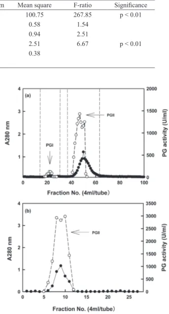
purificationof exo-pg
The crude enzyme solution separated on a
Sepharose CL-6B column, afforded two peaks
of exo-PG activity (designated as PGI and PGII) suggesting two isoforms (Fig. 1a). Because the PGII had higher amount and was stronger activity
than PGI, PGII was selected for further purification.
when the PGII solution (tube no. 38 to 64 as shown in Fig. 1a) was combined, concentrated and loaded
on a DEAE-Sepharose FFcolumn, one peak was
eluted (Fig. 1b). The results were in agreement with the reports of Mohamed et al. (2006), who observed two polygalacturonases PGI and PGII
separated from Trichoderma harzianum by two
chromatography steps using DEAE-Sepharose and Sephacryl S-200.
characterizationof exo-pg
The homogeneity of the purified PGII was demonstrated by the presence of one single protein band on polyacrylamide gel and its molar mass
was estimated to be 66.2 kDa as a single subunit
(Fig. 2). This observation was in the range reported for PGs from several fungi, which have molecular
masses ranging from 20 to 95 kDa (Gomes et al.
2009, Silva et al. 2007). In comparison with other
Penicillium sp., lower molecular weights (24.1
kDa) were detected for PG from P. viridicatum (Mohamed et al. 2003).
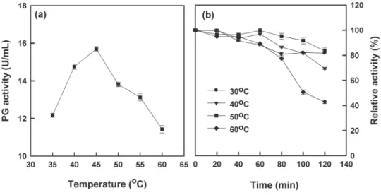
The effect of pH on the P. janthinellum
PGII activity toward polygalacturonic acid was examined at 45 oC. As shown in Figure 3a, the
enzyme showed hydrolase activity from pH 3.0 to 8.0, and maximum enzymatic activity (15.72 U/ mL) at pH 5.0. The same pH optimum was reported for PGs from Asergillus niger (Behere et al. 1993),
T. harzianum (Mohamed et al. 2006). The effect
of pH on the stability of P. janthinellum PGII was
Figure 1 - Elution of exo-PG from Sepharose CL-6B gel filtration column previously equilibrated with 13 mM Na2HPO4-citric acid buffer (pH 5.0) (a) and DEAE-Sepharose FF column based anion exchangers column equilibrated with 20 mM Na2HPO4-citric acid buffer (pH 6.5) (b). Protein (●); exo-PG activity (○).
TaBLe iii
Variance analysis of L9(34) orthogonal experiment on the optimization of exo-PG production. Variance source Sum of squares Degree of freedom Mean square F-ratio Significance
A:glucose 201.50 2 100.75 267.85 p < 0.01
B:yeast extract 1.16 2 0.58 1.54
C:KH2PO4 1.89 2 0.94 2.51
D:Citrus pectin 5.02 2 2.51 6.67 p < 0.01
Errors 6.77 18 0.38
investigated by incubating the enzyme at 45 oC at different pH’s for 4 h. The results showed that the enzyme was the most stable in pH of 6.0, with 90–100% of the full activity in a broader pH range of 4.0–8.0 (Fig. 3b). This is in agreement with the results reported by Kobayashi et al. (2001) for
Bacillus sp. PG. They reported that PG activity was
very stable in various 50 mM buffers between pH 6 and 12 when incubated at 30 oC for 1 h.
with regard to temperature, optimal P. janthi
nellum exo-PG activity was observed at 45 oC (Fig.
4a). Similarly, temperature optima for PGs from T.
reesei (Mohamed et al. 2003), Mucor flaves (Grade
et al. 2003), Bacillus sp. (Kobayashi et al. 2001) and
T. harzianum (Mohamed et al. 2006) were around
40 and 50 oC. The effect of temperature on thermal stability of P. janthinellum PGII was investigated by incubation the enzyme for 2 h in 13 mM Na2HPO4 -citric acid buffer, pH 5.0 at different temperature ranging from 30 to 60 oC prior to substrate addition
(Fig. 4b). In the absence of substrate for 1 h, PGII showed 83–100% of the original activity at 30–60
oC. After 2 h, PGII showed 68–88% of the original
activity at 30–50 oC, while at 60 oC, the enzyme lost 58% of its initial activity. Similarly, Kobayashi et al. (2001) found that Bacillus sp. PG was stable up to 50–55 oC in the presence of CaCl2.
Figure 2 - SDS-PAGE of exo-PG (a), purified PGII (b) and standard proteins (c) from DEAE-Sepharose FF chromato-graphy.
The kinetic parameters of P. janthinellum
PGII affinity for citrus pectin and different pectins
at pH 5.0 and 45 oC were determined by a typical
double reciprocal Lineweaver-Burk plot (Fig. 5).
The Km and Vmax for the enzyme was 1.74 mg/mL
and 18.08 μmol/ (mL•min), respectively. The Km
values of P. janthinellum PGII were lower than Kms ranged from 2.5 to 14.08 mg/mL of PGs from A.
niger (Parenicova et al. 1998), Neurospora crassa
(Polizeli et al. 1991), T. harzianum (Mohamed et al. 2006) and Saccharomyces cerevisiae (Blanco et al. 1997). Low Km indicated that P. janthinellum PGII
had high affinity using citrus pectin as substrate.
Vmax of P. janthinellum PGII was in the range of
Vmaxs, i.e. 13.0 to 2600 μmol/ (mL•min) from above organisms. The Kcat for the enzyme was calculated as 301.33/s. The Kcat values of P. janthinellum PGII were in the range of Kcats, i.e. 21.0 to 592/s from above organisms.
ConCLuSionS
The fungal strain Penicillium janthinellum sw09 produced an exo-polygalacturonase able to release galacturonic acid as a hydrolysis product from natural D.E. pectin. This enzyme exhibited the pH
and temperature profiles for optimum activity and
stability that differed from other fungal PGs, which
kept the stability of the enzyme in a pH range of
4.0-8.0 and at a temperature range of 30-60 oC. A thermostable nature with a high pH range for
activity makes it possible to have wide range of industrial applications. Further works on
scale-up fermentation optimization (i.e. aeration rate and agitation speed) in bioreactor and industrial application are in progress in our laboratory.
ReSuMo
Penicillium janthinellum sw09, isolado do solo, produz quantidades significativas de uma pectinase extracelular que foi posteriormente caracterizada como exo-poliga-lacturonase (exo-PG). Ao otimizar as condições de crescimento, P. janthinellum sw09 produz quantidades
elevadas de exo-PG (16,54 unidades/ml). A enzima im-pura foi purificada por cromatografia de filtração em gel e dois picos com atividade elevada de exo-PG (designa-dos como PGI e PGII) foram revela(designa-dos. Por análise em SDS-PAGE, PGII foi purificada utilizando uma coluna de DEAE-Sepharose FF, observou ser formada por uma única banda com uma massa molecular de 66,2 kDa. PGII purificada exibiu uma actividade máxima a uma temperatura de 45 °C e pH 5,0. Os perfis de estabilidade mostram que PGII é mais estável numa faixa de pH de 4,0-8,0 e abaixo de 60 ° C. O Km e Vmax para a enzima foi de 1,74 mg / ml e 18,08 μmol / (mL • min), resvamente. Devido a esta caracterização enzimática, pecti-nase é um candidato atrativo para ser aplicado na degra-dação da pectina.
Palavras-chave: Penicillium janthinellum, poligalactu-ronase, purificação, caracterização.
ReFeRenCeS
Behere a, satyanarayan v and padwal-desai sr. 1993. Separation and limited characterization of three polygalacturonases of Aspergillus niger.Enzyme Microb Tech 15: 158-161.
Blanco p, sieiro c, reBoredo nM and villa tg. 1997. Genetic determination of polygalacturonase production in wild-type and laboratory strains of Saccharomyces cerevisiae. Arch Microbiol 167: 284-288.
BorinMDF, said s and fonsecaMJV. 1996. Purification and biochemical characterization of an extracellular endopolygalacturonase from Penicillium frequentons. J Agr Food Chem 44: 1616-1620.
Bradford MM. 1976. A rapid and sensitive method for the quantitation of microgram quantities of protein utilizing the principle of protein-dye binding. Anal Biochem 72: 248-254.
de oliveira d and alvesTLM. 2000. A kinetic study of lipase-catalyzed alcoholysis of palm kernel oil. Appl Biochem Biotech84-86: 59-68.
devi na and raoAGA. 1996. Fractionation, purification, and preliminary characterization of polygalacturonases produced by Aspergillus carbonarius. Enzyme Microbiol Tech 18: 59-65.
goMes e, leiteRSR, da silva r and silva d. 2009. Purification of an Exopolygalacturonase from Penicillium viridicatum RFC3 Produced in Submerged Fermentation. Int J Microbiol 2009: 8.
grade rv, van driessche g, van BeeuMen J and Bhat
Mk. 2003. Purification, characterization and mode of action of an endopolygalacturonase from the psychrophilic fungus Mucor flavus. Enzyme Microbiol Technol 32: 321-330.
hedrick Jl and sMith aJ. 1968. Determination of the molecular weight of proteins by electrophoresis in slab gels with a transverse pore gradient of crosslinked polyacrylamide in the absence of denaturing agents. Arch Biochem Biophys 126: 155-164.
Joslyn na, Mist s and laMBart e. 1952. Clarification of apple juice by fungal PG preparations. Food Technol 6: 133-139.
kashyap dr, vohra pk, chopra s and tewari r. 2001. Applications of pectinases in the commercial sector: a review. Bioresource Technol 77: 215-227.
koBayashi t, higaki n, suzuMatsu a, sawada
k, hagihara h and kawai s. 2001. Purification and properties of a highmolecular-weight, alkaline exopolygalacturonase from strain of Bacillus. Enzyme Microbiol Technol 29: 70-75.
laeMMli u. 1970. Cleavage of structural proteins during the assembly of the head of bacteriophage T4. Nature 227: 680-685.
Martins es, silva d, da silva r and goMes e. 2002. Solid state production of thermostable pectinases from thermophilic Thermoascus aurantiacus. Process Biochem 37: 949-954.
Mathew a, eldo an and Molly ag. 2008. Optimization of culture conditions for the production of thermostable polygalacturonase by Penicillium SPC-F 20. J Ind Microbiol Biotechnol 35: 1001-1005.
Miller gl. 1959. Use of dinitrosalicylic acid reagent for determination of reducing sugar. Anal Chem 31: 426-428. MohaMed sa, christensen TMIE and Mikkelsen
Jd. 2003. New polygalacturonases from Trichoderma reesei: characterization and their specificities to partially methylated and acetylated pectins. Carbohydr Res 338: 515-524.
MohaMed sa, farid nM, hossiny en and Bassuiny
ri. 2006. Biochemical characterization of an extracellular polygalacturonase from Trichoderma harzianum. J Biotechnol 127: 54-64.
niture sk. 2008. Comparative biochemical and structural characterization of fungal polygalacturonases. Biologia 63: 1-19.
niture sk and rao a. 2004. Purification and biochemical characterization of polygalacturonase II produced in semi-solid medium by a strain of Fusarium moniliforme. Microbiological Res 159: 305-314.
pandey a, selvakuMar p, carlos rs and poonaM
n. 1999. Solid state fermentation for the production of industrial enzymes. Curr Sci 77: 149-1620.
parenicova l, Benen JAE, kester HCM and
visser J. 1998. pgaE encodes a fourth member of the endopolygalacturonase gene family from Aspergillus niger. Eur J Biochem 251: 72-80.
biochemical characterization of extracellular polygalac-turonase activity. J Gen Microbiol 137: 1815-1823. sanderson pg and spotts ra. 1995. Postharvest decay
of winter pear and apple fruit caused by species of
Penicillium. Phytopathol 85: 103-110.
silva d, Martins es, leiteRSR, da silva r, ferreira
v and goMes e. 2007. Purification and characterization of an exo-polygalacturonase produced by Penicillium viridicatum RFC3 in solid-state fermentation. Process Biochem 42: 1237-1243.
taguchi g. 1986. Introduction to Quality Engineering, UNIPUB/Kraus International, white Plains, NY, USA, vol. 1 & 2.
torres-favela e, aguilar c, esquivel-contreras
cJ and gustavo gv. 2003. Pectinase. In Enzyme Technology. Asiatech Publisher Inc. Delhi, p. 273-296. xu cp, kiM sw, hwang hJ, choi Jw and yun Jw. 2003.