Vol.57, n.4: pp. 587-594, July-August 2014 http://dx.doi.org/10.1590/S1516-8913201402214
ISSN 1516-8913 Printed in Brazil
BRAZILIAN ARCHIVES OF BIOLOGY AND TECHNOLOGY
A N I N T E R N A T I O N A L J O U R N A L
Purification and Characterization of a Polygalacturonase
Produced by
Wickerhamomyces anomalus
María Alicia Martos
1*, Ana Paula Butiuk
1, Natalia Lorena Rojas
2and Roque Alberto
Hours
21Laboratorio de Microbiología de Alimentos y Biotecnología Dr. Fernando O. Benassi; Facultad de Ciencias
Exactas Químicas y Naturales; Universidad Nacional de Misiones; Misiones - Argentina. 2Centro de Investigación y Desarrollo en Fermentaciones Industriales; Universidad Nacional de La Plata - CONICET; La Plata - Argentina
ABSTRACT
The aim of this work was to study the purification and physicochemical properties of an endo-polygalacturonase (PG) produced by Wickerhamomyces anomalus isolated from the citrus fruit peels. The enzyme was purified to homogeneity from the culture filtrate of W. anomalus grown on the yeast nitrogen base medium with glucose as carbon and energy source and citrus pectin as inductor. After anion-exchange chromatography and gel filtration chromatography, PG activity was eluted as a single peak, yielding 21% of the original activity. After dialysis and cation-exchange chromatography, only one fraction with PG activity was obtained, recovering 56% of initial enzyme activity and 1.3-fold increase in specific activity. The molecular weight of the enzyme was estimated as 43 kDa by the SDS-PAGE. The enzyme exhibited maximal activity at pH 4.2 and was stable over a pH range from 3.5 to 6.0 and up to 49ºC for 10 h. The Vmax and Km values with polygalacturonic acid as substrate were 0.26
mmol/L.min and 0.173 mg/mL, respectively. Cations such as Cu+2, Fe+3, Mg+2, Mn+2 and Zn+2 did not show any significant effect on PG activity but K+ and Ca+2 reduced it. The purified PG was able to macerate cassava tissues.
Key words:Wickerhamomyces anomalus, polygalacturonase, purification, characterization
*Author for correspondence: [email protected]
INTRODUCTION
Enzymes hydrolyzing the pectic substances are known as pectinolytic enzymes or pectinases. These enzymes are responsible for the degradation of pectic substances that occur as structural polysaccharides in the middle lamella and the primary cell walls of plant tissues. Based on their mode of action, these enzymes are classified as polygalacturonase (PG), pectin esterase (PE), pectin lyase (PL) and pectate lyase (PAL). PG, PL and PAL are depolymerizing enzymes, which split the α-(1,4)-glycosidic bonds between galacturonic monomers in pectic substances either by the hydrolysis (PG) or by β-elimination (PL, PAL). PE
catalyzes the de-esterification of the methoxyl group of pectin, forming pectic acid (Jayani et al. 2005; Tari et al. 2007).
damage to adjacent secondary cell walls, to help avoid cell lysis, keeping the nutritional properties of food (Costa et al. 2007).
A pectinolytic yeast strain was isolated from the citrus fruit peels in the province of Misiones, Argentina and it was identified as Wickerhamomyces anomalus, re-classification of the species Pichia anomala (Kurtzman 2008; Martos et al. 2013a). This wild yeast strain, grown on a yeast nitrogen base medium with glucose and citrus pectin produced an endo-PG with maceration activity of vegetable tissues. The supernatant of W. anomalus was able to macerate cassava and potato tissues (Martos et al. 2013a, b). In view of the potential applicability of PG secreated by W. anomalus in food technology, the present work reports the purification and some relevant physicochemical properties of the enzyme.
MATERIALS AND METHODS
Microorganism
W. anomalus, isolated from the citrus fruit peels in the province of Misiones, Argentina (Martos et al. 2013a) was used in this study.
Culture media
YM medium contained (g/L) yeast extract (Sigma Chemical Co., St. Louis, Mo, USA) 5, tryptone (Difco-Becton Dickinson & Co.,Sparks, MD, USA) 5, glucose (Britania, Buenos Aires, Argentina) 10, agar (Britania),15 and the pH was 5.0.
YNB medium contained (g/L) yeast nitrogen base (YNB, Difco) 6.7, glucose (Britania) 5, citrus pectin (Parafarm, Buenos Aires, Argentina) 5 and the pH was 5.0. Citrus pectin was washed with 70% (v/v) ethanol-HCl (0.05 N) solution to remove the soluble sugars (Cavalitto et al. 1996). All the components of media were autoclaved (121ºC, 15 min), except in the case of YNB solution, which was sterilized separately by filtration through a cellulosic filter paper (0.22 µm, Sartorius).
Enzyme production
Five hundred millilitre Erlenmeyer flasks with 95 mL of YNB medium were inoculated with 5.0 mL of an appropriate dilution of a suspension of the microorganism (OD620 = 0.96), grown in YM
medium (30ºC, 24 h). The Erlenmeyer flasks were
incubated at 30ºC for 10 h on a rotary shaker at 180 rpm. The biomass was separated by centrifugation at 2350 × g at 5ºC for 10 min. The culture medium supernatant, named enzymatic extract (EE), was frozen at -18ºC and used as source of extracellular enzyme (Martos et al. 2013a).
Enzyme purification procedures
The EE (500 mL) was concentrated (2 ×) on a vacuum rotary evaporator at 38ºC. The resulting solution was lyophilized to dryness and re-suspended (5×) in sodium acetate buffer (AcB 0.2 M, pH 5.0). The concentrated enzymatic extract (CEE) obtained was subject to two purification procedures (Procedure I and Procedure II). All the chromatographic steps were carried out on an Amersham FPLC system (ÄKTA FPLC-U900 Chromatographyc System, GE Healthcare). After each chromatographic step, fractions with PG activity were applied to a PD-10 desalting column (GE Healthcare) and then loaded onto a single well 10% SDS-PAGE minigel, in a Mighty Small II Unit (Hoefer SE 260, GE). Proteins were visualized by Coomassie Brilliant Blue G-250 staining (Smith 1984).
Procedure I
PG activity were pooled, concentrated by lyophilization and kept refrigerated until use.
Procedure II
The CEE (18 mL) was dialyzed overnight at 5ºC against buffer AcB (0.2 M, pH 5.0). After dialysis, the solution was freeze-dried, re-suspended in AcB and loaded onto a SP-Sepharose FF cation-exchange column (XK 26/20, GE Healthcare) equilibrated with the same buffer. The bound proteins were then eluted with a linear gradient of NaCl (0–0.5M) over four column volumes at a flow rate of 3.0 mL/min during 150 min. Fractions of 6.0 mL were collected and those with PG activity were pooled, concentrated by lyophilization and kept refrigerated until use.
Enzyme characterization Molecular weight
The molecular weight of the purified enzyme was determined by the SDS-PAGE, using a low molecular weight marker (14.5 a 94 kDa, GE Healthcare).
Effect of pH on the purified polygalacturonase activity and stability
The effect of pH on the purified PG activity was determined by incubating the reaction mixture at pH values ranging from 2.6 to 6.0 under the standard enzyme assay conditions. The pH stability of the enzyme was evaluated by measuring the residual activity under standard enzyme assay conditions after incubating the pure enzyme without substrate for 24 h at 5ºC at various pH from 2.6 to 7.0. The buffer employed in these measurements was citric phosphate buffer (CPB, 0.1M citric acid and 0.2 M Na2HPO4). The
pH was initially adjusted to pH 5.0 with NaOH and then recalibrated to the desired pH with HCl or NaOH. All the experiments were conducted in triplicate and the results showed the mean values of the activities.
Thermal stability of the purified PG
For the determination of the thermal stability, the enzyme was pre-incubated over a range of temperature from 37ºC to 60ºC in the CPB at pH optimum. Residual activity was measured during 10 h, under the standard enzyme assay conditions. All the experiments were conducted in triplicate and the results showed the mean values of the activities.
Enzyme kinetics
The Michaelis constant (Km) and Vmax values of
purified PG were determined from Lineweaver-Burk plots of enzyme activity measured under the standard enzyme assay conditions with polygalacturonic acid (PGA; Sigma) as the substrate at concentrations between 0.05 to 2.5 g/L in BCP at pH optimum.
Influences of metal ions on the purified PG activity Before the assay, the enzyme solution was dialized in order to remove all the interfering substances. The effect of cations such as Cu+2, Ca+2, Fe+3, K+,
Mg+2, Mn+2 and Zn+2 on enzyme activity was
tested in the reaction medium under the standard enzyme assay conditions.
Analytical techniques Enzyme activity assays
PG activity was assayed by measuring the reducing groups released after incubation of reaction medium at 30ºC for 10 min (pH 5.0) by dinitrosalicylic acid method using PGA as the substrate (Miller 1959). A calibration curve was made with galacturonic acid (GA, Sigma) as standard. One unit of PG was defined as the amount of enzyme which releases 1 µmol of GA per minute.
Protein estimation
Protein concentrations were determined by the Bradford method using bovine serum albumin (Sigma) as standard (Bradford 1976).
Assay of maceration activity
To evaluate the maceration capacity of the purified PG on cassava tissues, cassava was peeled and cut into pieces (3-4 mm on each side), placed in Petri dishes, submerged in a purified PG solution in AcB (0.2 M, pH 5.0) and incubated at 30ºC for 2 h. Maceration activity was estimated from the loss of coherence of tissue and by microscopic observations. Blanks were prepared with heat-denatured enzyme.
RESULTS AND DISCUSSION
Enzyme purification
column and Sephacryl S-100 gel filtration column are shown in Figures 1 and 2, respectively.
Table 1 - Summary of W. anomalus PG purification by
Procedure I.
Purification step
Total activity
(U)
Total protein
(mg)
Specific activity (U/mg protein)
Recovery (%)
CEE (10 ×). 731.3 4.93 148 100 Sephadex G-2 488 2.604 188 66 Sepharose Q 324.5 1.26 258 44 Sephacryl S-100 159 ND ND 21
ND: not detected, CEE: concentrated enzymatic extract.
Figure 1- Elution profile of W. anomalus PG on Q
Sepharose anion-exchange column (XK 26/20), equilibrated with AcB. Elution of bound proteins with a linear gradient of NaCl (0 - 0.5 M) in AcB, column volume: 7.5, flow rate: 1.5 mL/min, time of elution: 180 min.
Figure 2 - Elution profile of W. anomalus PG on
Sephacryl S-100 gel filtration column (XK 16/100), equilibrated with AcB (0.2 M, pH 5.0). Elution of proteins with AcB, column volume: 1.9, flow rate: 1.5 mL/min, time of elution: 150 min.
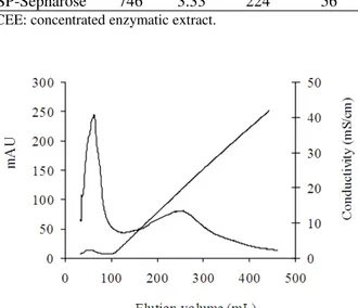
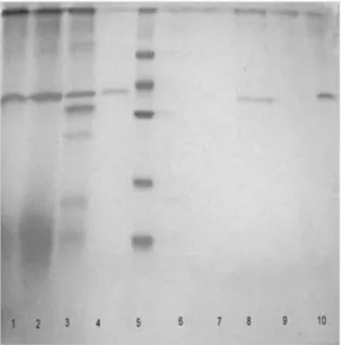
From Sephadex G-25 column, a fraction 6-13, free of salts, was collected, recovering 66% of initial enzyme activity (Table 1). During anion-exchange chromatography, the protein with PG activity did not bind to Q-Sepharose FF column; others contaminating proteins were eluted at high ionic strength (Fig. 1). In this step, 44% of the initial enzyme activity was recovered (Table 1). After the subsequent purification step using Sephacryl S-100 gel filtration column, PG activity was eluted as a single peak yielding 21% of the original PG activity (Fig. 2). A summary of the purification process of PG by Procedure II is presented in Table 2. Elution profiles of PG on SP-Sepharose FF cation-exchange column is shown in Figure 3. Figure 4 shows the SDS-PAGE of proteins obtained during the different steps of purification.
Table 2 - Summary of W.anomalus PG purification
process by Procedure II.
Purification step
Total activity
(U)
Total protein
(mg)
Specific activity (U/mg protein)
Recovery (%)
CEE 1.341 8.12 165 100
Dialysis 903 5.06 179 67
SP-Sepharose 746 3.33 224 56
CEE: concentrated enzymatic extract.
Figure 3 -Elution profile of W. anomalus PG on
Figure 4 - SDS-PAGE with Coomassie staining of the purification steps of W. anomalusPG. Lane 1:
concentrated enzymatic extract (CEE). Lane 2: after dyalisis. Lane 3: after desalted on a Sephadex G-25 column. Lane 4: after Q Sepharose chromatography. Lane 5: low molecular weight marker. Lane 6: after Q Sepharose chromatography (without PG activity). Lane 7: after Sephacryl S-100 chromatography (fraction 26-40, without PG activity). Lane 8: after Sephacryl S-100 chromatography (fraction 50-70). Lane. 9: after SP-Sepharose chromatography (fraction 7-13), without activity. Lane 10: after SP-Sepharose chromatography (fraction 26-70).
After dialysis, 67% of the initial enzymatic activity was recovered (Table 2). From cation-exchange chromatography a fraction (33-54) with PG activity was obtained, recovering 56% of initial enzyme activity and 1.3-fold increase in PG specific activity (Fig. 3). The protein in lane 4 (after Q Sepharose chromatography), lane 8 (after Sephacryl S-100 chromatography) and lane 10 (after SP-Sepharose chromatography) showed a single band on 10% SDS-PAGE (Fig. 4). PG produced by S. cerevisiae CECT1389, grown on YNB medium composed of glucose and PGA, was purified in a single step by size-exclusion chromatography from the culture filtrate (Blanco et al. 1994). Pedrolli et al. (2009) reported that pectic enzymes purifications were performed mainly by the chromatographic techniques. A. giganteus PG was purified after two simple steps: protein precipitation and anion-exchange
chromatography. Most of methods for purifying the fungal and bacterial PGases that have been published are associated with considerable enzyme losses, mainly caused by the relatively high number of steps required during the purification process (Pedrolli and Carmona 2010). The purification process described for W. anomalus in the present study, using procedure II, could be useful for future industrial scale application. By means of this process, high yields of the enzyme could be obtained in few stages.
Molecular weight
The SDS-PAGE revealed an apparent molecular weight of 43 kDa for W. anomalus PG (Fig.4). It has been reported that the molecular weight of PGases from different sources usually ranged between 40 and 60 kDa (Jayani et al. 2005; Pedrolli and Carmona 2010).
Effect of pH on PG activity and stability
The effect of pH on purified PG activity is shown in Figure 5. Figure 6 represents the values of enzymatic residual activity after incubating the enzyme at different pH for 24 h at 5°C.
Figure 5 - Effect of pH on purified PG activity produced
by W. anomalus. PG activity was determined
as a percentage of the enzyme activity under standard enzyme conditions.
the pH range of 4.5 to 6.0 (Blanco et al. 1994). Blanco et al. (1999) reported that yeasts PGases exhibited an optimum pH in the acidic region between 3.5 and 5.5. The optimum pH for PG produced by S. cerevisiae UCLMS-39 after a purification process was 3.5 (Fernández-González et al. 2004).
Figure 6 - Effect of pH on purified PG stability produced by W. anomalus. PG activity was
determined as a percentage of the enzyme activity under standard enzyme conditions.
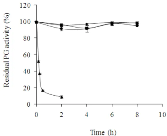
Thermal stability of PG
The effect of temperature on the purified PG stability produced by W. anomalus is shown in Figure 7.
Figure 7 showed that in the absence of substrate, purified PG was stable at 37, 43 and 49ºC for 10 h of incubation at optimum pH. The analysis of variance revealed no significant differences between these values (p < 0.05). At 55ºC, the enzymatic activity decreased and retained only 37% of the initial activity after 15 min of incubation. The thermal stability of PG produced by W. anomalus was similar to that reported for PGases from others yeasts such as S. cerevisiae IM1-8b and S. cerevisiae 1389, which were quite stable in the 20-50ºC temperature range but were inactivated (80%) within 5 min at 55ºC (Blanco 1997). Two PGases of Cryptococcus sp. (named p36 and p40) remained stable up to 40°C for 30 min (Miura et al. 2001). The optimum temperature for PG produced by S. cerevisiae UCLMS-39 was 50°C; higher from this temperature, the activity gradually decreased (Fernandez-Gonzalez et al. 2004). The knowledge of enzyme stability is
important to maintain the desired level of enzyme activity over a long period of time and improve its stability for an efficient application in an industrial process (Martins et al. 2007). Besides after any application, the enzyme has to be inactivated, so the knowledge of thermal inactivation has great importance too (Tari et al. 2008).
Some authors reported that during the purification procedure, some protein stabilizing factors might be lost (Naidu and Panda 2003; Martins et al. 2007; Pedrolli and Carmona 2010). Data published previously with crude PG from W. anomalus obtained in YNB medium showed that enzyme maintained 78% of its residual activity of after 30 min at 55ºC (Martos et al. 2013b). These results suggested that PG lost stability after the purification process.
Figure 7 - Effect of temperature on purified PG stability produced by W. anomalus. Symbol: ♦37ºC, ■
43ºC, ●49ºC, ▲ 55°C. PG activity was
determined as a percentage of the enzyme activity under standard enzyme conditions.
Kinetic parameters
Kinetic parameters such as Vmax and Km were
determined from the regression lines of Lineweaver-Burk plots. The Vmax and Km values
obtained for the PGA were 0.26 mmol/L.min and 0.173mg/mL (R2: 0.901), respectively. Similar K
M
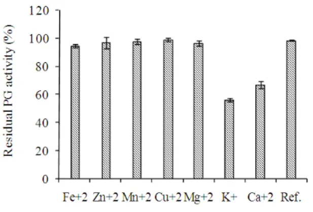
Effects of various cations on PG activity
The effects of various cations on purified PG activity is shown in Figure 8.
Cu+2, Fe+3, Mg+2, Mn+2 and Zn+2did not show any
significant effect on the purified PG activity under the assay conditions, whereas the residual PG activity was 67 and 56% in the presence of Ca+2 and K+, respectively. It was reported that PGases
tended to undergo changes in their physical and chemical properties in the presence of some ions. Therefore, it was necessary to test the effects of a number of representative cations in the reaction medium on PG activity (Miura et al. 2001).
Figure 8 - Effects of cations on purified PG activity produced by W. anomalus. PG activity was
determined as a percentage of the enzyme activity under standard enzyme conditions.
Assay of maceration activity
Purified PG was able to macerate the cassava tissue as shown by the loss of coherence of the tissue. Microscopic examination of the maceration product showed single cells. These results confirmed the maceration capacity of PG as it was reported previously with the supernatant of W. anomalus (Martos et al. 2013a).
CONCLUSIONS
Polygalacturonase from the culture supernatant of
W. anomalus, grown on YNB medium with
glucose and pectin, was efficiently purified using a two-step procedure with a high recovery (dialysis and cation-exchange chromatography). PG exhibited an optimum pH in the acidic region, a high stability over a large pH range and was stable
up to 50ºC, suited to most fruit and vegetable processing applications. These properties suggested that this PG could be a potential candidate for different applications in food industry, mainly in the maceration of cassava tissues of regional interest in the province of Misiones, Argentina.
REFERENCES
Blanco P, Sieiro C, Diaz A, Villa TG. Production and partial characterization of an endopolygalacturonase from Saccharomyces cerevisiae. Can J Microbiol.
1994; 40: 974-977.
Blanco P, Sieiro C, Diaz A, Villa TG. Differences between pectic enzymes produced by laboratory and wild-type strains of Saccharomyces cerevisiae. World J Microbiol Biotechnol. 1997; 13:711-712.
Blanco P, Sieiro C, Villa TG. Production of pectic enzymes in yeasts. Mini Review. FEMS Microbiol Lett. 1999; 175: 1-9.
Bradford MM. A rapid and sensitive method for the quantification of microgram quantities of protein utilizing the principle of protein-dye binding. Anal Biochem. 1976; 72:248-254.
Cavalitto SF, Arcas JA, Hours RA. Pectinase production profile of Aspergillus foetidus in solid
state cultures at different acidities. Biotechnol Lett.
1996; 18: 251-256.
Costa JAV, Colla E, Magagnin G, Oliveria dos Santos L, Vendruscolo M, Bertolin TE. Simultaneous amyloglucosidase and exo-polygalacturonase production by Aspergillus niger using solid-state
fermentation. Braz Arch Biol Technol. 2007; 50 (5):
759-766.
Croak S, Corredig M. The role of pectin in orange juice stabilization: Effect of pectin methylesterase and pectinase activity on the size of cloud particles. Food Hydrocolloid. 2006; 20:961-965.
Federici F. Production, purification and partial characterization of an endopolygalacturonase from
Cryptococcus albidus var. albidus. Antonie Van Leeuwenhoek. 1985; 51:139-150.
Fernández González M, Úbeda JF, Vasudevan TG, Cordero Otero RR, Briones AI. Evaluation of polygalacturonase activity in Saccharomyces cerevisiae wine strains. FEMS Microbiol Lett. 2004;
237: 261-266.
Gummadi SN, Panda T. Purification and biochemical properties of microbial pectinases: a review. Process Biochem. 2003; 38 (7): 987-996.
Jayani RS, Saxena S, Gupta R. Microbial pectinolytic enzymes: A review. Process Biochem. 2005; 40:
Kurtzman CP, Robnett CJ, Basehoar-Powers E. Relationships among species of Pichia, Issatchenkia
and Williopsis determined from multigene
phylogenetic analysis and the proposal of
Barnettozyma gen. nov., Lindnera gen. nov. and Wickerhamomyces gen. nov. FEMS Yeast Res. 2008;
8: 939-954.
Martins ES, Silva D, Leite RS, Gomes E. Purification and characterization of polygalacturonase produced by thermophilic Thermoascus aurantiacus
CBMAI-756 in submerged fermentation. Antonie Van Leeuwenhoek. 2007; 91: 291-299.
Martos MA, Zubreski ER, Combina M, Garro OA, Hours RA. Isolation of a yeast strain able to produce a polygalacturonase with maceration activity of cassava tissues. Food Sci Technol. 2013a; 33(2):
332-338.
Martos MA, Zubreski ER, Garro OA, Hours RA. Production of Pectinolytic Enzymes by the Yeast
Wickerhanomyces anomalus Isolated from Citrus
Fruits Peels. Biotechnol Res Int. 2013b; 1:1-7.
Miller GL. Use of dinitrosalicylic acid reagent for determination of reducing sugar. Anal Chem. 1959;
31: 426-428.
Miura T, Abe F, Inoue A, Usami R, Horikoshi K. Purification and characterization of novel extracellular endopolygalacturonases from a deep-sea yeast, Cryptococcus sp. Nº 6, isolated from the Japan
Trench. Biotechnol Lett. 2001; 23:1735-1739.
Naidu GSN, Panda T. Studies on pH and thermal inactivation of pectolytic enzymes from Aspergillus niger. Biochem Eng J. 2003; 16:57-67.
Nighojkar S, Phanse Y, Sinha D, Nighojkar A, Kumar A. Production of polygalacturonase by immobilized cells of Aspergillus niger using orange peel as
inducer. Process Biochem. 2006; 41:1136-1140.
Pedrolli DB, Carmona EC. Purification and characterization of the exopolygalacturonase produced by Aspergillus giganteus in submerged
cultures. J Ind Microbiol Biotechnol. 2010;
37:567-573.
Pedrolli DB, Monteiro AC, Gomes E, Carmona EC. Pectin and pectinases production, characterization and industrial application of microbial pectinolytic enzymes. Open Biotechnol J. 2009; 3:9-18.
Smith BJ. Quantification of proteins on polyacrylamide gels. Methods Mol Biol. 1984; 1:119-125.
Tari C, Dogan N, Gogus N. Biochemical and thermal characterization of crude exo-polygalacturonase produced by Aspergillus sojae. Food Chem. 2008;
111(4): 824-829.
Tari C, Gögus N, Tokatli F. Optimization of biomass, pellet size and polygalacturonase production by
Aspergillus sojae ATCC 20235 using response
surface methodology. Enzyme Microbial Technol.
2007; 40: 1108-1116.