J. Braz. Chem. Soc., Vol. 16, No. 2, 280-283, 2005. Printed in Brazil - ©2005 Sociedade Brasileira de Química 0103 - 5053 $6.00+0.00
Short Report
* e-mail: [email protected]
Biologically Active Polyketides Produced by
Penicillium
janthinellum
Isolated as an Endophytic
Fungus from Fruits of
Melia azedarach
Andrey M. do Rosário Marinhoa, Edson Rodrigues-Filho.*,a, Maria da Luz R. Moitinhob and Lourivaldo S. Santosc
a
Departamento de Química, Universidade Federal de São Carlos, CP 676, 13.565-905 São Carlos - SP, Brazil b
Departamento de Análises Clínicas, Universidade Estadual de Maringá, Av. Colombo, 5790, 87020-900 Maringá - PR, Brazil
c
Departamento de Química, Universidade Federal do Pará, Av. Augusto Corrêa, 1, 66075-970 Belém - PA, Brazil
Penicillium janthinellum, isolado como um fungo endofítico dos frutos de Melia azedarach, foi cultivado por 20 dias em milho branco triturado e autoclavado, onde os policetídeos conhecidos citrinina, emodina, 1,6,8-triidróxi-3-hidroximetilantraquinona, e uma nova antraquinona modificada, denominada janthinona, foram produzidos e isolados por procedimentos cromatográficos clássicos e identificados por extensivos estudos espectroscópicos, principalmente RMN 1D e 2D e EM. Essas substâncias foram ensaiadas contra diversas bactérias. Citrinina foi ensaiada pela primeira vez contra
Leishmania e inibiu 100% o crescimento de cepas depois de 48h a uma concentração de 40 µg mL-1.
Penicillium janthinellum, isolated as an endophytic fungus from fruits of Melia azedarach, was cultivated over 20 days on ground and autoclaved white corn, where the known polyketides citrinin, emodin, 1,6,8-trihydroxy-3-hydroxymethylanthraquinone, and a new modified anthraquinone, named janthinone, were produced and isolated by classical chromatographic procedures and identified by MS and 1D and 2D NMR spectroscopic data. The antibacterial properties of these polyketides were investigated. Citrinin inhibited 100% of Leishmania growth after 48h at a concentration of 40 µg mL-1.
Keywords: endophytic, Melia azedarach, Leishmania, Penicillium, polyketide
Introduction
Endophytic microorganisms are isolated from internal plant tissue and can be present even at the cell level.1,2 The
biochemistry of these associations is a fascinating and novel field of research. The colonization of the host plant by endophytes may be mediated by interesting secondary metabolites.3 In general these compounds show insecticidal
and antimicrobial activities, which may give the host some protection against further invasion.1,3
We have been studying the chemistry of microorganisms associated with Meliaceae plants. A large collection of fungi have been isolated from the fruits, leaves, stems and roots of Meliaazedarach.4 Species of
Penicillium isolated from this host plant have shown the ability to produce chemically interesting and biologically active compounds.5-7 Herein we report our chemical
investigations of another Penicillium, identified as P.
janthinellum, cultivated on sterilized white corn. This fungus produced polyketides, basically hydroxyanthra-quinones, ergosterol and poliols. Citrinin was tested against bacteria and Leishmania mexicana, showing promising inhibition properties.
Results and Discussion
The compounds 1-2 (Figure 1) were obtained from methanol extracts of the biomass produced by P. janthinellum on white-corn. These substances are yellow-orange pigments and exhibited typical characteristics of hydroxyanthraquinones. Their UV, NMR and MS physical data are in perfect agreement with those reported for 1,6,8-trihydroxy-3-methylanthraquinone8 (emodin, 1) and
1,6,8-trihydroxy-3-hydroxymethylanthraquinone9 (citreorosein,
2), respectively.
281 Biologically Active Polyketides Produced by Penicilliumjanthinellum
Vol. 16, No. 2, 2005
with the 1H and 13C NMR data, suggested C
16H12O5 (284 Da)
as the molecular formula of 3. The UV (λmax 236, 258, 304 and 364 nm) and IR (n 3463, 1742 and 1653 cm-1) data of
this compound are characteristic of hydroxyanthraquinones. The 1H NMR spectrum of 3, analyzed with the aid of 2D
NMR data (COSY, HMBC), showed the presence of hydrogen signals for two benzene rings [δ 6.74 (d, 1.8 Hz, 2); δ 6.62 (d, 1.8 Hz, 4); δ 7.51 (dd, 8.2 and 2.0 Hz, H-5); δ 7.74 (dd, 8.2 and 7.3 Hz, H-6); and δ 7.30 (dd, 7.3 and 2.0 Hz, H-7)]. The signals for H-2 and H-4 could be easily assigned due to a long range coupling (J 0.4 Hz) with the benzylic methyl hydrogens at δ 2.43 (brs, H-11); this methyl group occurs frequently in fungal hydroxyanthraquinones. The IR (νmax 1653 and 1742 cm
-1) and 13C NMR (δ 180.0
and 169.6) spectroscopic data indicated that compound 3
has a conjugated ketone and a conjugated lactone in its molecular structure. The position of the lactone function was based on the HMBC correlation detected between H-5 (whose signal was unequivocally ascribed by 1H-1H COSY)
and C-10. Other important HMBC are shown below in Figure 2. The NMR signal of the hydrogen H-7 (δ 7.30) showed a nuclear Overhauser effect (NOE) with the methoxyl at δ 4.03 confirming structure 3 (Figure 1) for this modified anthraquinone. It was named janthinone, and appears to be a new natural product. Similar lactones (with
C-11 oxidized) were produced by a Brazilian strain of Aspergillus versicolor.10
Compound 4 (citrinin) is a well known mycotoxin produced mainly by Penicillium citrinum and several Aspergillus species.11 This polyketide was produced in
good yields by P. janthinellum obtained from the host plant M. azedarach. The identification of 4 was achieved mainly by 1H and 13C NMR, which showed that it occurs in
the p-quinone form.12
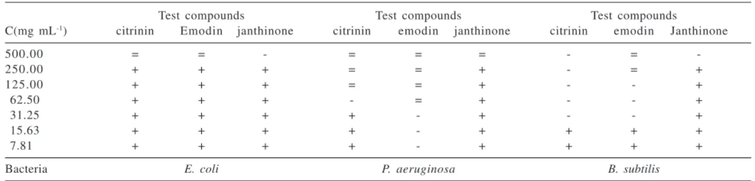
The antibacterial activity of citrinin (4) and the anthraquinones 1-3 was examined in the presence of Escherichia coli, Pseudomonas aeruginosa and Bacillus subtilis, and the results are shown in Table 1. Emodin and citrinin were almost completely inactive against E. coli, but shown promising activity against P. aeruginosa and B. subtilis. Compound 3, the new lactone janthinone, was considered inactive in all tests (>500 µg mL-1). The growth
inhibition property of citrinin (4) against Leishmania mexicana was also studied (Table 2). Inhibition rates at very low concentration of citrinin (5 µg mL-1) were
observed. The highest growth inhibition was observed after 48 h of inoculation of the microorganism in the medium containing 40 µg mL-1 of citrinin.
Experimental
General procedures
UV spectra were obtained in CH2Cl2 solution on a
Table 1. Growth behavior of bacteria in the presence of different concentrations of polyketides produced by P. janthinellum
Test compounds Test compounds Test compounds
C(mg mL-1) citrinin Emodin janthinone citrinin emodin janthinone citrinin emodin Janthinone
500.00 = = - = = = - =
-250.00 + + + = = + - = +
125.00 + + + = = + - - +
62.50 + + + - = + - - +
31.25 + + + + - + - - +
15.63 + + + + - + + + +
7.81 + + + + - + + + +
Bacteria E. coli P. aeruginosa B. subtilis
“+” : Growth like in the negative control; “-“ : bacteriostatic effect (growth in subculture); “=” : bactericidal effect (not grow in subculture).
Figure 1. Polyketides isolated from the fungi biomass produced by
P. janthinellum over white-corn.
282 Marinho et al. J. Braz. Chem. Soc.
HEWLETT PACKARD 8452-A spectrophotometer, and IR spectra were measured with a BOMEN MB-102 spectrophotometer in KBr pellets. Low-resolution APCIMS and ESIMS data were acquired in positive ion mode, using a MICROMASS QUATTRO-LC instrument equipped with an ESI/APCI “Z-spray” ion source. 1H and 13C NMR
experiments were recorded on a BRUKER DRX-400 spectrometer with Acetone-d6 or CDCl3 as the solvent and TMS as the internal standard.
Plant material
Fruits of Melia azedarach were collected on the campus of the Federal University of São Carlos, São Carlos, Brazil. Samples (dried leaves and fruits) of the plant are deposited in the Herbarium of Department of Botanic at the University to be included in their catalogue.
Microorganism
P. janthinellum was obtained from the collection of the Laboratório de Bioquímica Micromolecular of the Chemistry Department at Universidade Federal de São Carlos. This collection contains isolates recently obtained4
from Melia azedarach. P. janthinellum is identified by the number LaBioMi-018.
White corn culture of P. janthinellum and isolation of the polyketides
45 Erlenmeyer flasks (500 mL), containing 90g of white corn (“Yoki”) and 75 mL of distilled water per flask, were autoclaved twice at 121oC for 40 min. A small disc of the PDA
medium from the Petri dish containing mycelium of P. janthinellum was transferred under sterile conditions to 42 Erlenmeyer flasks containing sterilized corn. Three flasks were kept for control purposes. After 20 days of growth at 25 °C, methanol (200 mL) was added to each flask and allowed to stand for 5 h, and then it was filtered by gravity. The methanol was evaporated under reduced pressure, producing a yellowish residue (14.2 g). Part of this residue (10.0 g), was subjected to a low-pressure silica gel CC eluted with n-hexane, ethyl
acetate and methanol gradient. The medium polarity fractions eluted with ethyl acetate were repeatedly chromatographed on silica gel CC [n-hexane:ethyl acetate:methanol (60:35:05), isocratic ) and preparative TLC [chloroform:methanol (96:04)] and the polyketides, emodin (80.1 mg) (1), 1,6,8-trihydroxy-3-hydroxymethylanthraquinone (10.2 mg) (2), and janthinone (80.1 mg) (3), were finally purified. Citrinin (180.2 mg) (4) crystallized from a methanol solution of a fraction eluted with ethyl acetate:methanol (95:05) from the first silica gel CC.
Antibacterial bioassay
The susceptibilities of microorganisms to the test polyketides were determined by microbroth dilution assay as recommended13 by the Subcommittee on
Antifungal Susceptibility Testing of the US National Committee for Clinical Laboratory Standards (NCCLS). They were performed on 96 well plates with 100 µL of Mueller Hinton Broth (MHB), 100 µL of test compound and 5 µL of test bacteria at 1.0 x 10⁷ UFC mL-1, followed
by incubation at 37 oC (24h). The test substances obtained
from the fungal culture were dissolved in dimethylsulfoxide at initial concentration 500 µg mL-1.
The test microorganisms were Escherichia coli, Pseudomonas aeruginosa and Bacillus subtilis (obtained from Universidade de Maringá - PR, Brazil). Bioactivity was recorded as absence of red coloration in the wells after addition of 10 µL of 2,3,5-triphenyltetrazolium chloride. The tested microorganisms were subcultured on MHB plates. The activities of the test compounds were classified as bacteriostatic or bactericidal effects according the behavior of the microorganisms in these subcultures. Penicillin, vancomycin and tetracycline (25 µg mL-1 each) were used as positive controls; the
cultivation medium (MHB only) was used as negative control.
Leishmanicidal bioassay
Auxenic culture of Leishmania mexicana (MNYC/BZ/ 62/M379, Universidade de Maringá - PR, Brazil) in exponential phase of growth was incubated at 33 oC in
modified UM-54 medium along with 5, 10, 20, and 40 mg mL-1 of citrinin (4), dissolved in DMSO (0.1 % final
concentration), in a 24 wells ELISA plate. The inhibitions of growth, reported as percentage of death14 (Table 2), were
measured in a Neubauer chamber, by counting dead microorganisms after 24, 48 and 72 h compared with the controls (only medium without the test compound).
Table 2. Growth inhibition rate of Leishmania mexicana in the
presence of different concentrations of citrinin (4) Concentration (µg mL-1)
Time 5 1 0 2 0 4 0
24 h 1.2 % 25.0 % 56.2 % 90.2 %
48 h 0.3 % 27.5 % 81.2% 100.0 %
283 Biologically Active Polyketides Produced by Penicilliumjanthinellum
Vol. 16, No. 2, 2005 Emodin (1)
Orange amorphous powder; mp 253 – 256 °C (methanol); UV λmax/nm (CH2Cl2): 235, 259, 305 and 362;
IR νmax/cm
-1: 3415, 1670, 1629, 1480, 1384, and 758 (KBr); 1H NMR (400 MHz, acetone-d
6): δ 2.26 (s, CH3-11), δ 6.66
(d, J 2.2 Hz, H-7), δ 7.13 (brs, H-2), δ 7.24 (d, J 2.2 Hz, H-5), δ 7.56 (brs, H-4); APCIMS (Daughter ions, 40 eV): m/z 269 ([M-H]-, 50 %), 241 (41), 225 (100), 210 (32), 197
(49), 182 (66), 171 (22), 105 (21).
1,6,8-Trihydroxy-3-hydroxymethylantraquinone (2)
Orange amorphous powder; mp 280-287 °C (methanol); UV λmax/nm (CH2Cl2): 232, 260, 308 and 363; IR νmax/cm
-1:
3419, 1675, 1627, 1476, 1399 and 758 (KBr); 1H NMR
(400 MHz, acetone-d6): δ 4.78 (s, CH2-11), δ 6.67 (d, J 2.0 Hz, H-7), δ 7.27 (d, J 2.0 Hz, H-5), δ 7.31 (brs, H-2), δ 7.75 (brs, H-4); APCIMS (Full scan): m/z 285 ([M-H]-, 100 %).
Janthinone (3)
Yellow plates; mp 188 – 190 °C (methanol); UV λmax/
nm (CH2Cl2): 236, 258, 304 and 364; IR νmax/cm -1: 3463,
1742, 1653, 1488, 1437, 1368, 1200 (KBr); 1H NMR (400
MHz, CDCl3): δ 2.43 (brs, CH3-11), δ 4.03 (s, OCH3), δ
6.62 (d, J 1.8, H-4), δ 6.74 (d, J 1.8, H-2), δ 7.30 (dd, J 2.0 and 7.3, H-7), δ 7.51 (dd, J 2.0 and 8.2 Hz, H-5), δ 7.74 (dd, J 7.3 and 8.2 Hz, H-6); ESIMS (Full scan): 283 ([M-H]-,
100 %); EIMS (70 eV): m/z 284 ([M]+., 81%), 252 (100), 223 (63), 168 (25), 139 (60), 115 (37), 75 (22).
Citrinin (4)
Yellow needles; mp 158 – 163 °C (methanol/water); UV λmax/nm (CH2Cl2): 333; IR νmax/cm
-1: 3417, 2974, 2922,
1670, 1635, 1491, 1377, 1265 and 818 (KBr); 1H NMR
(400 MHz, CDCl3): δ 1.23 (d, J 7.1 Hz, CH3-10), δ 1.35 (d, J 6.7 Hz, CH3-9), δ 2.02 (s, CH3-11), δ 2.98 (q, J 7.1 Hz, H-4), δ 4.80 (q, J 6.7 Hz, H-3), δ 8.25 (s, H-1), δ 15.13 (s, OH), δ 15.90 (s, OH); ESIMS (Daughter ions, 20 eV): m/z 249 ([M-H]-, 21 %), 231 (10), 205 (100), 177 (61), 161
(36).
Acknowledgement
The authors are grateful to Fundação de Amparo à Pesquisa do Estado de São Paulo (FAPESP), Conselho Nacional de Desenvolvimento Científico e Tecnológico (CNPq) and Fundação Coordenação de Aperfeiçoamento de Pessoal de Ensino Superior (CAPES) for financial support and research fellowships.
References
1. Rizzo, I.; Varsavky, M.; Haidukowski, M.; Frade, H.; Toxicon
1997,35, 753.
2. Stierle, A.; Strobel, G.; Stierle, D.; Science1993, 260, 214. 3. Huang, E. X.; Huang, T. L.; Wildung, M. R.; Croteau, R.;
Scott, A. I.; Protein Expression Purif. 1998, 13, 90. 4. Santos, R.M.G; Rodrigues-Fo, E; Rocha, W.C.; Teixeira, M.F.S.;
World J. Microbiol. Biotechnol. 2003, 19, 767.
5. Santos, R.M.G; Rodrigues-Fo, E; J. Braz. Chem. Soc. 2003,
14, 722.
6. Santos, R.M.G; Rodrigues-Fo, E; Phytochemistry2002, 61, 907.
7. Santos, R.M.G; Rodrigues-Fo, E; Z. Naturforsch. 2003, 58c, 663.
8. Wells, J.M.; Cole, R.J.; Kirksey, J.W.; App. Microbiol. 1975,
30, 26.
9. Murakami, H.; Kobayashi, J.; Masuda, T.; Morooka, N.; Ueno, Y.; Mutat. Res. 1987, 180, 147.
10. Carvalho, M.R.; Barbosa, L.C.A.; Queiroz, J.H., Howarth, O.W.; Tetrahedron Lett.2001, 42, 809.
11. Vrabcheva, T.; Usleber, E.; Dietrich, R.; Martlbauer, E.; J. Agric. Food. Chem.2000, 48, 2483.
12. Poupko, R.; Luz, Z.; Destro, R.; J. Phys. Chem.1997, 101, 5097.
13. National Committee for Clinical Laboratory Standards;
Methods for Dilution and Antimicrobial Susceptibility Tests
for Bacteria that Grow Aerobically, NCCLS Approved Standard M7-A4: Wayne, PA, 1997.
14. Jorge, A.S.; Silveira, T.G.V.; Lonardoni, M.V.C.; Arraes, S.M.A.A.; Zanzarini, P.D.; Silva, C.M.; Mello, J.C.P.; Bertolini, D.A.; Mem. Inst. Oswaldo Cruz 1998, 93 (suppl.), 298.
Received: January 9, 2004
Published on the web: February 17, 2005