ACTA RADIOLÓGICA PORTUGUESA Maio-Agosto 2017 Vol 29 nº 2 29-33
Pleural Sarcomatoid Mesothelioma: a Rare Type of Malignant
Mesothelioma
Mesotelioma Sarcomatóide da Pleura: um Tipo Raro de Mesotelioma Maligno
Patrícia Leitão1, André Carvalho1, Bruno Araújo2, José Gonçalves31 Radiologist in trainee 2 Assistent Radiologist
3 Graduate Assistent in Radiology
Centro Hospitalar São João, Porto, Portugal Director of Radiology Department: Prof. Dr. Isabel Ramos
Correspondência Patrícia Leitão Radiology Department Centro Hospitalar São João Alameda Prof. Hernâni Monteiro 4200-319 Porto, Portugal e-mail: [email protected]
Resumo
O mesotelioma maligno da pleura é o tumor primário da pleura mais comum e apresenta um prognóstico sombrio. A TC continua a ser o método complementar de diagnóstico de eleição no diagnóstico, estadiamento e seguimento desta patologia, embora a RM e a PET/TC com 2-[fluorine-18]fluoro-2-deoxy-d-glucose (FDG) tenham surgido como estudos complementares importantes. Apresentamos um caso de um mesotelioma sarcomatóide da pleura, o tipo mais raro de mesotelioma, dando ênfase aos achados tomodensitométricos e à correlação anatomopatológica que permitiu chegar ao diagnóstico final.
Palavras-chave
Mesotelioma sarcomatóide; Tumores primários da pleura; Tomografia computadorizada.
Abstract
Malignant pleural mesothelioma is the most common primary tumor of the pleura and carries a poor prognosis. CT remains the method of choice to diagnosis, staging and follow-up this pathology alt-hough MR imaging and PET/CT with 2-[fluorine-18]fluoro-2-deoxy-d-glucose (FDG) have emerged as complementary studies. We present a case of pleural sarcomatoid mesothelioma, the rarest type of mesothelioma, with focus in thoracic CT findings and anatomopathological correlation that helped to reach the final diagnosis.
Keywords
Sarcomatoid mesothelioma; Primary tumors of pleura; Computed tomography.
Caso Clínico / Radiological Case Report
Introdution
Primary tumors of the pleura are rare and among them the most common is the diffuse malignant mes-othelioma. Unlike the benign form of focal mesothelioma, the majority of cases of malignant mesothe-lioma occur in patients with previous exposure to asbestos fibers, with a mean time from exposure to the development of the disease of about 40 years.1,2
Histological major types of malignant mesothelioma are epithelial, sarcomatoid and biphasic types, with sacomatoid being the rarest (only about 10-15% of cases3) and having the
worst prognosis (median survival time of 6 months).4
Usually clinical findings are not specific, with dyspnea, cough and weight loss being the most com-mon. CT has a key role in the diagnosis of the disease.
Clinical Case
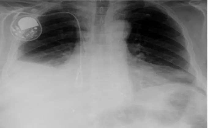
An 83-year-old man without previous respiratory symptoms was admitted at the hospital at December 2014 for evaluation of shortness of breath and weight loss for several months. He is medical history is unremarkable besides previous exposure to asbestos fibers many years before (former carpenter). Thoracic x-ray at admission showed right pleural effusion (fig.1) and CT was ordered for further eval-uation, revealing simple effusion with water density and no pleural thickening or contrast enhance-ment were seen. Lung evaluation showed no abnormalities (fig.2).
Symptomatic treatment with diuretics was carried out and imaging follow-up six months later was de-cided, demonstrating emergence of small pleural calcifications and persistence of right pleural effusion (fig.3). Sequential CT´s revealed rapid progression of the calcifications and development of an aggres-sive soft tissue mass extending to the right hilum and mediastinum (fig. 4, 5 and 6). Transthoracic lung biopsy was performed, showing a moderately cellular neoplasia of spindle to oval cells arranged in trabeculae within a collagenous matrix, with frequent mitotic figures and the immunohistochemistry study revealed positivity for GLUT-1 and negative for
Figure 1 – Initial thoracic x ray at admission on December 2014 shows large right pleural effusion and enlarge cardiac silhouette in a patient with cardiac pacemaker.
all other markers used, namely, WT1, CK5, CK7, CD34, podoplanin and calretinin (fig. 7).
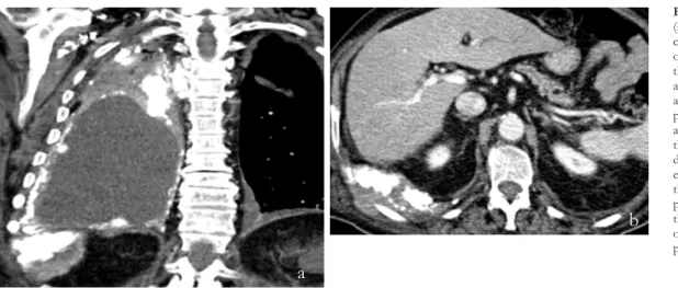
Follow-up CT after 7 months, there was also rib metastasis but no distant metastasis (fig.8). The patient died 2 months later of respiratory failure.
Figure 2 – First thoracic CT two days later. (a) Axial an d (b) coronal images show a large pleural effusion with water density and no pleural wall enhancement or pleural nodules. No pleural calcification is seen. Lung window didn´t show any alterations.
Figure 3 – Follow-up contrast-enhanced thoracic CT at August 2015 (6 months after the initial examination) continue to reveal right water-density pleural effusion and “de novo” small, coarse pleural calci-fications (arrowheads). There is some pleural thickening but no pleural nodules were seen.
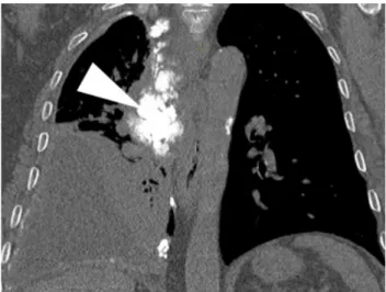
Figure 4 – Coronal non-enhanced thoracic CT at December 2015 shows substantial growth of the calcified mass at the mediastinal pleural (arrowhead), which invades and compresses the mediastinal structures. No pleural nodules or distant metastasis were seen.
Figure 5 – Coronal (a) and axial (b) contrast-enhanced CT at February 2016 shows the calcified mass to continue to grow and become even more ossified, causing collapse of the superior lobe of the ipsilateral lung. The other pleural calcifications also show increase in size and number. Note also left sided small pleural effusion.
Discussion
Pleural malignant mesothelioma is usually diagnosed late in disease course and has a poor prognosis. Clinical presentation of malignant mesothelioma is usually insidious with chest pain, dyspnea, fatigue and weight loss. At physical
a
b
Figure 6 – Sequencial CT images showing the rapid evolution of the pleural cacifications (arrowheads). (a) December 2014; (b) July 2015; (c) December 2015; (d) February 2016. Note that in image (c) there is significant narrowing of the right main bronchus and in (d) there is complete obstruction of the bronchus with lung collapse.
Figure 7 - Histology: moderately cellular neoplasia of spindle to oval cells arranged in trabeculae within a collagenous matrix. Frequent mitotic figures, apoptotic debris and focal areas of chondroid diferentiation were also present.
Immunohistochemistry: the neoplastic cells were diffusely positive for GLUT-1 and negative for all other markers used, namely, WT1, CK5, CK7, CD34, podoplanin and calretinin.
a
b
examination absence of respiratory sounds at auscultation and clubbing can be found but are nonspecific. Laboratory tests may show hypogammaglobulinemia, anemia and thrombocytosis.3 Chest CT is the imaging modality of choice
to diagnose, evaluate the extent of the disease and is often sufficient for staging and treatment planning. Thoracic MRI is not routinely used but can be useful in specific cases to evaluate the extent of the disease. PET can be useful in distin-guishing benign from malignant pleural calcifications and thickening (since malignant lesions have a higher uptake of FDG) and in staging of malignant mesothelioma (with an emerging role in detecting mediastinal node metastases and occult extrathoracic disease). Combined with spatial resolution pro-vided by CT, PET has a promising role in diagnosis, staging and surgery planning of malignant meso-thelioma.6
It is well recognized that the presence of calcified pleural plaques is a common benign finding associ-ated with asbestos exposure.5 On the contrary, typical features worrisome for
malignant mesotheli-oma are unilateral pleural effusion, nodular pleural thickening and interlobar fissure thickening.3,6
The presence of coarse pleural calcification is not always synonym of benign disease and radiologists must be aware of the histological sarcomatoid subtype, which presents with depositions of extensively and densely calcified soft tissue masses on the pleural layers. Unilateral, clumped, irregular, densely calcified pleural masses distributed fairly evenly throughout the substance of the thickened pleura are also signs that should alert for the presence of malignancy.7
Imaging findings in sarcomatoid malignant mesothelioma are similar to other malignant mesothelio-mas but with the presence of coarse calcified masses in the pleura. It is important not to confuse the sarcomatoid type of mesothelioma with development of a non-sarcomatoid mesothelioma that engulfs the preexisting benign calcified pleural plaques.7
Histologically, sarcomatoid mesothelioma is characterized by absence of epithelial elements in the biopsy material or <10% of epithelial tissue and the presence of spindle cells with nuclear atypia.3,4 The list of differential diagnosis of
sarcomatoid pleural mesothelioma includes sarcomas originat-ing in the chest wall, lung, abdominal wall or peritoneum with extension to the pleura, sarcomatoid carcinoma of the lung, metastases from osteosarcoma, breast, thyroid, ovary or thymoma and local-ized pleural mesothelioma (solitary fibrous tumor of pleura).1,5,8,9,10 Primary or secondary pleural 32
involvement by a tumor of the surrounding structures can be difficult to assess, being a crucial step to narrow the differential diagnosis list. Pulmonary neoplasms usually have acute angles with the chest wall, are centered in the lung, and engulf the pulmonary vasculature, whereas a pleural neo-plasm shows obtuse angles with the lateral chest wall and displaces the pulmonary vasculature. On the other hand features that helps differentiating extrapleural neoplasms with invasion of the pleura from primary pleural lesions is that the latter do not cause erosion of ribs and displace the extrapleural fat outward while extrapleural neoplasms displace the extrapleural fat inward.2
Although secondary involvement of pleura is more common than primary tumors, pleural metastases from osteogenic sarcomas are extremely rare and normally occur as lung metastases that migrated to the parietal pleura due to contact.10,11 A known history of a primary tumor producer
of osteoid is an important clue to the diagnosis of secondary involvement.
Solitary fibrous tumor of the pleura (localized pleural mesothelioma) is an uncommon pleural tumor of mesenchymal origin and can be either benign or malignant. There are no characteristic imaging find-ings but usually it appears as a peripheral, smooth, well-demarcated soft-tissue-density mass, often with a pedunculated stalk extending to the extending into the pleura/interlobar fissure/hilum;1,5,12
calcifications and pleural effusion are uncommon but if present can complicate the differ-ential to sarcomatoid malignant mesothelioma. Definite differentiation from malignant mesothelioma is achieved by immunohistochemical staining, in which solitary fibrous tumor is positive for CD34, un-like malignant mesothelioma.5,12
Conclusion
Sarcomatoid mesothelioma is the rarest form of malignant mesothelioma and is characterized by the presence of coarse pleural calcifications. It is important that the radiologist is acquainted with this tu-mor subtype in order to include it in the differential diagnosis list of malignant pleural calcifications and not to confound it with benign pleural calcified plaques.
Figure 8 – Coronal (a) and axial (b) contrast-enhanced CT one month later shows the development of a soft tissue mass at the site of the pleural calcifications at lung base, invading the ribs and direct diaphragmatic extension of the tumor to the peritoneum. Also note the marked thickening of the contralateral pleura.
a
Recebido / Received 16/10/2016 Aceite / Acceptance 18/05/2017 Divulgações Éticas / Ethical disclosures
Conflitos de interesse: Os autores declaram não possuir conflitos de interesse. Conflicts of interest: The authors have no conflicts of interest to declare. Suporte financeiro: O presente trabalho não foi suportado por nenhum
subsídio ou bolsa.
Financing Support: This work has not received any contribution, grant or
scholarship.
Confidencialidade dos dados: Os autores declaram ter seguido os protocolos do
seu centro de trabalho acerca da publicação dos dados de doentes.
Confidentiality of data: The authors declare that they have followed the
protocols of their work center on the publication of data from patients.
Protecção de pessoas e animais: Os autores declaram que os procedimentos
seguidos estavam de acordo com os regulamentos estabelecidos pelos responsáveis da Comissão de Investigação Clínica e Ética e de acordo com a Declaração de Helsínquia da Associação Médica Mundial
Protection of human and animal subjects: The authors declare that the procedures
followed were in accordance with the regulations of the relevant clinical research ethics committee and with those of the Code of Ethics of the World Medical Association (Declaration of Helsinki).
Refereces
1. Inai K. Pathology of mesothelioma. Environmental Health and Preventive Medicine. 2008;13:60-4.
2. Sureka B, Thukral BB, Sinha M. Radiological review of pleural tumors. The Indian Journal of Radiology & Imaging. 2013;23:313-20.
3. Nickell LT, Lichtenberger III JP, Khorashadi L, Abbott GF, Carter BW. Multimodality imaging for characterization, classification, and staging of malignant pleural mesothelioma. RadioGraphics. 2014;34:1692-706. 4. Klebe S, Brownlee NA, Mahar A, Burchette JL, Sporn TA, Vollmer RT, Roggli VL. Sarcomatoid mesothelioma: a clinical–pathologic correlation of 326 cases. Modern Pathology. 2010;23:470-9.
5. Kim KC, Vo HP. Localized malignant pleural sarcomatoid mesothelioma misdiagnosed as benign localized fibrous tumor. Journal of Thoracic Disease. 2016;8:E379-E84.
6. Wang ZJ, Reddy JP, Gotway MB, Higgins CB, Jablons DM, Ramaswamy M, Hawkins RA, Webb WR. Malignant pleural mesothelioma: evaluation with CT, MR imaging, and PET. RadioGraphics. 2004;24:105-19.
7. Mortimer AM, Rowlands J, Murphy P. Coarse pleural calcification in a mesothelioma patient raises the possibility of a rare tumour subtype: osteoblastic sarcomatoid mesothelioma. The British Journal of Radiology. 2011;84:e106-e8.
8. Lucas DR, Pass HI, Madan SK, Adsay NV, Wali A, Tabaczka P, Lonardo F. Sarcomatoid mesothelioma and its histological mimics: a comparative immunohistochemical study. Histopathology. 2003;42:270-9.
9. Verbeke N, Verstraete K, Sys G, Forsyth R, Kluyskens D, Denys H, Uyttendaele D, Rottey S. Osteosarcoma with extensive calcified pleural metastases at diagnosis. Acta Clin Belg. 2008;63:325-8.
10. Mori T, Yoshioka M, Iwatani K, Kobayashi H, Yoshimoto K, Nomori H. Kissing pleural metastases from metastatic osteosarcoma of the lung. Ann Thorac Cardiovasc Surg. 2006;12:129-32.
11. Saha D, Saha K, Banerjee A, Jash D. Osteosarcoma relapse as pleural metastasis. South Asian Journal of Cancer. 2013;2:56.
12. Ginat DT, Bokhari A, Bhatt S, Dogra V. Imaging features of solitary fibrous tumors. American Journal of Roentgenology. 2011;196:487-95.